Starfish Dissection Guide - Mr.E Science
Starfish Dissection Guide - Mr.E Science
Starfish Dissection Guide - Mr.E Science

You also want an ePaper? Increase the reach of your titles
YUMPU automatically turns print PDFs into web optimized ePapers that Google loves.
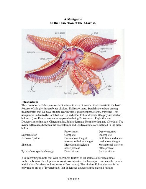
A Miniguideto the <strong>Dissection</strong> of the <strong>Starfish</strong>Introduction:The common starfish is an excellent animal to dissect in order to demonstrate the basicfeatures of a higher invertebrate phylum, Echinodermata. <strong>Starfish</strong> are unique amonginvertebrates that we have studied (earthworms, grasshoppers, clams, crayfish). Thisuniqueness is due to the fact that starfish and other Echinodermata (the phylum starfishbelong to) are Deuterostomes as opposed to being Protostomes. Phyla that areDeuterostomes include: Chaetognatha, Echinodermata, Hemichordata and Chordata. Themajor differences between the Protostomes and Deuterostomes are outlined in the tablebelow.ProtostomesDeuterostomesSegmentation Complete IncompleteNervous System Brain above the gut, Both brain and nervenerve cord below the gut cord above the gutSkeleton Mesodermal skeleton Mesodermal skeletonnever presentoften presentType of embryonic cleavage Determinate IndeterminateIt is interesting to note that well over three-fourths of all animals are Protostomes.In the embryonic development of most invertebrates, the blastopore becomes the mouthwhich classifies them as Protostomia (first mouth). The phylum Echinodermata is theonly major group of invertebrates that undergoes deuterostomic (second mouth)Page 1 of 5
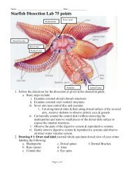
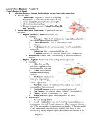
development, in that the embryonic blastopore becomes the anus and the "second mouth"forms as a new opening. This embryological evidence indicates that the phyllumChordata (subphyla: Cephalochordata, Urochordata and vertebrate) evolved from theEchinodermata, as this is the only invertebrate phylum with deuterostomic development.The phylum Echinodermata is composed of the most familiar marine animals and isdistinguished by (1)pentamerous radial symmetry, (2) a calcareous endoskeleton withprojecting spines, and also (3) a system of coelomic canals and tube feet (the watervascularsystem) that is used in slow locomotion and manipulation of food. Possession ofa system of coelomic canals differentiates the Echinoderms from their earlier acoelomicancestors, the flatworms (phylum Platyhelminthes). The echinoderms are divided intotwo subphyla. The subphyllum Pelmatozoa contains one class, Crinoidea (sea lilies). Thesecond subphyllum, Eleutherozoa, contains four classes: Ophiuroidea (brittle stars),Holothuroidea (sea cucumbers), Echinoidea (sea urchins and sand dollars), andAsteroidea (starfish).The unifying features of the class Asteroidea include 1) a star-shaped body with multiplesof five radially symmetrical arms that are not sharply set off from the central disk and 2)the water-vascular system. The starfish utilized in this dissection exercise is of the order:Forcipulata, family: Asteriidae, and genus: Asterias or Pisaster.About 1700 species of starfish have been described living in marine coastal waters fromthe north to the south poles. An interesting characteristic of Asteroids is the ability tocompletely regenerate a whole organism from only one fifth of the central disk that has atleast one arm remaining attached. Regeneration is slow, requiring up to a year forcomplete reformation.Attention to the details of the dissection procedure along with reading each section of thismanual before you proceed will help you to avoid damaging the starfish specimen. Inorder to perform the dissection the following tools are recommended:1. <strong>Dissection</strong> scissors2. Forceps3. Scalpel4. Dissecting needleIn order to keep your specimen moist during dissection, we recommend that the specimenbe sprayed with Delta-sol, a non-toxic odorless liquid that preserves the tissue and is nonirritatingto the skin; see our catalog for details.External FeaturesThe opening of the water-vascular system, the madreporite, is a large button-likestructure that is located off center on the disc between two arms. The inconspicuous anusis in the center of the disk, and both structures are located on the upper, dorsal or aboralsurface of the starfish.Page 2 of 5
aglike cardiac stomach, which protrudes through the mouth while feeding. The evertedstomach engulfs the prey and can insert itself into a slit in a shellfish only 0.1 mm wide.Digestion can begin outside the body until the cardiac stomach is retracted by five pairsof retractor muscles, one pair in each arm. Each of the structures mentioned here can beidentified in Figure 1.Reproductive: <strong>Starfish</strong> are dioecious, meaning the sexes are separate, though thelocation and number of gonads is the same in both sexes. Remove the digestive system bylifting the pyloric ceca from the arms and by cutting through the 10 retractor muscles tothe stomach, two from each arm. Now snip out the stomach at its opening to the oralsurface, the mouth. This will reveal pairs of gonads in each arm, lying under the hepaticcaeca, alongside the ambulacral groove, and yet close to the disk (Figures 1,3).The size of the gonads depends upon the phase of the breeding cycle at the time thespecimen was collected. The gonads are branched lobulaled sacs that open to the exteriorby small genital pores located on the periphery of the aboral disk (Figure 1). Eggs andsperm are released into the sea where fertilization takes place. Because the starfish canregenerate individuals from fragments, and thus increase the population, regeneration is aform of asexual reproduction.Water-vascular: The water-vascular system is an internal water pressure system. Waterenters the system through the sievelike madreporite in the aboral surface and passesthrough the stone canal into the ring canal which encircles the mouth. Nine sacs,Tiedemann's bodies, are located on the inner edges of the ring canal. These sacs are thebreeding grounds of amoeboid cells that are found in the fluid of the water-vascularsystem. Find these 3 structures in Figure 4 and in your specimen. Extending out from thering canal are five radial canals, one into each arm. Lying in the groove of the dorsal sideof the ambulacral ossicles is the radial canal. It is best seen by applying slight pressure toseparate the ossicles so that one can see into the groove, or in cross section of a severedarm. At regular intervals lateral canals branch off the radial canal and lead to muscularbulbs, ampulla, as well as opening to the tube feet (Figures 1,2,3). The ampullae areobvious rows of buff-colored sacs along both sides of the ambulacral ossicles. Continuedissecting the ambulacral ossicles of an arm to locate the lateral canals that lead to theampulla. The tube feet are about 1 cm long, and at the tip of each is a sucker. Theampulla contracts and water is forced into the tube foot, extending it and enabling it toattach to the substratum with its sucker. The muscles of the foot then contract and forcewater back into the ampulla, thus shortening the foot. Valves and reservoirs within thewater-vascular system control the internal pressures that operate the tube feet for stowlocomotion and remarkable, prolonged sucking action that opens oysters and clams forfood. The starfish is therefore considered to be a problem to the shellfish industry.Nervous: Nerves coordinate the arm movements for directional locomotion of thestarfish. The nerves are difficult to discern during dissection, so we will discuss themonly briefly. There is an oral nerve ring around the mouth from which extends five radialnerves along the ventral side of the ambulacral grooves, one into each arm. The armmovement is coordinated by ganglion cells located at the junction of each radial nervePage 4 of 5
and the oral nerve ring. Other local nerve centers control the stomach, tube feet andampulla. A network of nerves in the body wall controls the structures on the externalsurface of the starfish. These structures are not noted in the figures.There is a non functional blood-vascular or hemal system present in starfish. Circulationof coelomic fluids is accomplished by the action of cilia that line the body cavity. Theentire hemal system consists of an axial sinus located near the stone canal and a hemalring with branching radial hemal canals into the arms. These structures are not noted inthe figures.Natural History.<strong>Starfish</strong> are mainly coastal animals, being most numerous in tropical waters associatedwith coral reefs. Some species have been collected at ocean depths of up to 6000 meters.<strong>Starfish</strong> range in color from yellow and orange to red, blue and brown. The number ofarms may reach as high as 50 in some species.<strong>Starfish</strong> are predaceous, feeding on any animal inhabitant of the seashore waters that thestarfish can swallow or engulf such as mollusks, worms, Crustacea, fish and otherechinoderms. The pull of the tube feet is used to open clams and oysters and then the preyis engulfed by the protruded stomach. <strong>Starfish</strong> may be pests to the shellfish industry, butthey are treasures of the sea to the beachcomber.Page 5 of 5