Multimodal Preclinical Imaging Solutions
Multimodal Preclinical Imaging Solutions
Multimodal Preclinical Imaging Solutions

You also want an ePaper? Increase the reach of your titles
YUMPU automatically turns print PDFs into web optimized ePapers that Google loves.
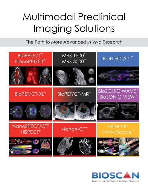
<strong>Multimodal</strong> <strong>Preclinical</strong><strong>Imaging</strong> <strong>Solutions</strong>The Path to More Advanced In Vivo ResearchBioPET/CT NanoPET/CT ®MRS 1500 MRS 3000 BioFLECT/CT BioPET/CT-XL BioPET/CT-MR BioSONIC WAVE BioSONIC VIEW NanoSPECT/CT ®HiSPECT ® NanoX-CT Minerve ®InVivoScope <strong>Multimodal</strong> <strong>Preclinical</strong> <strong>Imaging</strong> <strong>Solutions</strong>
Uncompromising PerformanceDistinctive imaging systems that work in concertBioscan provides differentiated, multimodal imaging solutions designed for the preclinical market:• Sub-mm PET, SPECT and X-Ray CT• True 360° Optical tomography• Translational MRI and PET/MRI• Feature-rich, translational Ultrasound• Common platforms for Animal Handling & Image AnalysisFeatures & Benefits:• Coregistration of multiple modalities made easy - All systems use the same post-processing softwareand animal handling equipment.• Efficiency in your daily research - Modalities can operate simultaneously, so there are no limitationson research workflow.• Truly translational - Transfer preclinical study protocols directly to the clinic.• Wide range of animal models - Accommodates animals from mice to rabbits and monkeys.• Compact design, small footprint - Bioscan multimodality solutions will fit in imaging facilities as smallas 9m 2 . The systems are internally shielded and can be installed side-by-side or even back-to-back.• Upgradable platforms - As your research needs change, upgrade your Bioscan systems to addthroughput and new capabilities.<strong>Multimodal</strong> imaging...BioPET/CTBioPET/CT-XLNanoPET/CTMRS 1.5T &3.0TBioSONICInVivoScopeMinerveNanoSPECT/CTNanoX-CTBioFLECT/CT...One common animal handling platform and analysis software2
Bioscan <strong>Multimodal</strong>ity CombinationsSuperior alternatives to the standard trimodality solutionResearchers interested in pursuing both PET and SPECT imaging often consider purchasing a trimodalitysystem that combines PET, SPECT and X-Ray CT in a single cabinet. Bioscan has developed solutions for thesecustomers that rely on two or more discrete systems, because this approach offers significant advantages forsystem performance, research efficiency and workflow.Efficiency Without Performance Compromises• Better efficiency - Dual systems (e.g., PET/CT and SPECT/CT) enable acquisitions to be completed simultaneously,increasing throughput and improving study workflow• No performance compromises - Achieve industry-leadingperformance with each of the modalities you purchase• Small footprint - Two Bioscan systems can fit in less than 9m2,less than many trimodality systems• Upgradable - Bioscan’s modular architecture can enableyou to upgrade your systems to add new modalities or addthroughputF-18 and Tc-99mF-18 (FDG):Heart andBladder (Pink)Tc-99m: Thyroidand Stomach(Green)Research ApplicationsSmall animal imaging - Keys to successMolecular imaging has become a widely-established procedure for diagnosing various disease states andmonitoring the effects of therapeutic treatments in humans. In recent years, molecular imaging has alsobecome a valuable preclinical research tool for studying the development and progression of disease inmouse and rat models, as well as the efficacy of potential therapeutic treatments. Dedicated small animalimaging systems, which include MRI, PET, SPECT, Fluorescence/Bioluminescence, X-Ray CT, and Ultrasound,allow for the characterization of biological, molecular and cellular processes in vivo, with exceptional levels ofresolution and sensitivity.OncologyCNSCardiologyDisease BiologyADME/Tox• Tumor biology• Metastasis• Therapeutic effect• Receptor density• Occupancy studies• Functional studies• Stem cell repair• Metabolic function• Gene expression• Cell trafficking• Biodistribution• Time activity curves(TAC)Performance• Sensitivity• Resolution• UniformityFunctionality• Flexibility• Anatomical localization• Absolute quantificationProductivity• Ease of use• Quality Assurance• ThroughputCost• Standard Configurations• Optional features• Serviceability3
BioPET/CT Small animal PET with sub-mm resolution throughout the FOVBioscan offers next-generation LYSO/GSO phoswich technology featuring true Depth-Of-Interaction (tDOI) withfour lines of response, eliminating the degradation of resolution away from the center of the imaging chamberthat results from PET detector parallax effects. The parallax effect is mitigated using information captured asgammas pass through the double crystal layers, enabling sub-millimeter PET resolution even at the edge of thefield of view. With its thick, dual-crystal layer (15mm: 7mm LYSO + 8mm GSO), the BioPET/CT also provides thehighest sensitivity per axial cm in the industry.Technical specificationsPET detector specifications:Crystals: Double-layer 15mm LYSO/GSOResolution: 0.8mmDynamic axial FOV: 180mmAxial FOV: 47mmTransaxial FOV: 68mmBore size: 80mmAbs center point sensitivity @350-700 keV: 3.1%Timing resolution: 1.5nsReconstruction time: FBP ≤ 10 seconds,2DOSEM ≤ 2 minutes, 3DOSEM 40kHz (NEMA NU2 2007)CT specifications:CT detector: 8x1.25 mm detectorMaximum technique: 140kV 7mADetector channels: 3264Max rotational speed: 60 RPMViews/second: 1440Resolution: 0.65mmAnimals:Rabbits, pigs, cats, dogs, monkeys, primatebrain imagingAcquisition:Static & Moving PET acquisitionList mode acquisitionRebinning: Single slice rebinning(SSRB), Fourier rebinning (FORE)DOI native modeCardiac & respiratory gating(optional, available 2013)Reconstruction:Filtered Back Projection (FBP)2D MLEM/OSEM3D MLEM/OSEM (available 2013)Portability:Powered transport wheels (optional)Self-shielded CTHuman BrainF-18 AV-133Human BrainF-18 AV-454
NanoPET/CT ®Sub-mm translational PET imagingNanoPET/CT uses twelve detector arrays of 81 x 39 LYSO crystals of 1.12 x 1.12 x 13mm 3 at a packing fractionof 92% to ensure PET performance with exceptional spatial resolution. This cutting-edge detector technologyis further complemented by a state-of-the-art data acquisition and processing architecture with 12-bithigh-speed digital to analog converters that fully digitize the detector signals. The NanoPET/CT’s advanceddetection technology and large field of view provide very high absolute sensitivity, together with the flexibilityto image mice, rats and larger animals such as rabbits and monkeys.Technical specificationsPET detector specifications:Crystals: 13mm LYSOResolution: 0.8mmDynamic axial FOV: 300mmAxial FOV: 95mmTransaxial FOV: 123mmBore size: 160mmAbs center point sensitivity @250-750 kev: 8.3%Timing resolution: 1.5nsReconstruction: Fast 3DOSEM reconstructionCT specifications:Dynamic field of view: 80 x 300mmDetector active area: 50 x 175mmPixel size: 11, 34, 100µmFocal spot: 9µmX-Ray peak energy: 35-80kv (Variable)Max current/power: 180µA/ 8WattsGPU-based, real-time CT reconstructionAnimals:Mice, rats, marmosets and rabbits 3kgHigh-spatialresolution systemMouse thyroid(F18-NIS)Dynamic & biodistribution studies -Whole body mouse & ratsRat Brain(FDG)NanoSPECT/CT ® & HiSPECT ®The gold standard multi-pinhole system in preclinical SPECTBioscan’s NanoSPECT/CT combines up to four large, proprietary Broadband Gamma Ray Detectors withpatented Multiplexed Multi-Pinhole SPECT (MMP-SPECT) to eliminate many of the sensitivity versus resolutiontrade-offs associated with traditional SPECT systems. Unlike any other preclinical SPECT system, Bioscan’sNanoSPECT/CT offers high-resolution and high-efficiency detection with a large field of view. As a result,the system can perform fast and dynamic whole-body scans in a single-step procedure, with high isotropicresolution. The NanoSPECT’s reconstruction software and aperture plates are available separately as theHiSPECT ® upgrade to clinical gamma cameras.Technical specificationsSPECT Detector specifications:Detectors: Na(Tl)-SpectroscopyFrom 25Kev to 365KeVResolution: from 350µm (whole body)Number of pinholes: up to 64Stationary, semi-stationary & rotationalimagingDynamic Axial FOV: 270mmTransaxial FOV: up to 230mm (for 5 kgrabbits)Bore size: 215mm x 230mmCT specificationDynamic Field of view: 80 x 300mmDetector Active Area: 100 x 100mmPixel Size: 50µmFocal Spot: 9µmX-Ray Peak Energy: 35-80kv (Variable)Max Current/power: 180µA/ 8 WattsMulti-isotope imaginge.g. 4 isotopes & CTFast Dynamic and Kinetic SPECT (4D-SPECT)Cardiac and respiratory gatingAnimals:Mice, rats, marmosets, rabbits (3kg) &rabbits (5kg)5Tc-99m, I-123, In-111and Tl-201, plus X-rayCTNanoparticle imaging with nano-liter precisionand pico-molar sensitivity
MRS 1500 & MRS 3000 Cryogen-free, superconducting MRI at 1.5T & 3.0TOur preclinical MRI systems, the MRS 1500 & MRS 3000, are state-of-the-art, cost-effective translationalMRI instruments with low running costs and no special site requirements. The innovative, cryogen-freesuperconducting magnets virtually have no fringe field, enabling safe use of the system in any facility andby any operator. Designed to complement Bioscan’s other molecular imaging modalities, these MRI systemsdeliver powerful performance and ease-of-use. The systems feature a small footprint and can be placed within2m of another imaging system. MRS instruments are in use worldwide for a wide range of life sciences researchapplications including, functional, molecular, anatomical and multi-modality imaging.Technical specificationsMagnetMagnet type: Cryogen-free, superconductingField strength: 1.5T and 3.0TFOV: 70mm DSVHomogeneity: DSV 30mm +/- 0.10ppmDSV 60mm +/- 1.0ppmClear bore diameter: 160mmExample Pulse SequencesSpin Echo Based SequencesFast SequencesEPI SequencesGradient Echo Based SequencesMagnetic Resonance Angiography2D & 3D Time of Flight sequenceBulk Spectroscopy SequencesIVS SequencesSTEAM spectroscopy sequencePoint Resolved Spectroscopy SequenceFat/water suppression sequenceISIS sequenceAnimals:Mice and RatsMouse Sagittal Image; Spin Echo TE 36ms,Resolution 93 x 93 µ x 1.0mm 1.5T (singleloop surface coil)Mouse Whole Body Image; Spin Echo TR 1200ms,FOV 65mm, THK 1.2mm, 3 averages, Scan Time7m 50s, 130x256, TE = 20 msBioPET/CT-MR Translational multimodality PET/CT/MRI (1.5T/3.0T)Bioscan’s entry into the PET/MR imaging segment combines superior PET performance with translational MRI at1.5T and 3.0T field strengths. Coupling the PET and MRI modalities while retaining superior imaging performanceis a significant technical challenge: As the two modalities are brought into closer proximity, the magnetic fieldinterferes with the PET detectors, creating a trade-off between field strength and PET resolution. To address thechallenge, Bioscan has developed a three-phase approach* to preclinical PET/MRI development, moving fromsequential imaging to integrated inline imaging and finally to simultaneous/dynamic imaging.Phase 1 (available today): Side-By-Side PET/CT and MRISequential imaging with the BioPET/CT positioned next to the MRS 1500 or 3000 system.Sub-mm PET performance, high-resolution, high-contrast MRI at translational fieldstrengths, seamless integration with PET-CT-MRI compatible animal beds andautomated fusion of PET, CT and MRI images with Bioscan’s InVivoScope software. Thisenables both parallel and sequential imaging with the PET, CT and MRI modalities.Phase 2 (Work-In-Progress): Integrated In-line PET/MRIIn-line PET/MRI system similar in configuration to current preclinical PET/CT systems.This phase will combine magnetic field-tolerant PET detection technology with abenchtop cryogen-free superconducting MRI system of minimum 3.0T.Phase 3 (Work-In-Progress): Simultaneous PET/MRINew approach to functional preclinical PET/MRI imaging providing sub-mm PETresolution at fMRI field strengths. Enabled by combining the tDOI technologyfrom Bioscan’s BioPET detectors with Digital Photon Counting (PDPC**) powered PETdetection.* Work supported by IMAPPI (ANR - France)** PDPC= Philips Digital Photon Counting and Digital SiPM Technology (Philips Corporate Technologies, Koninklijke Philips Electronics N.V.)SHAMProtocol Details: 208 g Wistar RatLDA Ligation- chronic infarct SHAM vs. MICardiac Gating: 30 min 18F-FDG uptake25 min acquistion Dose: 1 mCiDigital SiPM: the key todynamic PET/MR imagingMI6
BioFLECT/CT True 360° optical tomography with optional integrated CTWith the launch of BioFLECT, Bioscan enables researchers to realize the full potential of in vivo optical imaging.With BioFLECT’s true 360° tomographic capabilities, researchers will no longer have to choose betweenimaging only near the surface of the subject or partial tomography solutions that distort the subject or createartifacts. BioFLECT provides uniform sensitivity and resolution throughout the subject volume. For the first time,researchers will be able to examine deep tissue phenomena such as spontaneous tumors and metastaseswithout sacrificing image quality or quantitative accuracy. With the optional upgrade to integrated, one-passX-Ray CT, researchers will gain anatomical correlation data that will significantly improve the segmentation andresolution of the optical image.Technical specifications<strong>Imaging</strong> performance:Maximum resolution: ~1mm 3Sensitivity: 800fM/mm 3Uniform resolution and sensitivity throughoutFOVField-of-view:Axial FOV: 100mm (mice), 120mm (rats)Transaxial FOV: 38mm (mice), 42mm (rats)Bore size: 45 mmCT specificationDynamic Field of view: 50 x 150 mmDetector Active Area: 115 x 65 mmPixel Size: 75, 150, 300 µmFocal Spot: 18 µmX-Ray Peak Energy: 0-50 kv (Variable)Max Current/power: 800µA/50 WattsUniform resolution andsensitivity throughout thesubject<strong>Multimodal</strong>ity imaging(BioFLECT/NanoSPECT/X-Ray CT)<strong>Multimodal</strong>ity imaging(BioFLECT/NanoX-CT)Animals:Mice and ratsBioSONIC WAVE & VIEW Feature-rich preclinical ultrasound with customizable touchscreen interfaceThe BioSONIC product line offers researchers access to advanced high-frequency ultrasound imagingcapabilities in a package appropriate for today’s challenging funding environment. These compact,economical systems are fully customizable to support imaging techniques such as harmonic & compoundimaging, plane-wave imaging, panoramic imaging, free-hand 3D imaging, 3D GPS-guided injection & biopsy,and photoacoustics. The systems’ flexibility enables imaging of mice as well as larger animals with a single,portable system. Because BioSONIC systems are part of the Bioscan family, users can also gain the advantageof effortless co-registration of 3D ultrasound data with data from other imaging modalities via InVivoScope.Technical Specifications<strong>Imaging</strong> Modes:RF collectionB-ModeM-Mode: Regular, Anatomical, ColorDoppler: Color, Power, Pulsed Wave, ContinuousWave, Tissue - Triplex3D freehand registration4D with mechanical transducerContrast imaging - Harmonics & Pulse InversionElastographyPanoramic imagingTransducers:Bandwidth/FOV: 14-5 MHz/38mmBandwidth/FOV: 40-8 MHz/12mmTransducer ports: 3(Wave) 2(View)Data Export:DICOM compatible, supports export to DICOM serversand PACS applications7Transmit:Amplitude Maximum: +/- 47VChannels: 128Beamforming: Programmable delays, steeringPulse length: 96 bits@ 80MHz or 40MhzReceive:ADC: 14bitsChannels: 64Sampling Rate: 40MHzRF Sampling Max (DAQ): 80MHzBeamforming: Programmable delays, steering,dynamic focusing, apodizationPost-Processing Features:Speckle reductionTGC, compression, mapForm Factor:Footprint: 26” x 20” (Wave)18” x 7” (View)MousebreasttumorMousebladderMousekidney
NanoX-CT High resolution, high contrast CT upgradable to PET or SPECTBioscan’s NanoX-CT is upgradable to a NanoPET/CT or a NanoSPECT/CT system, depending on the initiallyselected configuration. The system acquires X-Ray CT images using helical scanning with volumetric conebeamdetection. The NanoX-CT uses a micro-focus source with variable magnification. The combination ofthis source with a large bore and detector allow the user to optimize resolution of CT images. Voxel sizes rangefrom 11-34 microns (
InVivoScope Version 2.0<strong>Multimodal</strong>ity image processing softwareBioscan’s InVivoScope (IVS) image processing software features the powerful fully 3D ROI tool developed bysoftware partner inviCRO. You will be able to classify each individual voxel of a three-dimensional volumedataset. IVS supports segmentation efforts with an easy-to-use, powerful toolbox for manual, semi-automaticand fully automatic segmentation. IVS fully supports multimodal and dynamic data. You only have to do thesegmentation for a fixed ROI once and it will be evaluated for all loaded datasets automatically. InVivoScopesupports the import and export of DICOM 3.0 compatible data, as well as providing raw and native imageformats (some with full DICOM conversion). Up to 3 modalities, such as Optical, SPECT, CT, PET or MR, can bedisplayed at any time, while even more data can be co-registered, analyzed and modified simultaneously.One software - Five modalities: PET-SPECT-CT-OPTICAL-MRAutomatic SegmentationMulti-planarreconstructionComprehensive tools for post-processingSoftware AccessoriesSoftware packages to amplify your dataBioscan offers a spectrum of software accessories and modules to expand your data management,quantification and analysis. Featured accessories include:FlowQuant © - A complete cardiac SPECT & PET image analysis packageIVS: Brain Atlas & Quantification Module - Mouse and rat brain modelsIVS: 3D Segmentation - Powerful 3D ROI tooliPACS © - Pre-Clinical <strong>Imaging</strong> Study ManagementImalytics - Advanced image analysis, quantification, and visualizationFlowQuant- Tracer kineticmodeling- Myocardial BloodFlow (MBF) analysis- Myocardial ViabilityAnalysis- Cardiac functionusing gated imageanalysisIVS: Brain Atlas &Quantification- Single-click coregistrationroutine- Single-click atlasdeformation routine- Spreadsheet readoutof radioactivityIVS: 3DSegmentation- Classify eachindividual voxel of 3Dvolume dataset- Powerful toolbox forsegmentation.- IVS supports 3D ROItooliPACS- Divide data inmeaningful domains- Set domain accesspolicies- Stores other fileformats, with versioncontrol & electronicsignaturesImalytics- Advancedfunctionality fortranslational research- Specializedapplications forpharmacokineticmodeling, dosimetryand dementia analysis9
Service & SupportGoing beyond products... providing solutionsTechnical Services &SupportField ApplicationsSupportBioscan’s technical services and support teamprovides exceptional support and service toBioscan’s diverse portfolio of small animal, invivo preclinical imaging products: BioFLECT/CT,BioSONIC, BioPET/CT, BioPET/CT-XL, NanoPET/CT,NanoX-CT, NanoSPECT/CT and the MRS 1500 &3000.These imaging products are sophisticated,precision instruments. In order to keep theseinstruments operating at their peak productivitylevels, our global network of factory-trained FieldService Engineers (FSEs) and Field ApplicationsScientists (FASs) are available to assist you withinstallation, preventative maintenance, calibration,technical guidance, and emergency repairservices.We provide fast and knowledgeable support toensure your system uptime and performance ismaximized. We also understand that our customershave their own unique requirements for service,which is why we offer a variety of service levelagreements. You can select the level of coveragethat best suits your needs.Bioscan’s support teams are committed toproviding the highest level of onsite and remotesupport in the industry with our expert team ofhighly trained and courteous staff.Bioscan’s Field Applications Scientist (FAS) Teamdelivers technical education and training forBioscan’s diverse portfolio of small animal, invivo preclinical imaging products. These trainingsessions are intended to educate the customer inthe expert operation of their new equipment, inorder for them to generate reliable, reproducibleand quantitative results to solve their researchqueries of their biological models.Our onsite training courses, with proven methodsfor training and delivery, are guaranteed toreward the end-user with a successful educationalexperience. Each training session is designedto enable the user to expertly operate his/herpreclinical imaging system. Bioscan has severalon-site training and applications optimizationofferings with the purchase of any new system. Inthe event that any customer requires more handsontraining and/or specialized customization ofacquisition protocols, Bioscan offers additionalcustomized onsite training packages ranging from1 to 3 days. These flexible training sessions areintended to be performed at the customer site. Thetraining content is developed specifically with thecustomer to help customize the training to supportspecific areas or applications of interest. If smallertime frames are required, the FAS Team also offershourly remote training sessions using TeamViewersoftware.Bioscan’s FAS Team is comprised of highly trainedand experienced research scientists focused oneducation excellence. Our goal is to exceedcustomer expectations and satisfaction bydelivering exceptional quality training that ismodality specific and has a measurable impact onresearch.FASTeamCustomersSalesTeamServiceTeamMarketingR&D10
<strong>Multimodal</strong> <strong>Preclinical</strong> <strong>Imaging</strong> <strong>Solutions</strong>Corporate Headquarters4590 MacArthur Blvd, NWWashington, DC, 20007 USATF: 1-800-255-7226Tel: 1-202-338-0974Email: info@bioscan.comBioscan Europe, Ltd.58 Avenue de WagramParis, France 75017Tel: +33 3 83 63 62 67Email: europe@bioscan.comWeb:www.bioscan.comwww.preclinical-imaging.comNanoPET®, NanoPET/CT®, NanoSPECT®, NanoSPECT/CT®, and HiSPECT® are registered trademarks of Bioscan Inc. Minerve is a registered trademark of Minerve SA. iPACS is a registered trademark ofinviCRO llc, Imalytics is a registered trademark of Philips, FlowQuant is copyright Ottawa Heart Institute. InVivoScope, MRS 1500, MRS 3000, BioPET/CT, BioPET/CT-MR, BioPET/CT-XL, BioCT, BioFLECT/CT, BioSONIC, BioSONIC WAVE, BioSONIC VIEW, NanoX-CT, Bioscan Inc are trademarks of Bioscan Inc. MMP-SPECT is a trademark of Bioscan Inc. under exclusive license from Scivis GmbH.BioPET/CT is designed and manufactured by Sedecal. BioPET/CT-XL is designed and manufactured by PDSI, Inc. NanoPET/CT is designed and manufactured by Mediso, Ltd. NanoSPECT/CT is manufactured byMediso, Ltd. NanoX-CT is manufactured by Mediso, Ltd.. © 2012 Bioscan Inc. All rights reserved.Rev-AK-08/18/2012BioPET/CT patents and applications: ES 2301303, EP 1941835, US 2009213983 and US 20090065699 MMP-SPECT patents: US 7,498,580 - EP 1421411 -US 7,199,371BioFLECT/CT Patents: US 5,952,664; 6,029,077; 6,044,288; 6,100,520; 6,130,958; 6,150,649; 6,211,512; 6,331,700; 6,681,130; 6,693,287; 6,693,287; 7,155,274; 7,212,848; Plus international equivalents