Hepatobiliary scan in Alagille syndrome - Hellenic Society of ...
Hepatobiliary scan in Alagille syndrome - Hellenic Society of ...
Hepatobiliary scan in Alagille syndrome - Hellenic Society of ...

You also want an ePaper? Increase the reach of your titles
YUMPU automatically turns print PDFs into web optimized ePapers that Google loves.
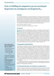
Case Reporttic right kidney. Ur<strong>in</strong>ary tract <strong>in</strong>fection (UTI) was diagnosed.The diagnostic work up also <strong>in</strong>cluded: magnetic resonancecholangio-pancreatography (MRCP), heart ultrasound, ophthalmologicalexam<strong>in</strong>ation and liver biopsy.The <strong>in</strong>fant was successfully treated with amikac<strong>in</strong> <strong>in</strong>travenously(i.v.) for ten days. Subsequent biochemistry showed thattransam<strong>in</strong>ases were persistently elevated: AST131-184U/L,ALT78-106UI/L and also hyperbilirub<strong>in</strong>aemia, ma<strong>in</strong>ly due to <strong>in</strong>creasedconjugated bilirub<strong>in</strong> between 6.8-8.1mg/dL (normalFigure 1. Static renal sc<strong>in</strong>tigraphy with 99m Tc-DMSA. Dysplastic rightkidney.values
Case Reportby the heart ultrasound. c) Embryotoxon found on slit lampopthalmological exam<strong>in</strong>ation. d) Dysplastic right kidney, bythe U/S and by 99m Tc-DMSA renal sc<strong>in</strong>tigraphy.The above fulfilled the criteria for the diagnosis <strong>of</strong> <strong>Alagille</strong><strong>syndrome</strong> with 4 major features and 1 m<strong>in</strong>or.The <strong>in</strong>fant was started on supplementation with lipid solublevitam<strong>in</strong>s (A, D, E, K) and treatment with cholestyram<strong>in</strong>eand ursodeoxycholic acid for cholestasis and cefaclor forprophylaxis aga<strong>in</strong>st UTI and followed up regularly by pediatricgastroenterologists. The child is still alive, with deteriorat<strong>in</strong>gliver function and <strong>in</strong> the list for liver transplantation.Discussion<strong>Alagille</strong> <strong>syndrome</strong> is an autosomal dom<strong>in</strong>ant disorder. Thedisease gene has been mapped at chromosome 20-band p12with a deletion <strong>of</strong> the short arm <strong>of</strong> chromosome 20 [7-9]. Thecl<strong>in</strong>ical spectrum <strong>of</strong> this <strong>syndrome</strong> <strong>in</strong>cludes: a) Chroniccholestasis due to <strong>in</strong>trahepatic bile duct hypoplasia. b) Characteristicfacies consisted <strong>of</strong> prom<strong>in</strong>ent forehead, moderatehypertelorism with deep-set eyes, small ch<strong>in</strong> po<strong>in</strong>ted anteriorlyand saddle or straight nose. c) Cardiovascular abnormalitieswith most common pulmonary artery stenosis. d) Vertebralarch defects with most common the non-fusion <strong>of</strong> theanterior arches <strong>of</strong> one or more dorsal vertebrae result<strong>in</strong>g abutterfly-like appearance. e) Posterior occular embryotoxon.f) Less associated abnormalities that are considered as m<strong>in</strong>orcriteria for diagnosis are, growth retardation, mental retardation,renal abnormalities, skeletal abnormalities and highpitchedvoice.Our patient had cholestasis due to paucity <strong>of</strong> <strong>in</strong>tralobularbile ducts, characteristic facies, pulmonary artery stenosis,embryotoxon and dysplastic right kidney, consistent with acomplete form <strong>of</strong> the disease.Recent reports <strong>in</strong>dicate that patients with this <strong>syndrome</strong>are at risk for serious cl<strong>in</strong>ical problems, <strong>in</strong>clud<strong>in</strong>g heart failure,liver failure and hepatocellular carc<strong>in</strong>oma [10-12]. Early diagnosis<strong>of</strong> the disease and appropriate follow up are important.It is imperative to differentiate surgically correctable lesionsfrom paucity <strong>of</strong> <strong>in</strong>terlobular bile ducts (like <strong>in</strong> Allagile <strong>syndrome</strong>).<strong>Hepatobiliary</strong> <strong>scan</strong>n<strong>in</strong>g is a useful diagnostic method fordifferentiat<strong>in</strong>g cholestatic jaundice <strong>in</strong> neonates, as non dra<strong>in</strong><strong>in</strong>g<strong>scan</strong>s may <strong>in</strong>dicate either severe neonatal hepatitis, or <strong>in</strong>terlobularbile duct paucity [13-15]. In patients with non dra<strong>in</strong><strong>in</strong>g<strong>scan</strong>s and possible Allagile <strong>syndrome</strong>, liver biopsy willconfirm or exclude cholestasis due to paucity <strong>of</strong> <strong>in</strong>trahepaticbile ducts.The prognosis <strong>of</strong> the disease is related to the severity andduration <strong>of</strong> cholestasis, the severity <strong>of</strong> cardiovascular abnormalitiesand the deterioration <strong>of</strong> liver function.Treatment consists <strong>of</strong> nutritional supplementation andantipruritic treatment [10]. When cirrhosis becomes uncompensated,liver transplantation is the treatment <strong>of</strong> choice [7,16, 17]. When the <strong>syndrome</strong> is complicated by liver tumour,transplantation is less likely to succeed and most patients diewith<strong>in</strong> 3 years as a result <strong>of</strong> tumour recurrence [16].In conclusion, <strong>Alagille</strong> <strong>syndrome</strong> is the second most commoncause <strong>of</strong> <strong>in</strong>trahepatic cholestasis. It is imperative to differentiatepaucity <strong>of</strong> <strong>in</strong>terlobular bile ducts from surgicallycorrectable lesions. <strong>Hepatobiliary</strong> <strong>scan</strong> was important for diagnosis.Bibliography1. Watson GH, Miller V. Arteriochepatic dysplasia: familial pulmonaryarterial stenosis with neonatal liver disease. Arch Dis Child 1973; 48:459-466.2. <strong>Alagille</strong> D, Odievre M, Gautier M, Dommergues JP. Hepatic ductularhypoplasia associated with characteristic faces, vertebral malformations,retarded physical, mental and sexual development, and cardiacmurmur. J Pediatr 1975; 86: 63-71.3. <strong>Alagille</strong> D, Estrada A, Hadcouel M et al. Syndromic paucity <strong>of</strong> <strong>in</strong>terlobularbile ducts (<strong>Alagille</strong> <strong>syndrome</strong> or arteriohepatic dysplasia):review <strong>of</strong> 80 cases. J Pediatr 1987; 110: 195-200.4. Dhorne-Pollet S, Deleuze JF, Hadchouel M, Bonaiti-Pellie C. Segregationanalysis <strong>of</strong> <strong>Alagille</strong> <strong>syndrome</strong>. J Med Genet 1994; 31: 453-457.5. Perperas A, Mandidis A, Stathopoulos E, Nikolopoulos N. Case <strong>of</strong> familiarpaucity <strong>of</strong> <strong>in</strong>terlobular bile ducts (<strong>Alagille</strong> <strong>syndrome</strong>). Iatriki1982; 42: 407-410. (Article <strong>in</strong> Greek)6. Balaska A, X<strong>in</strong>ias I, Papachristou F et al. <strong>Alagille</strong> <strong>syndrome</strong>: Description<strong>of</strong> two cases. Paediatr N Gr 1999; 11: 125-129.7. <strong>Alagille</strong> D. <strong>Alagille</strong> today. Cl<strong>in</strong> Invest Med 1996; 19 (5): 325-330.8. Krantz ID, Piccoli DA, Sp<strong>in</strong>ner NB. <strong>Alagille</strong> <strong>syndrome</strong>. J Med Genet1997; 34 (2): 152-157.9. Zhang F, Deleuze JF, Aurias A et al. Interstitial deletion <strong>of</strong> the shortarm <strong>of</strong> chromosome 20 <strong>in</strong> arteriohepatic dysplasia (<strong>Alagille</strong> <strong>syndrome</strong>).J Pediatr 1990; 116: 73-77.10. <strong>Alagille</strong> D. Management <strong>of</strong> paucity <strong>of</strong> <strong>in</strong>terlobular bile ducts. J Hepatol1985; 1: 561-565.11. Schwarzenberg SJ, Grothe R, Sharp H et al. Long-term complications<strong>of</strong> arteriohepatic dysplasia. Am J Med 1992; 93: 171-176.12. Silberbach M, Lashley D, Relier MD et al. Arteriohepatic dysplasiaand cardiovascular malformations. Am Heart J 1994; 127: 695-699.13. Torizuka T, Tamaki N, Fujita T et al. Focal liver hyperplasia <strong>in</strong> <strong>Alagille</strong><strong>syndrome</strong>: Assessment with hepatoreceptor and hepatobiliary imag<strong>in</strong>g.J Nucl Med 1996; 37: 1365-1367.14. Gilmour S, Hershkop M, Reifen R et al. Outcome <strong>of</strong> hepatobiliary<strong>scan</strong>n<strong>in</strong>g <strong>in</strong> neonatal hepatitis <strong>syndrome</strong> J Nucl Med 1997; 38: 1279-1282.15. Kam<strong>in</strong>ska A, Pawlowska J, Jankowska I. <strong>Hepatobiliary</strong> <strong>scan</strong>n<strong>in</strong>g <strong>in</strong>the diagnosis <strong>of</strong> biliary atresia. Med Sci Monit 2001; 7S1: 110-113.16. Keeffe EB, P<strong>in</strong>son CW, Ragsdale J, Zonana J. Hepatocellular carc<strong>in</strong>oma<strong>in</strong> arteriohepatic dysplasia. Am J Gaslroenlerol 1993; 88: 1446-1449.17. Iwatsuki S, Shaw BW Jr, Starzl TE. Five-year survival after liver transplantation.Transplant Proc 1985; 17: 259-263.[160<strong>Hellenic</strong> Journal <strong>of</strong> Nuclear Medic<strong>in</strong>e • May - August 2009www.nuclmed.gr