Dental cementum: the dynamic tissue covering of the root
Dental cementum: the dynamic tissue covering of the root
Dental cementum: the dynamic tissue covering of the root
Create successful ePaper yourself
Turn your PDF publications into a flip-book with our unique Google optimized e-Paper software.
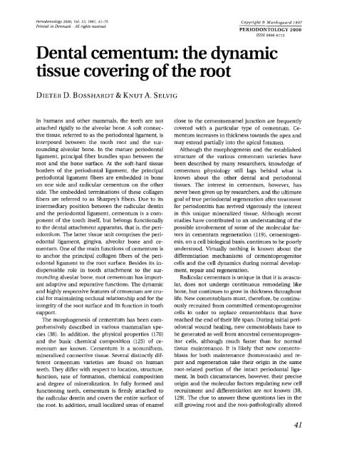
Periodontology 2000. Vol. 13, 1997, 41-75Printed in Denmark All rights reservedCopyright 0 Munkspaard 1997PERIODONTOLOGY 2000ISSN 0906-6713<strong>Dental</strong> <strong>cementum</strong>: <strong>the</strong> <strong>dynamic</strong><strong>tissue</strong> <strong>covering</strong> <strong>of</strong> <strong>the</strong> <strong>root</strong>DIETER D. BOSSHARDT & KNUT A. SELVIGIn humans and o<strong>the</strong>r mammals, <strong>the</strong> teeth are notattached rigidly to <strong>the</strong> alveolar bone. A s<strong>of</strong>t connective<strong>tissue</strong>, referred to as <strong>the</strong> periodontal ligament, isinterposed between <strong>the</strong> tooth <strong>root</strong> and <strong>the</strong> surroundingalveolar bone. In <strong>the</strong> mature periodontalligament, principal fiber bundles span between <strong>the</strong><strong>root</strong> and <strong>the</strong> bone surface. At <strong>the</strong> s<strong>of</strong>t-hard <strong>tissue</strong>borders <strong>of</strong> <strong>the</strong> periodontal ligament, <strong>the</strong> principalperiodontal ligament fibers are embedded in boneon one side and radicular <strong>cementum</strong> on <strong>the</strong> o<strong>the</strong>rside. The embedded terminations <strong>of</strong> <strong>the</strong>se collagenfibers are referred to as Sharpey’s fibers. Due to itsintermediary position between <strong>the</strong> radicular dentinand <strong>the</strong> periodontal ligament, <strong>cementum</strong> is a component<strong>of</strong> <strong>the</strong> tooth itself, but belongs functionallyto <strong>the</strong> dental attachment apparatus, that is, <strong>the</strong> periodontium.The latter <strong>tissue</strong> unit comprises <strong>the</strong> periodontalligament, gingiva, alveolar bone and <strong>cementum</strong>.One <strong>of</strong> <strong>the</strong> main functions <strong>of</strong> <strong>cementum</strong> isto anchor <strong>the</strong> principal collagen fibers <strong>of</strong> <strong>the</strong> periodontalligament to <strong>the</strong> <strong>root</strong> surface. Besides its indispensablerole in tooth attachment to <strong>the</strong> surroundingalveolar bone, <strong>root</strong> <strong>cementum</strong> has importantadaptive and reparative functions. The <strong>dynamic</strong>and highly responsive features <strong>of</strong> <strong>cementum</strong> are crucialfor maintaining occlusal relationship and for <strong>the</strong>integrity <strong>of</strong> <strong>the</strong> <strong>root</strong> surface and its function in toothsupport.The morphogenesis <strong>of</strong> <strong>cementum</strong> has been comprehensivelydescribed in various mammalian species(38). In addition, <strong>the</strong> physical properties (170)and <strong>the</strong> basic chemical composition (125) <strong>of</strong> <strong>cementum</strong>are known. Cementum is a nonuniform,mineralized connective <strong>tissue</strong>. Several distinctly different<strong>cementum</strong> varieties are found on humanteeth. They differ with respect to location, structure,function, rate <strong>of</strong> formation, chemical compositionand degree <strong>of</strong> mineralization. In fully formed andfunctioning teeth, <strong>cementum</strong> is firmly attached to<strong>the</strong> radicular dentin and covers <strong>the</strong> entire surface <strong>of</strong><strong>the</strong> <strong>root</strong>. In addition, small localized areas <strong>of</strong> enamelclose to <strong>the</strong> cementoenamel junction are frequentlycovered with a particular type <strong>of</strong> <strong>cementum</strong>. Cementumincreases in thickness towards <strong>the</strong> apex andmay extend partially into <strong>the</strong> apical foramen.Although <strong>the</strong> morphogenesis and <strong>the</strong> establishedstructure <strong>of</strong> <strong>the</strong> various <strong>cementum</strong> varieties havebeen described by many researchers, knowledge <strong>of</strong><strong>cementum</strong> physiology still lags behind what isknown about <strong>the</strong> o<strong>the</strong>r dental and periodontal<strong>tissue</strong>s. The interest in <strong>cementum</strong>, however, hasnever been given up by researchers, and <strong>the</strong> ultimategoal <strong>of</strong> true periodontal regeneration after treatmentfor periodontitis has revived vigorously <strong>the</strong> interestin this unique mineralized <strong>tissue</strong>. Although recentstudies have contributed to an understanding <strong>of</strong> <strong>the</strong>possible involvement <strong>of</strong> some <strong>of</strong> <strong>the</strong> molecular factorsin <strong>cementum</strong> regeneration (1 191, cementogenesis,on a cell biological basis, continues to be poorlyunderstood. Virtually nothing is known about <strong>the</strong>differentiation mechanisms <strong>of</strong> cementoprogenitorcells and <strong>the</strong> cell <strong>dynamic</strong>s during normal development,repair and regeneration.Radicular <strong>cementum</strong> is unique in that it is avascular,does not undergo continuous remodeling likebone, but continues to grow in thickness throughoutlife. New cementoblasts must, <strong>the</strong>refore, be continuouslyrecruited from committed cementoprogenitorcells in order to replace cementoblasts that havereached <strong>the</strong> end <strong>of</strong> <strong>the</strong>ir life span. During initial periodontalwound healing, new cementoblasts have tobe generated as well from ancestral cementoprogenitorcells, although much faster than for normal<strong>tissue</strong> maintenance. It is likely that new cementoblastsfor both maintenance (homeostasis) and repairand regeneration take <strong>the</strong>ir origin in <strong>the</strong> same<strong>root</strong>-related portion <strong>of</strong> <strong>the</strong> intact periodontal ligament.In both circumstances, however, <strong>the</strong>ir preciseorigin and <strong>the</strong> molecular factors regulating new cellrecruitment and differentiation are not known (38,129). The clue to answer <strong>the</strong>se questions lies in <strong>the</strong>still growing <strong>root</strong> and <strong>the</strong> non-pathologically altered41
Bosshardt & Selvig42
Human <strong>cementum</strong><strong>root</strong> surface. Basic knowledge <strong>of</strong> <strong>cementum</strong> developmentduring normalcy is <strong>the</strong>refore <strong>of</strong> utmost importance.The rodent has provided <strong>the</strong> most popular modelfor <strong>the</strong> study <strong>of</strong> tooth development in general. Although<strong>the</strong> tremendously fast growth rates <strong>of</strong> ratsand mice may render <strong>the</strong>se animals an excitingmodel for studying periodontal ligament developmentand tooth eruption, <strong>the</strong> rodent molar does notprovide a good parallel for <strong>the</strong> human situation withregard to cementogenesis (38). Over recent years, anincreasing quantity <strong>of</strong> data has accumulated thatallows human cementogenesis to be described exclusively.The aim <strong>of</strong> this chapter is to give a comprehensiveinsight into <strong>the</strong> structure, function, physicalproperties and chemical composition <strong>of</strong> human <strong>cementum</strong>during undisturbed development and repairas well as under some pathological conditions.DevelopmentThe formation <strong>of</strong> <strong>cementum</strong> can be subdivided intoa prefunctional and functional developmental stage.The prefunctional portion <strong>of</strong> <strong>cementum</strong> is formedduring <strong>root</strong> development. Since <strong>the</strong> formation <strong>of</strong> humantooth <strong>root</strong>s occurs over an extended period <strong>of</strong>time ranging between 3.75 and 7.75 years for permanentteeth, <strong>the</strong> prefunctional development <strong>of</strong> <strong>cementum</strong>is an extremely long-lasting process. Duringthis period <strong>of</strong> time, <strong>the</strong> primary distribution <strong>of</strong> <strong>the</strong>main <strong>cementum</strong> varieties is determined for each<strong>root</strong>. The functional development <strong>of</strong> <strong>cementum</strong>, on<strong>the</strong> o<strong>the</strong>r hand, commences when <strong>the</strong> tooth is aboutto reach <strong>the</strong> occlusal level, is associated with <strong>the</strong>attachment <strong>of</strong> <strong>the</strong> <strong>root</strong> to <strong>the</strong> surrounding bone andcontinues throughout life. It is mainly during <strong>the</strong>functional development that adaptive and reparativeprocesses are carried out by <strong>the</strong> biological responsiveness<strong>of</strong> <strong>cementum</strong>, which, in turn, influences <strong>the</strong>alterations in <strong>the</strong> distribution and appearance <strong>of</strong> <strong>the</strong><strong>cementum</strong> varieties on <strong>the</strong> <strong>root</strong> surface with time.Root formationFor an understanding <strong>of</strong> cementogenesis, <strong>the</strong> cellularevents occurring during <strong>root</strong> formation are <strong>of</strong> utmostimportance. Root formation commences when<strong>the</strong> enamel organ has reached its final size and <strong>the</strong>inner and outer cell layers <strong>of</strong> <strong>the</strong> enamel epi<strong>the</strong>lium,which delineate <strong>the</strong> enamel organ, proliferate from<strong>the</strong> cervical loop to form Hertwig’s epi<strong>the</strong>lial <strong>root</strong>sheath (59). Continuous cell mitotic activity at <strong>the</strong>apical termination <strong>of</strong> Hertwig’s <strong>root</strong> sheath leads toa coronoapical growth <strong>of</strong> this double cell layer. Itsmost apical portion, that is, <strong>the</strong> diaphragm, separates<strong>the</strong> dental papilla from <strong>the</strong> dental follicle (Fig.la,b). The inner and outer cell layer <strong>of</strong> Hertwig’s <strong>root</strong>sheath is surrounded by a basement membrane (Fig.lc,d). Similarly to <strong>the</strong> reciprocal epi<strong>the</strong>lial-mesenchymalinteractions occurring during crown formation(141, 158, 159, 209, 2121, cells originatingfrom <strong>the</strong> peripheral dental papilla differentiate along<strong>the</strong> internal basement membrane <strong>of</strong> <strong>the</strong> diaphragminto odontoblasts (Fig. Id) (8, 215). Once <strong>the</strong> firstmatrix <strong>of</strong> radicular mantle dentin is formed by <strong>the</strong>maturing odontoblasts and before <strong>the</strong> mineralization<strong>of</strong> <strong>the</strong> dentin matrix reaches <strong>the</strong> inner epi<strong>the</strong>lialcells, Hertwig’s <strong>root</strong> sheath becomes discontinuous.Epi<strong>the</strong>lial cell remnants <strong>of</strong> Hertwig’s <strong>root</strong> sheath persistin <strong>the</strong> still developing and, later in time, in <strong>the</strong>aging periodontal ligament at an approximate distance<strong>of</strong> 30-60 pm remote from <strong>the</strong> <strong>root</strong> surface,where <strong>the</strong>y are referred to as <strong>the</strong> epi<strong>the</strong>lial rests <strong>of</strong>Malassez (123). Although seen in longitudinal sectionsas isolated cell clusters surrounded by a basementmembrane, which separates <strong>the</strong>m from <strong>the</strong>surrounding connective <strong>tissue</strong>, <strong>the</strong>y apparently forma continuous network ensheathing <strong>the</strong> <strong>root</strong> at a certaindistance (21, 41, 53, 143, 167, 182, 183, 184). Although<strong>the</strong> number <strong>of</strong> epi<strong>the</strong>lial rests <strong>of</strong> Malassezdecreases with age (86, 151, 154, 199, 2201, cell mitoticactivity has also been observed (90, 107, 220).Their existence in <strong>the</strong> periodontal ligament throughoutlife implies that <strong>the</strong>y represent more than aFig. 1. Light (a) and transmission electron (b-d) micrographsshowing <strong>the</strong> most apical portion <strong>of</strong> two still growinghuman premolar <strong>root</strong>s developed to 50% (a) and 75%(b-d) <strong>of</strong> <strong>the</strong>ir final length. a, b. Hertwig’s <strong>root</strong> sheath(HRS) consists <strong>of</strong> an inner (IE) and outer epi<strong>the</strong>lial celllayer (OE). Cell mitotic activity at <strong>the</strong> apical loop (arrowin a points at a dividing cell) results in an apical growth<strong>of</strong> Hertwig’s <strong>root</strong> sheath, <strong>the</strong>reby separating <strong>the</strong> cells <strong>of</strong><strong>the</strong> dental follicle proper (DFP) from <strong>the</strong> pre-odontoblasts(pOB) <strong>of</strong> <strong>the</strong> dental papilla. Note <strong>the</strong> striking morphologicaldiversity <strong>of</strong> <strong>the</strong> epi<strong>the</strong>lial cells within Hertwig’s <strong>root</strong>sheath in b. Source: Bosshardt & Schroeder (32) with permissionfrom <strong>the</strong> publisher. c, d. A basement membrane(BM, arrowheads) separates <strong>the</strong> epi<strong>the</strong>lial cells <strong>of</strong> Hertwig’s<strong>root</strong> sheath from <strong>the</strong> surrounding mesenchyme.OB: odontoblasts; PD: predentin. Original magnification:a: x 800; b: x 2200; c, d x 7700.43
Human <strong>cementum</strong>merely vestigial structure. Their function, however,continues to be unknown.Cementoblast originThe differentiation <strong>of</strong> cementoblasts from cementoprogenitorcells and <strong>the</strong> formation <strong>of</strong> <strong>the</strong> dentinocementaljunction are temporally and spatiallyclosely related to dentin formation. The initiation <strong>of</strong>cementogenesis is, <strong>the</strong>refore, restricted to a narrowband encircling <strong>the</strong> forming <strong>root</strong> at its most apicalportion. This circular band extends only 200-300 pmcoronally from <strong>the</strong> advancing <strong>root</strong> edge and shifts in<strong>the</strong> apical direction while <strong>the</strong> <strong>root</strong> elongates (30, 32).Based on transplantation and 3H-thymidinestudies performed in mice (71, 96, 145, 201, 202, 204,234) and supported by ultrastructural indications fordirected cell migration towards <strong>the</strong> <strong>root</strong> surface inrat molars (46), it is widely accepted today that <strong>the</strong>cementoprogenitor cells arise from <strong>the</strong> dental follicleproper, which is <strong>of</strong> ectomesenchymal origin (that is,a derivative <strong>of</strong> <strong>the</strong> cranial neural crest). However, aspointed out by Thomas & Kollar (214), labeled cementoblastscould also be <strong>of</strong> epi<strong>the</strong>lial origin, sincecells <strong>of</strong> <strong>the</strong> enamel organ, which give rise to Hertwig’s<strong>root</strong> sheath, also incorporate 3H-thymidineprior to transplantation. Recent ultrastructural andimmunohistochemical studies support, indeed, <strong>the</strong>hypo<strong>the</strong>sis that <strong>the</strong> cementoblasts originate fromepi<strong>the</strong>lial cells <strong>of</strong> Hertwig’s <strong>root</strong> sheath when <strong>the</strong>yundergo an epi<strong>the</strong>lial-mesenchymal transformation(33, 38, 121, 214, 216). Such phenotypic transformationshave been well documented during embryonicdevelopment (93) and include, for instance,neural crest (110), sclerotome (191) and cardiaccushion mesenchyme (126). Ano<strong>the</strong>r example <strong>of</strong>phenotypic transformation has recently been shownduring palatogenesis when <strong>the</strong> ectodermal cells <strong>of</strong><strong>the</strong> palatal medial edge epi<strong>the</strong>lium transform intomesenchymal cells (66, 87). This example is <strong>of</strong> particularinterest in analogy to a possible epi<strong>the</strong>lialmesenchymaltransformation <strong>of</strong> Hertwig’s <strong>root</strong>sheath cells, since <strong>the</strong> epi<strong>the</strong>lial cells <strong>of</strong> <strong>the</strong> palatalmidline seam belong to <strong>the</strong> oral epi<strong>the</strong>lium and <strong>the</strong>underlying mesenchymal cells are believed to beneural crest in origin.<strong>the</strong> <strong>root</strong> surface, <strong>the</strong> mineralization front has reached andpartially passed <strong>the</strong> fibrillar dentinocemental junction.OB: odontoblasts. Original magnification: a-d: x 6250.Differentiation <strong>of</strong> cementoblastsFor <strong>the</strong> time being, <strong>the</strong> nature and origin <strong>of</strong> <strong>the</strong> moleculesthat trigger both a possible cell migration towards<strong>the</strong> <strong>root</strong> surface and cementoblast differentiationare not known. However, several possibilitieshave been suggested and, notably, all <strong>of</strong> <strong>the</strong>m havebeen derived from experiments in rats and mice. Achemical substance produced early in rat molar dentinogenesishas been suggested to act as a chemoattractantfor <strong>the</strong> cells <strong>of</strong> <strong>the</strong> dental follicle proper (46).Since it has been postulated that <strong>the</strong> disruption <strong>of</strong>Hertwig’s <strong>root</strong> sheath appears to be a consequence<strong>of</strong> this directed cell migration (461, such a chemoattractantmust have effect through <strong>the</strong> intact Hertwig’s<strong>root</strong> sheath, which is composed <strong>of</strong> two celllayers surrounded by a basement membrane oneach side. Although <strong>the</strong> dentin matrix is known toinduce in vitro cell migration (1681, it seems very unlikelythat a matrix component can be effectivethrough such a barrier.Although repeatedly suggested (96, 113, 143, 1651,interactions between <strong>the</strong> dental follicle proper andHertwig’s <strong>root</strong> sheath, which would eventually leadto cementoblast differentiation, have never beenshown. In analogy to <strong>the</strong> reciprocal epi<strong>the</strong>lial-mesenchymalinteractions between <strong>the</strong> inner epi<strong>the</strong>lialcells <strong>of</strong> Hertwig’s <strong>root</strong> sheath and <strong>the</strong> differentiatingodontoblasts, timed modulations in basement membranecomposition could possibly act as inductivesignals for cementoblast differentiation. Results fromrecombination experiments using murine molars indicateindeed that a mineralized <strong>tissue</strong> adhering to<strong>the</strong> developing dentinal <strong>root</strong> surface depends on <strong>the</strong>presence <strong>of</strong> basement membrane components (120).These experiments could, however, not clarifywhe<strong>the</strong>r <strong>the</strong> <strong>tissue</strong> formed on <strong>the</strong> <strong>root</strong> surface wasei<strong>the</strong>r bone or <strong>cementum</strong>. In addition, it remains tobe determined whe<strong>the</strong>r components <strong>of</strong> <strong>the</strong> basementmembrane induce <strong>the</strong> cells from <strong>the</strong> dentalfollicle proper to differentiate into cementoblasts.O<strong>the</strong>r extracellular matrix proteins that have beensuggested to play a role in cementoblast differentiationare noncollagenous proteins also found inbone. High expression <strong>of</strong> <strong>the</strong> two major noncollagenousproteins, bone sialoprotein (122, 193) andosteopontin (192, 194), has been detected on <strong>the</strong>surface <strong>of</strong> <strong>the</strong> forming molar <strong>root</strong>s in mice, and ithas been proposed that bone sialoprotein might beinvolved in precementoblast chemoattraction, adhesionto <strong>the</strong> <strong>root</strong> surface and cell differentiation(122). Since <strong>the</strong>se results were derived from immunohistochemicalstudies <strong>of</strong> thick sections, <strong>the</strong>y45
Bosshardt & Selvig46
Human <strong>cementum</strong>do not allow a precise immunolocalization. Thesequential appearance <strong>of</strong> bone sialoprotein and osteopontinduring <strong>root</strong> development and <strong>the</strong>ir preciseroles remain to be determined. The forming <strong>root</strong>comprises <strong>the</strong> initial mineralization <strong>of</strong> both dentinand <strong>cementum</strong>, a process that has to be preciselyharmonized in time and space. The high expressions<strong>of</strong> bone sialoprotein and osteopontin are likely to berelated to <strong>the</strong> mineralization process <strong>of</strong> mantle dentinand <strong>cementum</strong>, including <strong>the</strong>ir interface. Thesituation at this interfacial site is more complicatedin <strong>the</strong> rodent molar, since an interfacial layer, whichappears to be rich in glycoproteins, is frequently interposedbetween dentin and <strong>the</strong> <strong>cementum</strong> properand may significantly contribute to <strong>the</strong> highimmunoreaction on <strong>the</strong> <strong>root</strong> surface (see: The development<strong>of</strong> <strong>the</strong> dentinocemental junction). As a matter<strong>of</strong> fact, <strong>the</strong>re exists no <strong>tissue</strong> intermediate between<strong>cementum</strong> and dentin in human teeth, and itremains to be determined whe<strong>the</strong>r bone sialoproteinand/or osteopontin expression precede and <strong>the</strong>reforeinduce cementoblast differentiation.Ano<strong>the</strong>r group <strong>of</strong> proteins, that are immunologicallyrelated to enamel proteins have also been proposedto be involved in early cementogenesis (164,186, 187). They appear to be a normal feature on<strong>the</strong> <strong>root</strong>-analogue surface <strong>of</strong> rodent incisors and afrequent matrix constituent <strong>of</strong> <strong>the</strong> cervical <strong>root</strong> surface<strong>of</strong> rodent molars and have also been characterizedbiochemically from extracts <strong>of</strong> human <strong>cementum</strong>(188). The inner epi<strong>the</strong>lial cells <strong>of</strong> Hertwig’s<strong>root</strong> sheath, which are a derivative <strong>of</strong> <strong>the</strong> inner cells<strong>of</strong> <strong>the</strong> enamel organ, do, at least for a certain time,maintain <strong>the</strong> potential for <strong>the</strong> production and secretion<strong>of</strong> enamel or enamel-related proteins, as can beclearly seen in <strong>the</strong> extreme case <strong>of</strong> enamel drops andpearls occasionally <strong>covering</strong> <strong>the</strong> <strong>root</strong> surface. Althoughrepeatedly suggested, it is still not clearwhe<strong>the</strong>r and how <strong>the</strong>se proteins influence <strong>the</strong> initiation<strong>of</strong> cementogenesis.The development <strong>of</strong> <strong>the</strong> dentinocemental junctionNo matter what <strong>the</strong> factors are that trigger <strong>the</strong> differentiation<strong>of</strong> cementoprogenitor cells into fully activated<strong>cementum</strong>-producing cells, <strong>the</strong>y differentiatealong <strong>the</strong> newly deposited and not yet mineralizedmatrix <strong>of</strong> <strong>the</strong> radicular mantle dentin into cementoblasts.At <strong>the</strong> beginning <strong>of</strong> <strong>the</strong>ir maturation on <strong>the</strong><strong>root</strong> surface, <strong>the</strong>y extend numerous tiny cytoplasmicprocesses into <strong>the</strong> loosely arranged and not yet mineralizeddentinal matrix (Fig. 2a). This enables <strong>the</strong>cementoblasts to position <strong>the</strong> initially secreted collagenfibrils <strong>of</strong> <strong>the</strong> <strong>cementum</strong> matrix among those <strong>of</strong><strong>the</strong> dentinal matrix (Fig. 2b), and this crucial stepleads eventually to an intimate interdigitation <strong>of</strong> <strong>the</strong>two different fibril populations (Fig. 2c) (30, 32). Themineralization <strong>of</strong> <strong>the</strong> outermost layer <strong>of</strong> <strong>the</strong> dentinmatrix, that is, <strong>the</strong> mantle dentin, appears to be delayedand <strong>the</strong> mineralization front in dentin doesreach <strong>the</strong> future dentinocemental junction, not before<strong>the</strong> implantation <strong>of</strong> <strong>the</strong> <strong>cementum</strong> matrix is establishedand <strong>the</strong> dentinal matrix is completelycovered with <strong>the</strong> collagen fibrils <strong>of</strong> <strong>cementum</strong> (Fig.2d), The term intermediate <strong>cementum</strong> appears repeatedlyin <strong>the</strong> literature and in textbooks. Althoughoriginally described for <strong>the</strong> apical portion <strong>of</strong> humanteeth (191, <strong>the</strong>re exists no interfacial layer betweendentin and <strong>cementum</strong> in human teeth (38). In rodentmolars and incisors, on <strong>the</strong> o<strong>the</strong>r hand, an intermediatelayer has frequently been observed, particularlybetween acellular extrinsic fiber <strong>cementum</strong>and dentin (112, 144, 146, 172, 225, 226). This layerappears to be rich in glycoproteins but containssparsely distributed collagen fibrils (225, 226). Theorigin <strong>of</strong> this layer is still controversial. It is ei<strong>the</strong>rFig. 3. Light micrographs showing <strong>the</strong> different <strong>cementum</strong>varieties found on human teeth. a. The acellular afibrillar<strong>cementum</strong> (AAC) shown in this micrograph overlaps <strong>the</strong>enamel on <strong>the</strong> crown (ES; enamel space), apposes to <strong>the</strong>dentin (D) and merges into acellular extrinsic fiber <strong>cementum</strong>(AEFC). b, c. The acellular extrinsic fiber <strong>cementum</strong>is usually found on <strong>the</strong> cervical half <strong>of</strong> <strong>the</strong> <strong>root</strong>.The extrinsic matrix fibers remain short during <strong>the</strong> prefunctionaldevelopment (b) but become continuous with<strong>the</strong> principal fibers <strong>of</strong> <strong>the</strong> periodontal ligament (PL) when<strong>the</strong> tooth is in functional occlusion (c). d, e. Cellular intrinsicfiber <strong>cementum</strong> (CIFC) during its initial attachmentto <strong>the</strong> <strong>root</strong> dentin at <strong>the</strong> apical portion <strong>of</strong> a humanpremolar <strong>root</strong> developed to 75% <strong>of</strong> its final length (d)and at a more advanced stage <strong>of</strong> formation (e). f. Later indevelopment, <strong>the</strong> cervically located acellular extrinsicfiber <strong>cementum</strong> splits into several layers which interfacewith <strong>the</strong> more apically located cellular intrinsic fiber <strong>cementum</strong>layers to form <strong>the</strong> cellular mixed stratified <strong>cementum</strong>(CMSC). Source: Schroeder (169). g. Cellular intrinsicfiber <strong>cementum</strong> is also found as a reparative <strong>tissue</strong>filling resorptive defects <strong>of</strong> <strong>the</strong> <strong>root</strong>. Note <strong>the</strong> reversal line(RL) demarcating <strong>the</strong> junction between old and new<strong>tissue</strong>s. Source: Bosshardt (35) with permission from <strong>the</strong>publisher. HRS: Hertwig’s <strong>root</strong> sheath; OB odontoblasts;PD: predentin. Original magnification: a-d: ~440; e:X130; f: ~80; g: X230.47
Bosshardt & SeluigFig. 4. Transmission electron micrographs <strong>of</strong> acellularafibrillar <strong>cementum</strong> {AAC) at <strong>the</strong> cementoenamel junction.The area outlined in a corresponds to b. a. The acellularafibrillar <strong>cementum</strong> apposes to <strong>the</strong> enamel space(ES), which contains residual enamel proteins, and tapersin <strong>the</strong> coronal direction. b. The acellular afibrillar ce-mentum is characterized by numerous incremental layerswith varying electron density and texture. c. In <strong>the</strong> apicaldirection, <strong>the</strong> acellular afibrillar <strong>cementum</strong> abuts against<strong>the</strong> dentin (D) and <strong>the</strong> acellular extrinsic fiber <strong>cementum</strong>(AEFC). Original magnification: a: ~3200; b, c: x 13,000.formed by cementoblasts (47, 167, 225) or by <strong>the</strong> innerepi<strong>the</strong>lial <strong>root</strong> sheath cells (144, 146, 161, 188,194).The <strong>cementum</strong> varieties:<strong>the</strong>ir location, formation, structureand functionHuman teeth have three fundamentally differentvarieties <strong>of</strong> <strong>cementum</strong>. The location <strong>of</strong> <strong>the</strong>se varietiesshows tooth-type specific distribution patternsbut may also vary along and around <strong>the</strong> surface <strong>of</strong><strong>the</strong> same tooth. The following general rule appliesto human teeth: acellular afibrillar <strong>cementum</strong> coversminor areas <strong>of</strong> <strong>the</strong> enamel, particularly at and along<strong>the</strong> cementoenamel junction (Fig. 3a). Acellular extrinsicfiber <strong>cementum</strong> is mainly found on <strong>the</strong> cervi-cal and middle <strong>root</strong> portions (Fig. 3a-c). On frontteeth, it may also cover part <strong>of</strong> <strong>the</strong> apical <strong>root</strong> portion,since <strong>the</strong> apical extension <strong>of</strong> acellular extrinsicfiber <strong>cementum</strong> on <strong>the</strong> <strong>root</strong> surface increases fromposterior to anterior teeth. Cellular intrinsic fiber <strong>cementum</strong>is initially deposited on <strong>root</strong> surface areaswhere no acellular extrinsic fiber <strong>cementum</strong> hasbeen laid down on <strong>the</strong> dentin (Fig. 3d). This mayoccur in <strong>the</strong> furcations and on <strong>the</strong> apical <strong>root</strong> portions(Fig. 3d,e). Cellular intrinsic fiber <strong>cementum</strong>may overgrow layers <strong>of</strong> acellular extrinsic fiber <strong>cementum</strong>,and acellular extrinsic fiber <strong>cementum</strong>, inturn, can overlay cellular intrinsic fiber <strong>cementum</strong>.The so formed mingled <strong>cementum</strong> is called cellularmixed stratified <strong>cementum</strong> and is confined to <strong>the</strong>apical <strong>root</strong> portions and to <strong>the</strong> furcations (Fig. 3f).In addition, mainly cellular intrinsic fiber <strong>cementum</strong>participates in <strong>the</strong> repair process <strong>of</strong> previously resorbed<strong>root</strong>s (Fig. 3g).48
Acellular af5brillar <strong>cementum</strong>The acellular afibrillar <strong>cementum</strong> consists <strong>of</strong> a mineralizedmatrix, which appears similar to <strong>the</strong> interfibrillarmatrix <strong>of</strong> acellular extrinsic fiber <strong>cementum</strong>,but contains nei<strong>the</strong>r collagen fibrils nor embeddedcells. The lack <strong>of</strong> collagen fibrils indicates that this<strong>cementum</strong> variety has no function in tooth attachment.The acellular afibrillar <strong>cementum</strong> can be identifiedby light and electron microscopy. Under <strong>the</strong>light microscope, <strong>the</strong> acellular afibrillar <strong>cementum</strong>stands out by its basophilia and its more or less uniformappearance (Fig. 3a). In <strong>the</strong> electron microscope,however, <strong>the</strong> structure <strong>of</strong> acellular afibrillar<strong>cementum</strong> is less homogeneous (Fig. 4a-c). A variablenumber <strong>of</strong> layers with varying electron densityand different texture, which can be ei<strong>the</strong>r granularor reticular, give <strong>the</strong> acellular afibrillar <strong>cementum</strong> amultifarious appearance.Acellular afibrillar <strong>cementum</strong> is deposited as isolatedpatches over minor areas <strong>of</strong> enamel and dentin.Cementum islands represent isolated patches <strong>of</strong>acellular afibrillar <strong>cementum</strong> deposited on <strong>the</strong> enamelover small areas <strong>of</strong> <strong>the</strong> dental crown just coronalto <strong>the</strong> cementoenamel junction. Cementumspurs are found around <strong>the</strong> cementoenamel junction,where <strong>the</strong>y cover minor areas <strong>of</strong> <strong>the</strong> enameland <strong>the</strong> adjacent dentin <strong>of</strong> <strong>the</strong> <strong>root</strong> (Fig. 3a, 4a). Cementumspurs may be covered by acellular extrinsicfiber <strong>cementum</strong> and/or by junctional epi<strong>the</strong>lium.The areas and location <strong>of</strong> acellular afibrillar <strong>cementum</strong>vary from tooth to tooth and along <strong>the</strong>cementoenamel junction <strong>of</strong> <strong>the</strong> same tooth. This unpredictabledistribution pattern indicates that acel-Fig. 5. Schematic diagram depicting <strong>the</strong> gradual development<strong>of</strong> acellular extrinsic fiber <strong>cementum</strong> during its prefunctionalgenesis along a human premolar <strong>root</strong> developedto about 50% <strong>of</strong> its final length. 1. Attachment <strong>of</strong><strong>the</strong> acellular extrinsic fiber <strong>cementum</strong> matrix to <strong>the</strong> notyet mineralized matrix <strong>of</strong> <strong>the</strong> radicular mantle dentin(NMD). The latter is continuous with <strong>the</strong> pulpal predentinlayer (PD) and tapers in <strong>the</strong> coronal direction. Note thatHertwig’s epi<strong>the</strong>lial <strong>root</strong> sheath (HRS) is associated with<strong>the</strong> nonmineralized matrix <strong>of</strong> <strong>the</strong> radicular mantle dentinfor an extremely short distance at <strong>the</strong> advancing <strong>root</strong> edge(ARE); 2. Establishment <strong>of</strong> <strong>the</strong> acellular extrinsic fiber <strong>cementum</strong>matrix on <strong>the</strong> <strong>root</strong> surface in form <strong>of</strong> a shortcollagenous fiber fringe (FF); 3. When <strong>the</strong> fiber fringe hasattained its maximum numerical density, a cell-fiberfringe meshwork establishes on <strong>the</strong> <strong>root</strong> surface. 1-3. Theexternal mineralization front in dentin (mineralized dentin,MD) is gradually approaching <strong>the</strong> base <strong>of</strong> <strong>the</strong> fiberfringe implantation on <strong>the</strong> <strong>root</strong> surface. 4. The externalmineralization front in dentin has reached <strong>the</strong> future dentinocementaljunction. E: enamel; ERM: epi<strong>the</strong>lial cellrests <strong>of</strong> Malassez. Source: Bosshardt & Schroeder (30) withpermission from <strong>the</strong> publisher.49
Bosshardt & Seluig50
Human <strong>cementum</strong>lular afibrillar <strong>cementum</strong> formation is likely to be adevelopmental curiosity that deviates from <strong>the</strong> normra<strong>the</strong>r than an indispensable <strong>tissue</strong>.The cells responsible for <strong>the</strong> formation <strong>of</strong> acellularafibrillar <strong>cementum</strong> have still not been determinedwith precision. Its formation commences at <strong>the</strong> end<strong>of</strong> enamel maturation and continues for an unknownperiod <strong>of</strong> time. It is believed that connective<strong>tissue</strong> cells are responsible for <strong>the</strong> acellular afibrillar<strong>cementum</strong> formation when <strong>the</strong>y come in contactwith <strong>the</strong> enamel surface (166, 167). To make thispossible, cells <strong>of</strong> <strong>the</strong> reduced enamel epi<strong>the</strong>liummust be lost or detached from <strong>the</strong> enamel. On <strong>the</strong>o<strong>the</strong>r hand, it cannot be completely ruled out thatacellular afibrillar <strong>cementum</strong> is an epi<strong>the</strong>lia1 productinitially produced when <strong>the</strong> ameloblasts transforminto <strong>the</strong> reduced enamel epi<strong>the</strong>lium and when <strong>the</strong>cells <strong>of</strong> <strong>the</strong> inner enamel epi<strong>the</strong>lium are about togenerate <strong>the</strong> inner cells <strong>of</strong> Hertwig’s <strong>root</strong> sheath. Invitro experiments showed that calcified layers, whichmorphologically resembled acellular afibrillar <strong>cementum</strong>,formed around demineralized dentin slicesimmersed in serum-containing culture mediumsupplemented with alkaline phosphatase and an organicsource for phosphate such as <strong>the</strong> monophosphateester P-glycerophosphate (17). The notion thatsuch acellular afibrillar <strong>cementum</strong>-like layersformed also in <strong>the</strong> absence <strong>of</strong> cells suggests that thismatrix represents a co-precipitate <strong>of</strong> medium- orserum-derived components and mineral. However,<strong>the</strong> enzyme alkaline phosphatase is required formineralization to occur. In <strong>the</strong> in vivo situation, thisenzyme is particularly associated with periodontalligament cells in <strong>the</strong> vicinity to bone and <strong>cementum</strong>(88).Acellular extrinsic fiber <strong>cementum</strong>The acellular extrinsic fiber <strong>cementum</strong> is usuallyconfined to <strong>the</strong> coronal half <strong>of</strong> <strong>the</strong> <strong>root</strong>. Its formationcommences <strong>the</strong>refore shortly after crown formationis completed and always before cellular intrinsicfiber <strong>cementum</strong> starts to form on more apical <strong>root</strong>portions. The gradual development <strong>of</strong> acellular extrinsicfiber <strong>cementum</strong> can be followed along <strong>the</strong>forming <strong>root</strong> (Fig. 5, 6a,b) (30, 31, 33). The cementoblastsproducing acellular extrinsic fiber <strong>cementum</strong>commence <strong>the</strong>ir cell differentiation in closest proximityto <strong>the</strong> advancing <strong>root</strong> edge. This may occuronly about 20 to 30 pm coronal to <strong>the</strong> first depositeddentinal matrix. These cells resemble fibroblasts, reveala well-developed rough endoplasmic reticulum,are interconnected by desmosome-like junctionsand commence to produce and attach <strong>the</strong> collagenous<strong>cementum</strong> matrix as close as 50 pm coronalto <strong>the</strong> <strong>root</strong> edge (Fig. 6c). Fur<strong>the</strong>r collagen depositionresults in a complete <strong>covering</strong> <strong>of</strong> <strong>the</strong> not yetmineralized dentinal matrix along <strong>the</strong> next 100 pm<strong>of</strong> <strong>the</strong> <strong>root</strong> surface (Fig. 6d). About 200 to 300 pmcoronal to <strong>the</strong> advancing <strong>root</strong> edge, <strong>the</strong> initial acellularextrinsic fiber <strong>cementum</strong> matrix is established on<strong>the</strong> dentinal matrix (Fig. 6e). The acellular extrinsicfiber <strong>cementum</strong> matrix consists <strong>of</strong> a dense fringe <strong>of</strong>short collagenous fibers that are implanted into <strong>the</strong>dentinal matrix and oriented about perpendicularlyto <strong>the</strong> <strong>root</strong> surface (Fig. 6e, 7a). The outwardly progressingmineralization front in dentin (Fig. 6d) doesnot reach <strong>the</strong> future dentinocemental junction until<strong>the</strong> collagenous interdigitation <strong>of</strong> <strong>the</strong> two fibrilpopulations is established (Fig. 6e). Since <strong>the</strong> mineralization<strong>of</strong> <strong>the</strong> dentinal matrix commences about-Fig. 6. Light (a, b) and transmission electron (c-e) micrographsillustrating <strong>the</strong> initial attachment and gradual development<strong>of</strong> <strong>the</strong> acellular extrinsic fiber <strong>cementum</strong> matrix(that is, fiber fringe; FF) along <strong>the</strong> apical portion <strong>of</strong> ahuman premolar <strong>root</strong> developed to about 50% <strong>of</strong> its finallength. The area outlined in a corresponds to b. The positions<strong>of</strong> <strong>the</strong> micrographs c and d are indicated in b by<strong>the</strong> corresponding letters. a, b. Immediately coronal to <strong>the</strong>disintegration <strong>of</strong> Hertwig’s epi<strong>the</strong>lial <strong>root</strong> sheath (OE indicates<strong>the</strong> outer and IE <strong>the</strong> inner epi<strong>the</strong>lial cell layer), intenselystained cells distend <strong>the</strong> space between <strong>the</strong> <strong>root</strong>surface and <strong>the</strong> outer epi<strong>the</strong>lial cell layer. More coronally,<strong>the</strong> cells do no more reveal <strong>the</strong> same intense staining anda fiber fringe (arrows) appears on <strong>the</strong> <strong>root</strong> surface. Theonset <strong>of</strong> dentin mineralization is seen 100 pm coronal to<strong>the</strong> advancing <strong>root</strong> edge (ARE) and somewhat away from<strong>the</strong> external <strong>root</strong> surface (lowest arrowhead). In <strong>the</strong> cor-onal direction, <strong>the</strong> external mineralization front (MF, arrowheadspointing to <strong>the</strong> left) in dentin (D) is gradualiyapproaching <strong>the</strong> <strong>root</strong> surface. c. The cementoblasts (CB)on <strong>the</strong> <strong>root</strong> surface commence to implant <strong>the</strong> fiber fringeamong <strong>the</strong> collagenous matrix fibrils <strong>of</strong> predentin (PD) at<strong>the</strong> future dentinocemental junction (DCJ). Source: Bosshardt(35) with permission from <strong>the</strong> publisher. d. When<strong>the</strong> fiber fringe is <strong>covering</strong> almost <strong>the</strong> entire predentin,<strong>the</strong> external mineralization front in dentin is about toreach <strong>the</strong> fibrillar dentinocemental junction. Source:Bosshardt & Schroeder (30) with permission from <strong>the</strong>publisher. e. The established fiber fringe continues fora short distance into <strong>the</strong> space <strong>of</strong> <strong>the</strong> immature periodontalligament (PL). The mineralization front has completelyreached and partly passed <strong>the</strong> fibrillar dentinocementaljunction. OB: odontoblasts. Original magnification:a: x 130; b: ~440; c, d x6700; e: X4400.51
Bosshardt & SeluigFig. 7. Light micrographs demonstrating <strong>the</strong> growth <strong>of</strong>acellular extrinsic fiber <strong>cementum</strong> (AEFC) on human premolar<strong>root</strong>s. a. The acellular extrinsic fiber <strong>cementum</strong>matrix consists <strong>of</strong> short fringe fibers (FF) that emergefrom <strong>the</strong> <strong>root</strong> surface and appose to a fibrocellular meshworkoccupying <strong>the</strong> space <strong>of</strong> <strong>the</strong> immature periodontalligament (PL). A <strong>cementum</strong> layer is not yet visible. b. Mineralization<strong>of</strong> <strong>the</strong> fiber fringe, which is indicated by round,basophilic dots within <strong>the</strong> fiber fringe base (arrows), proceedsvery slowly. This acellular extrinsic fiber <strong>cementum</strong>layer has developed over approximately 2 years. Note that<strong>the</strong> fiber fringe is still short and apposes to a fibrocellularmeshwork running broadly parallel to <strong>the</strong> <strong>root</strong> surface.c. This 15-pm-thick acellular extrinsic fiber <strong>cementum</strong>layer has developed over approximately 5 years. Note <strong>the</strong>change in <strong>the</strong> orientation <strong>of</strong> <strong>the</strong> periodontal ligamentfibers and <strong>the</strong> continuation <strong>of</strong> some <strong>of</strong> <strong>the</strong> fringe fiberswith <strong>the</strong> developing principal periodontal ligament fibers.D dentin. Original magnification: a-c: X440.100 pm coronal to <strong>the</strong> advancing <strong>root</strong> edge andsomewhat underneath <strong>the</strong> <strong>root</strong> surface (Fig. 6b1, <strong>the</strong>mineralization <strong>of</strong> <strong>the</strong> mantle dentin seems apparentlyto be delayed. With <strong>the</strong> onset <strong>of</strong> <strong>cementum</strong>mineralization, <strong>the</strong> acellular extrinsic fiber <strong>cementum</strong>commences to grow in thickness (Fig. 7b,c).This growth is extremely slow but quite constant(Fig. 10a) (33, 179). The extrinsic fibers remain shortuntil <strong>the</strong> tooth is about to reach <strong>the</strong> occlusal level(Fig. 7c). On cervical <strong>root</strong> surfaces <strong>of</strong> human premolars,<strong>the</strong> prefunctional acellular extrinsic fiber <strong>cementum</strong>development may last 5 years or more, thatis, until acellular extrinsic fiber <strong>cementum</strong> hasreached a thickness <strong>of</strong> about 15 pm (Fig. 7c) (31, 33,38). How <strong>the</strong> short fiber fringe becomes elongatedand eventually continuous with <strong>the</strong> principal peri-odontal ligament fibers is still an open question. Theacellular extrinsic fiber <strong>cementum</strong> continues to growas long as <strong>the</strong> adjacent periodontal ligament remainsundisturbed. The extraordinarily high numericaldensity <strong>of</strong> fibers inserting into acellular extrinsicfiber <strong>cementum</strong> (approximately 30,000/mm2; (167))is a reflection <strong>of</strong> <strong>the</strong> significant function <strong>of</strong> this <strong>cementum</strong>variety for tooth anchorage to <strong>the</strong> surroundingbone. Due to posteruptive tooth movements,changes can occur in <strong>the</strong> direction <strong>of</strong> <strong>the</strong>Sharpey's fibers. These changes are accentuated byindividual acellular extrinsic fiber <strong>cementum</strong> layersinterfaced by growth lines, also known as resting orincremental lines. Although acellular extrinsic fiber<strong>cementum</strong> is a quite constantly growing <strong>tissue</strong>, <strong>the</strong>selines appear to represent <strong>the</strong> periodic deposition <strong>of</strong>52
Human <strong>cementum</strong><strong>cementum</strong> layers in frequent association with anabrupt change in <strong>the</strong> direction <strong>of</strong> Sharpey’s fibers.Moreover, as can be deduced from faster growthrates on distal (4.3 pm/year) than on mesial (1.4 pm/year) <strong>root</strong> surfaces (56), acellular extrinsic fiber <strong>cementum</strong>has <strong>the</strong> potential to adapt to functionallydictated alterations such as mesial tooth drift.Cellular and acellular intrinsic fiber <strong>cementum</strong>Although <strong>the</strong> intrinsic <strong>cementum</strong> alone has no immediatefunction in tooth attachment, its importantrole as an adaptive <strong>tissue</strong> (167) that brings and maintains<strong>the</strong> tooth in its proper position should not beunderestimated. In addition, only cellular intrinsicfiber <strong>cementum</strong> can repair a resorptive defect <strong>of</strong> <strong>the</strong><strong>root</strong> in a reasonable time due to its capacity to growmuch faster than any o<strong>the</strong>r known <strong>cementum</strong> type.Functional stimuli, that is, <strong>the</strong> force generated bytooth contact and mastication, are widely held responsiblefor <strong>the</strong> onset and appositional growth <strong>of</strong>cellular intrinsic fiber <strong>cementum</strong>. This assumptionprobably originates from observations showing that<strong>the</strong> genesis <strong>of</strong> this <strong>cementum</strong> variety coincides with<strong>the</strong> first occlusal tooth contact (51, 58, 95, 140) andthat functioning teeth appear to have thicker <strong>cementum</strong>layers than teeth which are not in function(84, 99). Several observations, however, are not inkeeping with this concept. As already suggested byKronfeld (105, 106) and Kellner (1041, mastication isapparently not a prerequisite for cellular intrinsicfiber <strong>cementum</strong> genesis, since i) <strong>the</strong> furcations <strong>of</strong>human teeth are covered with thick <strong>cementum</strong>layers before <strong>the</strong>y emerge into <strong>the</strong> oral cavity (1051,ii) impacted and erupted teeth without antagonistsappear to have thicker <strong>cementum</strong> layers than fullyerupted and functioning teeth (9, 103, 104, 106, 181)and iii) over-compression <strong>of</strong> <strong>the</strong> periodontal ligamentcauses <strong>root</strong> resorption. Thus, it seems that <strong>the</strong>initiation <strong>of</strong> cellular intrinsic fiber <strong>cementum</strong> genesisdoes not depend on stimuli transmitted by masticatoryforces and that influence by pressure mayreduce <strong>the</strong> rate <strong>of</strong> matrix deposition.As for acellular extrinsic fiber <strong>cementum</strong>, <strong>the</strong>initiation <strong>of</strong> cellular intrinsic fiber <strong>cementum</strong> genesison <strong>the</strong> forming <strong>root</strong> commences in closest proximityto <strong>the</strong> advancing <strong>root</strong> edge (32, 34). Precementoblastsdifferentiate along <strong>the</strong> not yet mineralizeddentinal matrix into large, basophilic cells (Fig.8a). They first project numerous cytoplasmic processesinto <strong>the</strong> loose dentinal matrix and immediatelycommence to implant <strong>the</strong> initial collagen fibrilsamong those <strong>of</strong> <strong>the</strong> dentinal matrix (Fig. 8b). Additionalcementoblasts, which are remote from <strong>the</strong>dentinal surface, deposit <strong>the</strong>ir <strong>cementum</strong> matrix atvarious locations around <strong>the</strong>mselves (Fig. 8c). Thismultipolar and fast matrix deposition, which occursin <strong>the</strong> space between deviating epi<strong>the</strong>lial cells <strong>of</strong>Hertwig’s <strong>root</strong> sheath and <strong>the</strong> dentinal surface (Fig.8a), appears to be <strong>the</strong> reason for <strong>the</strong> incorporation<strong>of</strong> some <strong>of</strong> <strong>the</strong> cementoblasts (32, 67, 146). The cellsentrapped in <strong>the</strong> mineralized <strong>cementum</strong> are referredto as cementocytes and occupy lacunae, which areinterconnected through canaliculi (see: Physiologicalactivity <strong>of</strong> cementocytes) (Fig. 16, 17). The cementoblastsattain <strong>the</strong>ir full syn<strong>the</strong>tic activity approximately100 pm coronal to <strong>the</strong> advancing <strong>root</strong> edge (Fig.9a). They are large, basophilic cells with an euchromatin-richnucleus and an abundant endoplasmicreticulum. The rapid matrix deposition slows downsoon, and fur<strong>the</strong>r collagen matrix is deposited in amore unipolar mode <strong>of</strong> secretion (Fig. 9b-e). In rarecases, <strong>the</strong> intrinsic <strong>cementum</strong> is formed in an extremelyunipolar mode <strong>of</strong> matrix deposition andcompletely lacks cementocytes (Fig. 8d, 9d,e). Thisparticular <strong>tissue</strong> consists <strong>of</strong> densely bundled collagenfibrils and is named acellular intrinsic fiber <strong>cementum</strong>(29). The collagen fibrils produced during<strong>the</strong> fast, multipolar cellular intrinsic fiber <strong>cementum</strong>initiation show a more random orientation thanthose <strong>of</strong> <strong>the</strong> subsequently deposited matrix. Therefore,<strong>the</strong> bulk <strong>of</strong> <strong>the</strong> intrinsic collagen fibrils formdiscrete bundles oriented mainly parallel to <strong>the</strong> <strong>root</strong>surface (Fig. 1 la). Polarized light microscopic investigationsin deciduous (1021, permanent (163)and impacted human teeth (99) suggest that <strong>the</strong>seintrinsic fibers are arranged in an orderly fashionaround <strong>the</strong> <strong>root</strong> (Fig. llb).Unlike in rat molars (227, 228), initial cellular intrinsicfiber <strong>cementum</strong> deposition on human tooth<strong>root</strong>s is not necessarily associated with <strong>the</strong> simultaneousformation <strong>of</strong> extrinsic fibers, that is, <strong>the</strong> futureSharpey’s fibers. In humans, <strong>the</strong> extrinsic fibersare oriented about perpendicularly to <strong>the</strong> <strong>root</strong> surfaceand traverse <strong>the</strong> intrinsic <strong>cementum</strong> varietyei<strong>the</strong>r sporadically or densely arrayed in parallel. Although<strong>the</strong> numerical density <strong>of</strong> <strong>the</strong>se highly aggregatedextrinsic fibers may be distinctly less than inpure acellular extrinsic fiber <strong>cementum</strong> (1671, <strong>the</strong>yare considered as <strong>the</strong> matrix <strong>of</strong> acellular extrinsicfiber <strong>cementum</strong> that intermingles or alternates with<strong>the</strong> intrinsic fibers. This mixed <strong>cementum</strong> is referredto as cellular mixed stratified <strong>cementum</strong>. When <strong>the</strong>extrinsic fibers are continuous with <strong>the</strong> functionallyoriented principal fibers <strong>of</strong> <strong>the</strong> periodontal ligament,53
Bosshardt & Selvig54
Human <strong>cementum</strong>Fig. 9. Light microscopic radioautographs demonstrating<strong>the</strong> prefimctional deposition <strong>of</strong> cellular intrinsic fiber <strong>cementum</strong>(CIFC) and acellular intrinsic fiber <strong>cementum</strong>(AIFC) close to (a) and more remote from <strong>the</strong> advancing<strong>root</strong> edge (ARE) (b-e). These human premolars with <strong>root</strong>sdeveloped to 75-95% <strong>of</strong> <strong>the</strong>ir final length were in vitropulse-labeled with 3H-proline for 15 min followed by achase incubation for 45 min (a, d), 1 h 45 min (b), 4 h 45min (e), and 23 h 45 min (c). Silver grains are, first, local-ized in clusters over <strong>the</strong> paranuclear cytoplasm <strong>of</strong> cementoblasts(arrowheads in a, d), appear, later, over <strong>the</strong>peripheral cytoplasm <strong>of</strong> cementoblasts and <strong>the</strong> subjacent<strong>cementum</strong> matrix (b), and eventually cover <strong>the</strong> peripheral<strong>cementum</strong> matrix (c, e). Source: Bosshardt & Schroeder(34) with permission from <strong>the</strong> publisher. D: dentin; HRS:Hertwig’s epi<strong>the</strong>lial <strong>root</strong> sheath; OB: odontoblasts; PD:predentin; PL periodontal ligament. Original magnification:a: x440; k. x700.Fig. 8. Light (a) and transmission electron (b-d) micrographsillustrating <strong>the</strong> genesis <strong>of</strong> cellular intrinsic fiber<strong>cementum</strong> (CIFC) and acellular intrinsic fiber <strong>cementum</strong>(AIFC) at <strong>the</strong> advancing <strong>root</strong> edge (ARE) (a-c) and on <strong>the</strong>established <strong>cementum</strong> layer (d), respectively Human premolar<strong>root</strong>s developed to 75% <strong>of</strong> <strong>the</strong>ir final length. a. At<strong>the</strong> advancing <strong>root</strong> edge, <strong>the</strong> inner cells <strong>of</strong> Hertwig’s epi<strong>the</strong>lial<strong>root</strong> sheath (HRS) cover not more than 20 pm <strong>of</strong><strong>the</strong> newly deposited predentin (PD), whereas <strong>the</strong> outercell layer <strong>of</strong> Hertwig’s epi<strong>the</strong>lial <strong>root</strong> sheath continues forabout 150 pm in <strong>the</strong> coronal direction. Cementoblasts(CB) distend <strong>the</strong> space between <strong>the</strong> <strong>root</strong> surface and <strong>the</strong>deviating outer epi<strong>the</strong>lial cell layer. The cellular intrinsicfiber <strong>cementum</strong> thickness increases rapidly in <strong>the</strong> coronaldirection. Arrowheads point at <strong>the</strong> mineralization front(MF) <strong>of</strong> both dentin (D) and cellular intrinsic fiber ce-mentum. The interrupted line corresponds to <strong>the</strong> dentinocementaljunction. b. Large cementoblasts commence toproduce and attach <strong>the</strong> initial cellular intrinsic fiber <strong>cementum</strong>matrix to <strong>the</strong> external predentin matrix about 50pm coronal to <strong>the</strong> advancing <strong>root</strong> edge. Note that <strong>the</strong>reis no intermediate layer between <strong>the</strong> two matrices at <strong>the</strong>dentinocemental junction (DCJ). c. The cellular intrinsicfiber <strong>cementum</strong> matrix is also produced away from <strong>the</strong>predentin among <strong>the</strong> cementoblasts. d. Fur<strong>the</strong>r matrix isdeposited by cementoblasts, which form a continuous celllayer when <strong>the</strong>y are associated with acellular intrinsicfiber <strong>cementum</strong>. Sources: a-c: Bosshardt & Schroeder(32); d Bosshardt & Schroeder (34) with permission from<strong>the</strong> publisher. OB: odontoblasts. Original magnification:a: x440; b: X7700; c: X3200; d X4400.55
Bosshardt & SelvigFig. 10. Fluorescence lines in <strong>cementum</strong>, dentin (D) andalveolar bone (AB) <strong>of</strong> a mandibular second deciduous molar<strong>of</strong> a Macaca fmcicularis monkey. The animal receivedsequential injections <strong>of</strong> calcein (green lines) and xylenolorange (orange lines) on average every 33 days. The tw<strong>of</strong>luorochromes bind to sites <strong>of</strong> ongoing mineralization andproduce clear fluorescence lines. Note <strong>the</strong> difference in<strong>the</strong> labeling pattern between acellular extrinsic fiber ce-mentum (AEFC) and cellular intrinsic fiber <strong>cementum</strong>(CIFC). The labeling pattern and <strong>the</strong> difference in <strong>the</strong>growth rates between acellular extrinsic fiber <strong>cementum</strong>and cellular intrinsic fiber <strong>cementum</strong> are basically <strong>the</strong>same in human teeth, albeit acellular extrinsic fiber <strong>cementum</strong>and cellular intrinsic fiber <strong>cementum</strong> grow moreslowly. Source: Bosshardt et al. (28). PL: periodontal ligament.Original magnification: a, b: x250.<strong>the</strong>y can be regarded as Sharpey’s fibers. As layers <strong>of</strong>acellular extrinsic fiber <strong>cementum</strong> and cellular andacellular intrinsic fiber <strong>cementum</strong> develop unpredictablyin time, space and thickness (167, 1691, particular<strong>root</strong> surface areas covered with cellular mixedstratified <strong>cementum</strong> may temporarily remain unsupportedby periodontal fibers.Like pure acellular extrinsic fiber <strong>cementum</strong> on<strong>the</strong> coronal half <strong>of</strong> <strong>the</strong> <strong>root</strong>, cellular mixed stratified<strong>cementum</strong> increases in thickness throughoutlife (236). However, <strong>the</strong> unpredictable <strong>dynamic</strong>s <strong>of</strong><strong>the</strong> <strong>tissue</strong> alternations renders growth rate determinationmuch more difficult for cellular mixedstratified <strong>cementum</strong>. Never<strong>the</strong>less, <strong>the</strong> initial cellu-lar intrinsic fiber <strong>cementum</strong> growth has beenmeasured in deciduous teeth <strong>of</strong> a non-human primate(28). It may be up to 30-fold faster than <strong>the</strong>more regular acellular extrinsic fiber <strong>cementum</strong>deposition (Fig. lob). The <strong>dynamic</strong> <strong>tissue</strong> alternationsand <strong>the</strong> variations in growth rates are reflectedby a <strong>tissue</strong> layering <strong>of</strong> cellular mixed stratified<strong>cementum</strong> with layers <strong>of</strong> acellular extrinsicfiber <strong>cementum</strong> and incremental lines (that is,growth lines or resting lines) interfacing layers <strong>of</strong>cellular and acellular intrinsic fiber <strong>cementum</strong>. Thepatch-wise deposition <strong>of</strong> cellular mixed stratified<strong>cementum</strong> results in great circumferential variationsin <strong>cementum</strong> thickness and reflects periods56
Human <strong>cementum</strong>Fig. 11. Polarized light photomicrographs <strong>of</strong> <strong>cementum</strong>.a. In a section made parallel to <strong>the</strong> <strong>root</strong> surface, intrinsiccollagen fibers in cellular intrinsic fiber <strong>cementum</strong> appearas wide sheets <strong>of</strong> alternating directions. b. Cementum onan impacted third molar. Collagen fibers are almost exclusively<strong>of</strong> <strong>the</strong> intrinsic type arranged in an orderlyfashion parallel to <strong>the</strong> <strong>root</strong> surface (arrow). D: dentin.Original magnification x 130.<strong>of</strong> accelerated deposition <strong>of</strong> cellular intrinsic fiber<strong>cementum</strong>, which are probably due to functionaldemands in order to reposition <strong>the</strong> tooth when itis shifting in its bony socket during its post-eruptivetooth movements.MineralizationMineralization begins in <strong>the</strong> depth <strong>of</strong> pre<strong>cementum</strong>.Fine hydroxyapatite crystals are deposited, first betweenand, secondly, within <strong>the</strong> collagen fibrils (Fig.12) by a process which, apparently, is identical to <strong>the</strong>mineralization <strong>of</strong> bone <strong>tissue</strong>. Zander & Hurzeler(236) examined <strong>the</strong> thickness <strong>of</strong> <strong>cementum</strong> on humanteeth extracted from individuals <strong>of</strong> varying ages.From <strong>the</strong>ir data it can be calculated that <strong>the</strong> mean,linear rate <strong>of</strong> <strong>cementum</strong> deposition on single-<strong>root</strong>edteeth is about 3 pm per year, but varying greatly withtooth type, <strong>root</strong> surface area, and type <strong>of</strong> <strong>cementum</strong>being formed. A similar rate has been found for acellularextrinsic fiber <strong>cementum</strong> in young human premolars(33) and in nonfunctioning, impacted teeth(9). Cementum forms at a much higher rate in deciduousteeth <strong>of</strong> <strong>the</strong> Macacafasciculuris monkey (0.1pm/day for acellular extrinsic fiber <strong>cementum</strong> andup to 3.1 pmlday for cellular intrinsic fiber <strong>cementum</strong>(2811, and, presumably, in most o<strong>the</strong>r mammals.The width <strong>of</strong> <strong>the</strong> pre<strong>cementum</strong> layer in <strong>the</strong> humanis about 3-5 pm (74, 173). The mineral crystalsreach mature size similar to mineral crystals in boneand dentin within 1 to 4 pm from <strong>the</strong> calcificationfront (173). Thus, it appears that <strong>the</strong> processes <strong>of</strong> establishing<strong>the</strong> appropriate condition for crystallizationas well as for <strong>the</strong> appositional growth <strong>of</strong> <strong>the</strong> individualcrystals in <strong>cementum</strong> normally are extremelyslow and extend over a period <strong>of</strong> severalmonths.The distribution <strong>of</strong> mineral within <strong>the</strong> mature<strong>tissue</strong> shows a great deal <strong>of</strong> variability. Studies by microradiography,using a technique that basically reflects<strong>the</strong> distribution <strong>of</strong> calcium in <strong>the</strong> <strong>tissue</strong>, haveshown that cellular mixed stratified <strong>cementum</strong> generallyhas a lower mineral content than acellular extrinsicfiber <strong>cementum</strong>. This difference can in partbe accounted for by <strong>the</strong> nonmineralized structurespresent in cellular intrinsic fiber <strong>cementum</strong>. Thesemay include cementocyte lacunae as well as largerinclusions <strong>of</strong> cellular elements. In addition, <strong>the</strong> Sharpey’sfibers <strong>of</strong> cellular mixed stratified <strong>cementum</strong>generally retain an unmineralized core (Fig. 13) (61,57
Bosshardt & SelvbFig. 12. a. Crystals at <strong>the</strong> calcification front <strong>of</strong> human <strong>cementum</strong>are plate-like structures that appear as fine, electron-densethreads when standing on edge. Crystals lyingflat in <strong>the</strong> plane <strong>of</strong> <strong>the</strong> section exhibit lower electron densityand are barely identifiable (arrows). The characteristiccross-banding pattern <strong>of</strong> <strong>the</strong> collagen fibrils inserting in<strong>the</strong> <strong>cementum</strong> can be seen. The crystals are generallyoriented with <strong>the</strong>ir long axis parallel to <strong>the</strong> direction <strong>of</strong><strong>the</strong> collagen fibrils, which course horizontally in <strong>the</strong> figure.b. Thin section parallel to <strong>the</strong> <strong>root</strong> surface at <strong>the</strong> calcificationfront illustrates cross-sectioned collagen fibrils(circular images) and individual mineral crystals (fine,electron-dense structures). As <strong>cementum</strong> deposition advances,crystals are deposited, first, within collagen fibrils(left) and, later, between <strong>the</strong> fibrils to form <strong>the</strong> completelycalcified <strong>tissue</strong> (right). c. Crystals in deep layer <strong>of</strong> <strong>cementum</strong>.Great variations in length and width among <strong>the</strong>crystals <strong>of</strong> low electron density are evident. Their thickness,as revealed by <strong>the</strong> crystals standing on edge, showsless variation. The organic matrix has been extracted, inorder to enhance electron contrast <strong>of</strong> <strong>the</strong> mineral component.Transmission electron micrographs. Originalmagnification: a, c: x 120,000; b: x 60,000.173). The latter feature is in contrast to <strong>the</strong> intrinsicfibers and to <strong>the</strong> Sharpey’s fibers in acellular extrinsicfiber <strong>cementum</strong> which exhibit a more completedegree <strong>of</strong> mineralization. A similar difference in mineralizationpattern between Sharpey’s fibers and intrinsicfibers is present in <strong>the</strong> alveolar bone as well.Selvig (173) has pointed out that Sharpey’s fibers arederived from periodontal fibers, which are not calcifiablein <strong>the</strong>ir original location and that, <strong>the</strong>refore,<strong>the</strong>se fibers will calcify after <strong>the</strong>y have become embeddedin bone and <strong>cementum</strong> only if <strong>the</strong>y have acquired<strong>the</strong> concentration <strong>of</strong> inorganic ions and o<strong>the</strong>rcomponents required for calcification, and if possibleinhibiting substances have been removed. Thisexchange would be less complete in regions wherehard <strong>tissue</strong> formation progresses at a more rapidrate, such as during formation <strong>of</strong> alveolar bone orcellular intrinsic fiber <strong>cementum</strong>. More recent dataseem to indicate that <strong>the</strong>se fibers have a coating <strong>of</strong>type I11 collagen, which may prevent mineralization<strong>of</strong> <strong>the</strong> type I collagen in <strong>the</strong> core (218).Although additional <strong>cementum</strong> is laid downthroughout life, <strong>the</strong> mineral content <strong>of</strong> this <strong>tissue</strong>,once formed, does not seem to change significantlywith age (136, 170, 197). This is in contrast to <strong>root</strong>dentin, which increases in mineral content and <strong>root</strong>transparency with age by obliteration <strong>of</strong> <strong>the</strong> dentinaltubules.58
Human <strong>cementum</strong>Fig. 13. a. Microradiograph illustrating uncalcified cores exhibits an irregularly shaped, uncalcified core, sur<strong>of</strong>Sharpey’s fibers (dark, radially oriented structures) and rounded by an electron-dense peripheral part. The fiberslacunae in cellular <strong>cementum</strong>. b. Thin section prepared in this section are 10 pm or less in diameter. Transmissionparallel to <strong>the</strong> <strong>root</strong> surface <strong>of</strong> cellular <strong>cementum</strong>, illustrat- electron micrograph. DCJ: dentinocemental junction.ing Sharpey’s fibers in cross section. Each Sharpey’s fiber Original magnification: a: X200; b: x4000.BiochemistrySince <strong>cementum</strong> is not a uniform, mineralized connective<strong>tissue</strong>, differences in <strong>the</strong> proportional composition<strong>of</strong> <strong>the</strong> chemical constituents exist between<strong>the</strong> various <strong>cementum</strong> varieties. Thus, <strong>the</strong> percentages<strong>of</strong> its chemical components may vary fromsample to sample, particularly so when <strong>the</strong>y originatefrom different species. Biochemical studieshave shown that “<strong>cementum</strong>” has a chemical compositionsimilar to bone. To about equal parts pervolume, <strong>cementum</strong> is composed <strong>of</strong> water, organicmatrix and mineral. About 50% <strong>of</strong> <strong>the</strong> dry mass isinorganic and consists <strong>of</strong> hydroxyapatite crystals.The remaining organic matrix contains largely collagensand to a lesser degree mainly glycoproteinsand proteoglycans.Organic matrixCollagens. The organic matrix <strong>of</strong> <strong>cementum</strong> consistsprimarily <strong>of</strong> collagens. Like in bone and periodontalligament, <strong>the</strong> two typical fibril-forming collagenstype I and I11 are also found in <strong>cementum</strong>. Biochemicalanalyses <strong>of</strong> bovine (26, 27) and human (48) <strong>cementum</strong>have revealed that approximately 90% <strong>of</strong><strong>the</strong> organic matrix is type I collagen and a minorproportion <strong>of</strong> approximately 5% accounts for type I11collagen. It has been suggested that <strong>the</strong> collagentype I fibrils are coated by type I11 collagen (218). On<strong>the</strong> o<strong>the</strong>r hand, immunocytochemical and biochemical(14, 83,94, 97, 101, 137) as well as in vim studies(108), using different <strong>tissue</strong>s, have shown that collagentype I is apparently co-localized with collagen/procollagen TVpe I11 in <strong>the</strong> same fibril ra<strong>the</strong>r thansurrounded by it. The collagens are composed <strong>of</strong>three polypeptide alpha chains coiled around eacho<strong>the</strong>r to form <strong>the</strong> classic triple helix configuration.The procollagen molecules are secreted and aggregateextracellularly to form cross-striated collagenfibrils with <strong>the</strong> typical 67 nm banding pattern. Thisstriking banding pattern stands out in electronmicrographs and is partly obscured when <strong>the</strong> collagenousmatrix is mineralized.59
Bosshardt & SeluigIn most <strong>tissue</strong>s, <strong>the</strong> collagens play importantstructural and morphogenic roles (93). In mineralized<strong>tissue</strong>s, <strong>the</strong>y provide also a scaffold for <strong>the</strong> mineralcrystals (49). Banded collagen fibrils are frequentlyobserved in membrane-bounded compartmentswithin <strong>the</strong> cementoblast cytoplasm (32). Theirrole is still a controversial subject (32, 109, 124).These compartments appear to be continuous with<strong>the</strong> extracellular space and serve to position newlyproduced fibril segments to already existing fibrilbundles (22-25). However, in most studies, primarilyconcerning fibroblasts from <strong>the</strong> periodontal ligamentand <strong>the</strong> gingiva, <strong>the</strong>se membrane-boundedcollagen fibrils have been associated with enzymaticcollagen degradation (57, 69, 77, 78, 162, 180, 203,205,206,229), whereas only a few authors associated<strong>the</strong>m with polymerization and secretion <strong>of</strong> collagen(43-45, 79).Noncollagenous proteins. Cementum is rich in glycoconjugates,which represent ei<strong>the</strong>r glycolipids, glycoproteinsor proteoglycans, and harbors a variety <strong>of</strong>o<strong>the</strong>r proteins.Like in bone, <strong>the</strong> predominant noncollagenousproteins are bone sialoprotein and osteopontin (seealso: Differentiation <strong>of</strong> cementoblasts). Both arephosphorylated (70, 150) and sulfated (62, 133) glycoproteins.They bind tightly to collagenous matricesand hydroxyapatite, participate in <strong>the</strong> mineralizationprocess and reveal cell attachment propertiesthrough <strong>the</strong> tripeptide sequence Arg-Gly-Asp (RGD)that binds to integrins (42, 65, 189). Since <strong>the</strong>ir definiteroles in <strong>the</strong> mineralization process are notknown, <strong>the</strong>ir concerted functions in mineralizationare currently being investigated by several researchgroups. As revealed by immunocytochemistry, acellularafibrillar <strong>cementum</strong> and acellular extrinsic fiber<strong>cementum</strong> appear to contain much more <strong>of</strong> <strong>the</strong>setwo glycoproteins than cellular intrinsic fiber <strong>cementum</strong>(37). The structural organization and <strong>the</strong>much slower rate <strong>of</strong> formation may possibly accountfor <strong>the</strong> higher immunoreaction and <strong>the</strong> higher degree<strong>of</strong> mineralization found in acellular extrinsicfiber <strong>cementum</strong>.Osteonectin is ano<strong>the</strong>r glycosylated protein foundin <strong>the</strong> extracellular matrix <strong>of</strong> mineralized <strong>tissue</strong>s. Asshown in bone, a close relationship between osteonectinand collagen seems to exist in <strong>the</strong> mineralizationprocess (60, 64, 157, 208). An immunohistochemicalstudy <strong>of</strong> human <strong>cementum</strong> showed thatosteonectin is expressed by acellular extrinsic fiber<strong>cementum</strong>- and cellular intrinsic fiber <strong>cementum</strong>producingcementoblasts and cementocytes,whereas <strong>the</strong> reaction in both <strong>cementum</strong> varietieswas negative (153).The two glycoproteins fibronectin (98) and tenascinare more widely distributed, high-molecularweightand multifunctional proteins <strong>of</strong> <strong>the</strong> extracellularmatrix. One function <strong>of</strong> fibronectin is to bindcells to components <strong>of</strong> <strong>the</strong> extracellular matrix. Duringtooth development, fibronectin and tenascin arepresent in <strong>the</strong> basement membrane <strong>of</strong> Hertwig’s <strong>root</strong>sheath at <strong>the</strong> time <strong>of</strong> odontoblast differentiation(111, 131, 210, 211, 213). Later in development, <strong>the</strong>yare also found at <strong>the</strong> attachment site <strong>of</strong> <strong>the</strong> periodontalligament to <strong>the</strong> <strong>cementum</strong> surface but notin <strong>the</strong> <strong>cementum</strong> layer itself (117).Enamel-related proteins have been detected in <strong>cementum</strong>by immunohistochemistry and biochemicalanalysis (see: Differentiation <strong>of</strong> cementoblasts).Since <strong>the</strong> presence <strong>of</strong> <strong>the</strong>se proteins in <strong>cementum</strong> iscontroversial, <strong>the</strong>ir functions in cementogenesisawait fur<strong>the</strong>r clarification by high-resolutionimmunodetection.The proteoglycans, which are widely distributed inmammalian <strong>tissue</strong>s, consist <strong>of</strong> a core protein towhich sulfated polysaccharides (that is, glycosaminoglycans)are covalently linked. Their functions in<strong>the</strong> extracellular matrix are manifold. The proteoglycans<strong>of</strong> <strong>cementum</strong> are small proteins (125). Biochemicalanalyses <strong>of</strong> extracts from human <strong>cementum</strong>have identified chondroitin sulfate, dermatansulfate and hyaluronic acid as <strong>the</strong> glycosaminoglycanconstituents <strong>of</strong> <strong>the</strong>se proteoglycans (1 1).Osteocalcin, a very small protein found in abundancein <strong>the</strong> extracellular matrices <strong>of</strong> bone, dentinand <strong>cementum</strong>, appears to be involved in <strong>the</strong> mineralizationprocess (92). An immunohistochemicalstudy <strong>of</strong> rat molars has shown that cellular intrinsicfiber <strong>cementum</strong> and associated cementoblasts andcementocytes stained for osteocalcin, whereas acellularextrinsic fiber <strong>cementum</strong> and its associated cementoblastsdid not (39). Ano<strong>the</strong>r immunohistochemicalstudy <strong>of</strong> <strong>the</strong> rat molar (2071, however,found that acellular extrinsic fiber <strong>cementum</strong> butnot <strong>the</strong> associated cementoblasts stained positivelyfor osteocalcin, whereas cellular intrinsic fiber <strong>cementum</strong>and its associated cementoblasts showedmoderate and weak staining, respectively. Although<strong>the</strong>se results are controversial, in both studies aphenotypic difference between cementoblasts hasbeen suggested, with <strong>the</strong> cellular intrinsic fiber <strong>cementum</strong>-producingcementoblasts and cementocytesexpressing a more osteoblast-like phenotype.The enzyme alkaline phosphatase is believed toparticipate in <strong>cementum</strong> mineralization (16). Super-60
Human <strong>cementum</strong>saturation <strong>of</strong> phosphate ions, released from organicphosphate esters, would result in <strong>the</strong> precipitation<strong>of</strong> calcium phosphate salts (15, 17, 18). Although alkalinephosphatase exists in a plasma membraneboundform, part <strong>of</strong> <strong>the</strong> enzyme may also be boundto <strong>the</strong> extracellular matrix (89). In rat molars, <strong>the</strong> enzymeis heterogeneously distributed in <strong>the</strong> periodontalligament, with <strong>the</strong> highest activity beingfound adjacent to alveolar bone and <strong>cementum</strong> (88).The enzyme activity adjacent to cellular intrinsicfiber <strong>cementum</strong> is higher than that to acellular extrinsicfiber <strong>cementum</strong>, and <strong>the</strong> thickness <strong>of</strong> <strong>the</strong> lattercorrelates positively with <strong>the</strong> enzyme activity(88).Mineral componentThe composition <strong>of</strong> dental <strong>cementum</strong> has not beenstudied to <strong>the</strong> same extent as that <strong>of</strong> o<strong>the</strong>r mineralized<strong>tissue</strong>s. Cementum is generally less mineralizedthan <strong>root</strong> dentin from <strong>the</strong> same teeth (50, 139, 170),although this may not be without exception (72).Acellular extrinsic fiber <strong>cementum</strong> appears morehighly mineralized than cellular intrinsic fiber <strong>cementum</strong>and cellular mixed stratified <strong>cementum</strong>(195). The difference can in part be explained by <strong>the</strong>presence <strong>of</strong> uncalcified spaces, such as lacunae andby <strong>the</strong> uncalcified core <strong>of</strong> Sharpey’s fibers. In addition,<strong>the</strong> matrix <strong>of</strong> acellular extrinsic fiber <strong>cementum</strong>may be more completely mineralized becauseits formation is a slow process that allowslonger direct contact <strong>of</strong> <strong>tissue</strong> fluids (195). Rockert(156) examined <strong>cementum</strong> <strong>of</strong> monkey teeth byquantitative X-ray microscopy and found that <strong>the</strong>concentration <strong>of</strong> Ca varied within a wide range, from0.10 to 0.83 mg Ca/mm3. This variation confirmsthat <strong>cementum</strong> is not a completely mineralized or ahomogeneously mineralized <strong>tissue</strong>.The region <strong>of</strong> <strong>the</strong> dentinocemental junction maydemonstrate a variable appearance but commonlyshows a zone <strong>of</strong> high mineral content and low organiccontent delineated by zones <strong>of</strong> low mineralcontent on <strong>the</strong> dentin side and sometimes on <strong>the</strong><strong>cementum</strong> side as well (2, 61, 73, 155, 223).Chemical analysis and physicochemical studiesindicate that <strong>the</strong> mineral component is <strong>the</strong> same asin o<strong>the</strong>r calcified <strong>tissue</strong>s; that is, hydroxyapatite (Ca-,,(P04)6(OH),), with small amounts <strong>of</strong> amorphouscalcium phosphates present. Transmission electronmicroscopy and electron diffraction analyses haveconfirmed that <strong>the</strong> mineral crystals are arrangedwith <strong>the</strong>ir crystallographic c-axis parallel to <strong>the</strong> longaxis <strong>of</strong> <strong>the</strong> collagen fibril with which <strong>the</strong>y are associ-ated (173) (Fig. 12). Such studies also indicate that<strong>the</strong> crystallinity <strong>of</strong> <strong>the</strong> mineral component is lowerin <strong>cementum</strong> than in <strong>the</strong> o<strong>the</strong>r hard <strong>tissue</strong>s. Theminute size <strong>of</strong> <strong>the</strong> mineral crystals compared wi<strong>the</strong>namel results in a much larger specific surface area<strong>of</strong> <strong>the</strong> mineral component. As a consequence, <strong>cementum</strong>has a greater capacity for adsorption <strong>of</strong>fluoride and o<strong>the</strong>r elements over time but also morereadily decalcifies in <strong>the</strong> presence <strong>of</strong> acidic conditions.As in o<strong>the</strong>r hard <strong>tissue</strong>s, <strong>the</strong> hydroxyapatite <strong>of</strong> <strong>cementum</strong>is not chemically pure but contains o<strong>the</strong>relements that have been taken up from <strong>the</strong> <strong>tissue</strong>fluid during initial crystallization. The amount <strong>of</strong><strong>the</strong>se ions initially incorporated into <strong>the</strong> mineralphase reflects <strong>the</strong>ir concentration in <strong>the</strong> fluid environmentduring mineralization. Over time, <strong>the</strong>concentration may change by additional uptake orsubstitution by o<strong>the</strong>r ions.Thus, <strong>cementum</strong> contains 0.5-0.9% magnesium(91, 136, 139). Both physicochemical considerationsand analyses <strong>of</strong> dental hard <strong>tissue</strong>s indicate that <strong>the</strong>Mg ion occupies <strong>the</strong> place <strong>of</strong> an equal number <strong>of</strong>Ca ions in <strong>the</strong> hydroxyapatite crystal lattice. The Mgcontent in <strong>cementum</strong> is about half that in dentin.The significance <strong>of</strong> <strong>the</strong> low Mg content remains obscure,but <strong>the</strong> finding is in harmony with <strong>the</strong> notionthat <strong>the</strong> composition <strong>of</strong> <strong>cementum</strong> is more similarto bone <strong>tissue</strong> than to dentin. The concentration <strong>of</strong>Mg appears to be lower at <strong>the</strong> surface than in deeperlayers <strong>of</strong> <strong>cementum</strong> (Fig. 14a).The distribution <strong>of</strong> fluoride shows <strong>the</strong> oppositetrend (91, 132, 134, 219, 232) (Fig. 14b). Cementumappears to have a high fluoride content comparedwith o<strong>the</strong>r hard <strong>tissue</strong>s. Concentrations up to 0.9%ash weight have been reported, but some authorshave found considerably lower values (40, 82, 134,135, 185, 198, 230, 232). The ash content <strong>of</strong> <strong>cementum</strong>is 45-65% <strong>of</strong> <strong>the</strong> dry weight. Previously reportedvalues must <strong>the</strong>refore be reduced correspondinglyin order to arrive at <strong>the</strong> dry weight (136).Fluoride concentration in <strong>cementum</strong> shows a generalincrease with age and varies with <strong>the</strong> nutritionalfluoride supply to <strong>the</strong> individual (134, 136, 219, 233).An increase in fluoride concentration with increasedfluoride exposure is found in bone, dentin and enamelas well (40, 81, 100, 219).Cementum contains 0.1-0.3% sulfur as a constituent<strong>of</strong> <strong>the</strong> organic matrix. Sulfur shows a more evendistribution than <strong>the</strong> inorganic components and doesnot exhibit consistent variation trends within <strong>the</strong> normal<strong>tissue</strong> (91) (Fig. 14b). This is consistent with <strong>the</strong>observation by microradiography <strong>of</strong> calcified and de-61
Bosshardt & SelvirraCalcium1i l1lPhosphorusMagnesiumcRoot surfaceDCJCalciumJRoot surfaceSulfurDC JFig. 14. 'Isrpical electron microprobe scan through normal<strong>cementum</strong> from <strong>the</strong> surface to <strong>the</strong> dentinocemental junction.a. The concentration <strong>of</strong> phosphorus closely parallelsthat <strong>of</strong> calcium. Magnesium exhibits lower concentrationnear <strong>the</strong> <strong>root</strong> surface than in <strong>the</strong> deep layer <strong>of</strong> <strong>the</strong> <strong>cementum</strong>and <strong>the</strong> underlying <strong>root</strong> dentin. b. Fluoride exhibitsa distinct concentration gradient. Sulfur, which illustrates<strong>the</strong> distribution <strong>of</strong> <strong>the</strong> organic matrix, shows lessvariation in concentration than <strong>the</strong> inorganic components.calcified sections that incremental lines do not correspondto variations in organic content (2).A number <strong>of</strong> trace elements may also be presentin normal <strong>cementum</strong> in concentrations detectableby electron microprobe analysis, in particular Cu, Znand Na (12, 40); however, <strong>the</strong>ir distribution and significancedo not seem to have been studied in anydetail.Metabolism (turnover) at <strong>the</strong> <strong>tissue</strong>and molecular levelsAlthough dental <strong>cementum</strong> is usually referred to asa bone-like <strong>tissue</strong>, <strong>the</strong>re are obvious differences inregard to vascularity, cellular components, and rate<strong>of</strong> turnover. Bone <strong>tissue</strong>, including alveolar bone,participates actively in <strong>the</strong> metabolism <strong>of</strong> <strong>the</strong> bodyand constitutes a reservoir <strong>of</strong> calcium and o<strong>the</strong>r elementsthat <strong>the</strong> body can draw upon as needed. The<strong>cementum</strong> is largely excluded from such processes.A variety <strong>of</strong> noncollagenous proteins are stored in<strong>the</strong> mineralized matrix <strong>of</strong> <strong>cementum</strong> (see: Organicmatrix). Whereas most <strong>of</strong> <strong>the</strong>se extracellular proteinsare typical matrix constituents <strong>of</strong> collagen-basedmineralized <strong>tissue</strong>s, o<strong>the</strong>rs appear specific for <strong>cementum</strong>.Among <strong>the</strong>se is a <strong>cementum</strong>-derivedattachment protein that mediates <strong>the</strong> attachment <strong>of</strong>connective <strong>tissue</strong> cells (128, 142, 148). Some data indicatethat this protein is different from both bonesialoprotein and osteopontin. On <strong>the</strong> o<strong>the</strong>r hand, <strong>the</strong>possibility cannot be ruled out that this protein mayrepresent a degradation product or a posttranslationalvariant <strong>of</strong> known proteins. Ano<strong>the</strong>r proteinbelieved to be specific for <strong>cementum</strong> is a <strong>cementum</strong>-derivedgrowth factor (231). During <strong>root</strong> resorptionand surgical instrumentation, proteins exposedto <strong>the</strong> <strong>root</strong> surface and/or released from <strong>cementum</strong>and dentin could possibly influence <strong>the</strong>initiation <strong>of</strong> <strong>the</strong> repair process by cell migration, division,attachment and differentiation.Fluoride accumulates in <strong>the</strong> surface layer <strong>of</strong> <strong>cementum</strong>,which is exposed to <strong>the</strong> circulating <strong>tissue</strong>fluids in <strong>the</strong> periodontal ligament (134, 200). Since<strong>the</strong> F ion reacts aggressively with hydroxyapatite,fluoride concentrates near <strong>the</strong> surface and showslimited diffusion into deeper layers <strong>of</strong> <strong>the</strong> <strong>tissue</strong>.Thus, <strong>the</strong> mean fluoride content <strong>of</strong> <strong>the</strong> fine layer <strong>of</strong>cervical <strong>cementum</strong> is higher than that <strong>of</strong> <strong>the</strong> thickerapical <strong>cementum</strong> (40, 198). As a consequence <strong>of</strong> itslongevity, <strong>cementum</strong> appears to be <strong>the</strong> most fluoride-rich<strong>tissue</strong> <strong>of</strong> <strong>the</strong> body (82, 185, 232). By contrast,<strong>the</strong> bone <strong>tissue</strong> facing <strong>the</strong> periodontal ligament isconstantly being remodeled and, <strong>the</strong>refore, has nochance to accumulate <strong>the</strong> same amount <strong>of</strong> fluoride.For <strong>the</strong> same reason, <strong>the</strong> fluoride concentration <strong>of</strong>alveolar bone is lower than that <strong>of</strong> <strong>the</strong> cortical lamellae<strong>of</strong> <strong>the</strong> maxilla and mandible (232) and <strong>of</strong> mosto<strong>the</strong>r bones in <strong>the</strong> body. The relatively high fluoridecontent <strong>of</strong> <strong>the</strong> surface layer compared with deeperlayers <strong>of</strong> <strong>cementum</strong> and <strong>root</strong> dentin may help explainwhy any <strong>root</strong> resorption tends to be <strong>of</strong> an underminingcharacter (Fig. 15). Root caries as wellseems to progress in a similar pattern.The two opposing hard <strong>tissue</strong>s, dental <strong>cementum</strong>and alveolar bone, are strikingly different in histologicalstructure, physiological activity, biochemicalcomposition, as well as in age and reactivity <strong>of</strong> <strong>the</strong>ir62
Human <strong>cementum</strong>mechanism would, in turn, maintain <strong>the</strong> tooth in itsproper position in relation to its antagonistic andneighboring teeth.Nonfunctioning, impacted teeth generally appearto have thicker <strong>cementum</strong> than functioning teeth(91, and <strong>the</strong> structural architecture is different. In <strong>the</strong><strong>cementum</strong> <strong>of</strong> impacted teeth, Sharpey’s fibers maybe nearly completely absent, and <strong>the</strong> <strong>cementum</strong> isbuilt up mainly by intrinsic fibers arranged parallelto <strong>the</strong> <strong>root</strong> surface (99) (Fig. 11). In <strong>the</strong> periodontalligament <strong>of</strong> such teeth as well, <strong>the</strong> fiber arrangementmay be predominantly parallel to <strong>the</strong> <strong>root</strong> surface.Physiological activity <strong>of</strong> cementocytesFig. 15. Root resorption following application <strong>of</strong> excessiveorthodontic force. After a 4-month remission period, <strong>the</strong>lesion has been partially repaired by cellular intrinsic fiber<strong>cementum</strong>. Photomicrograph. Original magnificationX 150.organic and inorganic components. These differencesmay explain why <strong>cementum</strong>, to a large extent,is excluded from <strong>the</strong> normal metabolism <strong>of</strong> <strong>the</strong> bodyand, consequently, remains unaffected by manypathological conditions which involve bone <strong>tissue</strong>.Age changesContinuous depositionCementum formation on <strong>the</strong> <strong>root</strong>s <strong>of</strong> human teethcontinues throughout life unless disturbed by periapicalor periodontal pathology. When mean values<strong>of</strong> <strong>cementum</strong> thickness are computed for a largesample, it appears that <strong>cementum</strong> is deposited at alinear rate (9, 236). More <strong>cementum</strong> is formed apicallythan cervically. In addition, <strong>cementum</strong> thicknessshows characteristic variations among tooth groupsand tooth surfaces (33, 190). There is a tendency for<strong>cementum</strong> to reduce <strong>root</strong> surface concavities. Thus,thicker layers <strong>of</strong> <strong>cementum</strong> may form in <strong>root</strong> surfacegrooves and in <strong>the</strong> furcations <strong>of</strong> multi<strong>root</strong>ed teeth.Also, great variations in width <strong>of</strong> incremental layersindicate that <strong>the</strong> rate <strong>of</strong> <strong>cementum</strong> formation mayvary from time to time. The reasons for <strong>the</strong>se variationsare not completely clear. However, changes intooth position may exert temporal and spatial variationsin pressure and tension on <strong>root</strong> and bone surfaces.The biological responsiveness <strong>of</strong> cementoblaststo <strong>the</strong>se stimuli may influence <strong>the</strong> rate as wellas pattern <strong>of</strong> <strong>cementum</strong> deposition. This regulatoryDeposition <strong>of</strong> cellular intrinsic fiber <strong>cementum</strong> ischaracterized by <strong>the</strong> entrapment <strong>of</strong> cementoblasts as<strong>the</strong>y become surrounded by <strong>the</strong> matrix which <strong>the</strong>yhave formed. It appears that <strong>the</strong> number <strong>of</strong> cells thatbecome incorporated is proportional to <strong>the</strong> rate <strong>of</strong><strong>cementum</strong> deposition (29, 67, 146). The density <strong>of</strong>cells in cellular intrinsic fiber <strong>cementum</strong> on humanteeth is, however, much lower than in bone <strong>tissue</strong>.Also, <strong>the</strong> system <strong>of</strong> interconnecting canaliculi thatmight serve to maintain nutritional supply and cellcontacts is more sparse. Cementocytes close to <strong>the</strong><strong>cementum</strong> surface may resemble cementoblasts;however, <strong>the</strong> amount <strong>of</strong> cytoplasm is reduced and<strong>the</strong>y contain less endoplasmic reticulum and fewermitochondria (72,74) (Fig. 16). Characteristically, <strong>the</strong>most well-developed cell processes <strong>of</strong> <strong>the</strong> cementocytespoint toward <strong>the</strong> <strong>root</strong> surface (167, 176)(Fig. 17). These observations indicate that <strong>the</strong> exchange<strong>of</strong> metabolites through cellular intrinsic fiber<strong>cementum</strong> is limited. In deeper layers <strong>of</strong> cellular intrinsicfiber <strong>cementum</strong>, more advanced nuclear andcytoplasmic changes may occur (72,74), or <strong>the</strong> lacunaemay appear empty. Whe<strong>the</strong>r <strong>the</strong> eventual celldeath is due to starvation or is a consequence <strong>of</strong> ageis, however, not known.Although early studies <strong>of</strong> <strong>root</strong> permeability haveindicated that <strong>the</strong> dentinocemental junction representsa barrier against permeation <strong>of</strong> substances experimentallyapplied to <strong>the</strong> <strong>root</strong> surface, Erasquin &Muruzabal(63) have observed necrosis <strong>of</strong> cells in <strong>the</strong>deep layers <strong>of</strong> <strong>cementum</strong> after <strong>root</strong> canal treatmentin <strong>the</strong> molar teeth <strong>of</strong> rats.Cementum reactions to physiological toothmovement and occlusal forcesThe distribution <strong>of</strong> <strong>cementum</strong> on impacted teethtends to indicate that occlusal forces are not63
Bosshardt & SelvigResorption and repair%es <strong>of</strong> resorptionFig. 16. Electron micrograph <strong>of</strong> cementocyte located approximately30 pm from <strong>the</strong> <strong>cementum</strong> surface. Note <strong>the</strong>paucity <strong>of</strong> organelles. A pyknotic nucleus (N), a few mitochondria(M) and traces <strong>of</strong> endoplasmic reticulum (ER)can be seen. The cytoplasm is filled with a structurelessmaterial. The lacunar wall is lined by scattered nonmineralizedcollagen fibrils (CF). Source: Furseth (74) withpermission from <strong>the</strong> publisher. Original magnificationX 14,000.necessary to stimulate <strong>cementum</strong> deposition. Inposterior teeth in <strong>the</strong> human, <strong>cementum</strong> is markedlythicker on <strong>the</strong> distal than on <strong>the</strong> mesial <strong>root</strong>surface, indicating a relationship to mesial drift(56). It has been suggested that <strong>cementum</strong> isthicker in areas exposed to tensional forces on labialand lingual surfaces <strong>of</strong> incisors (84, 167). Thedeposition <strong>of</strong> considerably more new <strong>cementum</strong>has been noted on <strong>the</strong> tension side compared with<strong>the</strong> pressure side <strong>of</strong> <strong>the</strong> <strong>root</strong> surface <strong>of</strong> teeth undergoingorthodontic tooth movement in rhesusmonkeys (149). This finding correlates with <strong>the</strong> observation<strong>of</strong> appositional layers <strong>of</strong> bone lining <strong>the</strong>distal wall <strong>of</strong> alveolar sockets, and indicates that<strong>cementum</strong>, like bone <strong>tissue</strong>, has <strong>the</strong> potential tobe <strong>dynamic</strong>ally responsive and that its growth maybe stimulated by tensional forces (56).Although physiological <strong>root</strong> resorption is a normalphenomenon <strong>of</strong> deciduous teeth during tooth shedding,permanent teeth do not undergo physiologicalresorption. A variety <strong>of</strong> o<strong>the</strong>r factors, however, caninduce <strong>root</strong> resorption on teeth <strong>of</strong> ei<strong>the</strong>r dentition.These factors can be ei<strong>the</strong>r pathological or not. In<strong>the</strong> former case, infectious and systemic diseases aswell as tumors may cause <strong>root</strong> resorption. Undernonpathological circumstances, trauma (ei<strong>the</strong>rmechanical, chemical or <strong>the</strong>rmal) or sustained overcompression<strong>of</strong> <strong>the</strong> periodontal ligament can resultin <strong>the</strong> resorption <strong>of</strong> <strong>cementum</strong> and dentin. In <strong>the</strong>vast majority <strong>of</strong> cases, however, idiopathic resorptiondoes occur (127). Root resorption can be fur<strong>the</strong>rclassified by location into internal and external andby <strong>the</strong> degree <strong>of</strong> persistence into transient or progressive.Although it is widely accepted that <strong>the</strong> <strong>root</strong> surfaceis more resistant to resorption than alveolarbone, it is also known that <strong>the</strong> number <strong>of</strong> teeth resorbedand <strong>the</strong> severity <strong>of</strong> resorption are markedlyincreased by orthodontic treatment (127). However,<strong>the</strong> frequency <strong>of</strong> resorptive defects is likely to behigher than generally believed, since very superficialresorptions are too small to be detected radiographically.In most cases, however, <strong>the</strong>se resorptions arereversible and <strong>the</strong>refore <strong>of</strong> minor clinical significance.When <strong>the</strong> resorptive activity <strong>of</strong> odontoclastshas ceased and <strong>the</strong> stimulus for new odontoclast recruitmentdisappears, <strong>the</strong>y become filled by repair<strong>cementum</strong>.RepairThe non-pathologically resorbed <strong>root</strong> (Fig. 18) is aparticularly good model for studying <strong>the</strong> repair processand <strong>the</strong> adaptation <strong>of</strong> <strong>the</strong> adjacent periodontalligament, because complications with an infectiousdisease process do not exist. It can be assumed that<strong>the</strong> origin and differentiation mechanism <strong>of</strong> <strong>the</strong> cellsinvolved in <strong>the</strong> repair process do not differ in <strong>the</strong>setwo situations.Morphological studies have shown that two differentrepair matrices become attached to <strong>the</strong> resorbed<strong>root</strong> surface (35, 36). Following <strong>the</strong> detachment<strong>of</strong> odontoclasts from <strong>the</strong> <strong>root</strong> surface, cementogeniccells repopulate <strong>the</strong> Howship’s lacunae(Fig. 18a) and attach <strong>the</strong> initial repair matrix to athin decalcified layer <strong>of</strong> residual and exposed col-64
Human <strong>cementum</strong>Fig. 17. Cementocyte lacunae and canaliculi as seen inground section. a. The cells incorporated in cellular <strong>cementum</strong>are more widely spaced than in bone <strong>tissue</strong> andexhibit few intercellular connections. Most cell processespoint toward <strong>the</strong> <strong>cementum</strong> surface (top). Out-<strong>of</strong>-focus lacunaeappear as dark dots. b. Detail illustrating canaliculiextending from two cementocyte lacunae toward <strong>the</strong> <strong>cementum</strong>surface which is just outside <strong>the</strong> top <strong>of</strong> <strong>the</strong>photomicrograph. c. Ground section prepared parallel to<strong>the</strong> <strong>cementum</strong> surface. In this projection <strong>the</strong> lacunaehave a symmetrical appearance. Original magnification:a: x 150; b, c: x400.lagen fibrils. These cells and <strong>the</strong>ir respective repair<strong>tissue</strong>s reveal remarkable homologies to <strong>the</strong> initialgenesis <strong>of</strong> <strong>the</strong> two major <strong>cementum</strong> varieties (thatis, acellular extrinsic fiber <strong>cementum</strong> and cellularintrinsic fiber <strong>cementum</strong>) on growing human <strong>root</strong>s(Fig. 18b,e,f and c,g, respectively). In analogy to <strong>the</strong>formation <strong>of</strong> <strong>the</strong> genuine dentinocemental junction,<strong>the</strong> interdigitation <strong>of</strong> <strong>the</strong> newly formed collagenfibrils with <strong>the</strong> residual dentinal matrix fibrilsoccurs before (Fig. 18e,g) <strong>the</strong> new attachment sitebecomes obscured by electron-dense material (Fig.18h) <strong>the</strong> globular accumulation <strong>of</strong> which is indicative<strong>of</strong> mineralization. Eventually, a basophilic andelectron-dense reversal line forms at <strong>the</strong> fibrillarjunction (Fig. 18d). Subsequently deposited repairmatrix usually resembles cellular intrinsic fiber <strong>cementum</strong>formed on nonresorbed <strong>root</strong>s (Fig. 18d).Since <strong>the</strong> cement line found between old and newbone appears to be laid down before <strong>the</strong> collagenousmatrix <strong>of</strong> new bone is deposited (10, 130,2371, <strong>the</strong> sequential steps <strong>of</strong> initial bone and <strong>root</strong>repair may be different.The strong resemblance <strong>of</strong> <strong>the</strong> initial formation <strong>of</strong><strong>the</strong> two repair matrices with <strong>the</strong> initiation <strong>of</strong> acellularextrinsic fiber <strong>cementum</strong> and cellular intrinsicfiber <strong>cementum</strong> on <strong>the</strong> forming <strong>root</strong> indicates thatrepair cementogenesis recapitulates <strong>the</strong> events occurringduring <strong>root</strong> development, a notion that is inline with <strong>the</strong> views <strong>of</strong> Aukhil(7) and MacNeil & Somerman(119) but in contrast with <strong>the</strong> concept holdby Pitaru et al. (147). However, since <strong>the</strong> precise origin<strong>of</strong> <strong>the</strong> cementoprogenitor cells and <strong>the</strong> molecularfactors that trigger <strong>the</strong>ir differentiation are notknown, future studies are still imperative to shedlight into <strong>the</strong> cell <strong>dynamic</strong>s occurring during <strong>root</strong> repair.Alterations resultingfrom periodontal pathologyEffect <strong>of</strong> gingival inflammationSubsurface alterations. Cementum may undergoalterations in structure as well as in <strong>the</strong> composition<strong>of</strong> its organic and inorganic components consequentialto pathological changes in <strong>the</strong> immediate environment.Several in-depth reviews <strong>of</strong> this subjecthave been presented (5,54, 80). The <strong>cementum</strong> mayalso become affected by pulpal pathology and <strong>root</strong>surface caries; however, <strong>the</strong>se processes are not discussedhere.The longstanding presence <strong>of</strong> an inflammatoryprocess in <strong>the</strong> gingival connective <strong>tissue</strong> results ina net loss <strong>of</strong> collagen and in breakdown <strong>of</strong> dentogingivalfibers. Although <strong>the</strong> enzymatic breakdown65
Bosshardt & Selvig66
Human <strong>cementum</strong><strong>of</strong> collagen fibers is obvious in <strong>the</strong> gingival s<strong>of</strong>t<strong>tissue</strong>, <strong>the</strong> extension <strong>of</strong> this process into <strong>the</strong> hard<strong>tissue</strong> <strong>of</strong> <strong>the</strong> <strong>root</strong>, with loss <strong>of</strong> collagen cross-bandingand dissolution <strong>of</strong> mineral crystals, has alsobeen described (174, 175). This process, however,is ra<strong>the</strong>r surface-limited with a diffuse transition tosubjacent unaffected <strong>tissue</strong>, which explains why ithas been detected and described only by electronmicroscopy.Cervical <strong>root</strong> resorption. The development <strong>of</strong> large<strong>root</strong> resorption defects in <strong>the</strong> cervical region is, mostlikely, also triggered by inflammatory processes in<strong>the</strong> adjacent connective <strong>tissue</strong>. Most frequently, cervicalresorption is seen in cases <strong>of</strong> hyperplastic gingivitis(152). Such resorption generally has an underminingcharacter.It has not been explained why alveolar bone<strong>of</strong>ten is resorbed before <strong>the</strong> teeth, as is <strong>the</strong> case inapical and marginal periodontitis, as well as in orthodontictooth movement. For <strong>the</strong> lack <strong>of</strong> moreplausible alternatives, <strong>the</strong> epi<strong>the</strong>lial rests <strong>of</strong> Malassezhave been ascribed a protective function (116,143, 196, 217). More commonly, immunity to resorptionhas been linked to <strong>the</strong> presence <strong>of</strong> an uncalcified,“vital” layer <strong>of</strong> pre<strong>cementum</strong> on <strong>the</strong> <strong>root</strong>surface (85, 181). This appears to be an attractive,although somewhat simplistic, explanation basedupon <strong>the</strong> observation that, on <strong>the</strong> periodontal surface<strong>of</strong> <strong>the</strong> alveolar bone, resorption occurs at siteswhere <strong>the</strong> bone surface is not covered by osteoid,that is, where bone apposition has ended or whereinflammatory breakdown <strong>of</strong> adjacent collagenfibers has occurred. Studies by Lindskog et al.(115) indicate that <strong>the</strong> cells lining <strong>the</strong> <strong>root</strong> surfacedo not respond to parathyroid hormone as do cellslining bone, thus reinforcing previous studies suggestinga protective function for <strong>the</strong>se cells against<strong>root</strong> resorption (1 14). One plausible explanationfor a delay in <strong>root</strong> resorption is <strong>the</strong> fact that <strong>cementum</strong>is not vascularized. As <strong>the</strong> odontoclastsmost likely take <strong>the</strong>ir origin from <strong>the</strong> bone marrow,<strong>the</strong>se cells cannot attack <strong>the</strong> <strong>root</strong> surface as fast as<strong>the</strong> osteoclasts reach <strong>the</strong> bone surface.Some authors have pointed out that resorption<strong>of</strong> alveolar bone <strong>of</strong>ten occurs adjacent to <strong>the</strong> orifice<strong>of</strong> vascular canals where branches <strong>of</strong> <strong>the</strong> interalveolarvessels enter <strong>the</strong> periodontal ligament, indicatingan association with “circulatory disturbances”(152). The latter hypo<strong>the</strong>sis is supported by<strong>the</strong> observation that <strong>root</strong> resorption followingclearing-up <strong>of</strong> a hyalinized zone consequent to <strong>the</strong>application <strong>of</strong> excessive orthodontic force is <strong>of</strong>tenassociated with <strong>the</strong> ingrowth <strong>of</strong> a large blood vesselclose to <strong>the</strong> <strong>root</strong> surface (152, and our own unpublishedmaterial).Exposure to <strong>the</strong> oral environmentBacterial contamination. More obvious alterationsmay occur following exposure <strong>of</strong> <strong>the</strong> <strong>cementum</strong>surface to <strong>the</strong> environment <strong>of</strong> <strong>the</strong> periodontalpocket or <strong>the</strong> oral cavity. This includes <strong>the</strong> development<strong>of</strong> overt <strong>cementum</strong> caries, which is notdescribed here. Even <strong>the</strong> noncarious <strong>cementum</strong> is,however, sufficiently porous to allow permeation <strong>of</strong>organic substances derived from saliva or bacterialplaque, as well as <strong>of</strong> inorganic ions. Permeation <strong>of</strong>particulate matter, including bacteria, is facilitatedby <strong>the</strong> occurrence <strong>of</strong> minifractures and cracks,Fig. 18. Light (a-d) and electron (e-h) micrographs illustrating<strong>the</strong> sequences <strong>of</strong> repair following <strong>the</strong> resorption<strong>of</strong> human premolar <strong>root</strong>s. ’ho different repair matricesbecome attached to <strong>the</strong> resorbed <strong>root</strong> surface, one resemblingacellular extrinsic fiber <strong>cementum</strong> (b, e, f) and <strong>the</strong>o<strong>the</strong>r one resembling cellular intrinsic fiber <strong>cementum</strong> (c,g). a. The resorption has penetrated deeply into <strong>the</strong> dentin(D). The pr<strong>of</strong>ile <strong>of</strong> Howship’s lacunae is seen. b, c. Theinitial repair matrix consists <strong>of</strong> ei<strong>the</strong>r a short fiber fringe(FF in b) or a thin layer <strong>of</strong> cementoid (arrowheads in c).Note that <strong>the</strong> attachment <strong>of</strong> <strong>the</strong> repair matrix can occurside-by-side with odontoclasts (OC in c). d. The bulk <strong>of</strong><strong>the</strong> resorptive defect is usually filled with cellular intrinsicfiber <strong>cementum</strong> (CIFC). Note that a reversal line (RL) isseen at <strong>the</strong> junction between dentin and cellular intrinsicfiber <strong>cementum</strong>. e. Cementoblasts (CB) attach <strong>the</strong> colla-gen fibrils <strong>of</strong> <strong>the</strong> fiber fringe matrix to a thin residual seam<strong>of</strong> exposed dentinal matrix (star). f. Fur<strong>the</strong>r development<strong>of</strong> this type <strong>of</strong> initial repair matrix results in a dense collagenousfiber fringe resembling <strong>the</strong> matrix <strong>of</strong> acellular extrinsicfiber <strong>cementum</strong>. Note that <strong>the</strong>re is no reversal lineseen interfacing <strong>the</strong> new fibrillar junction. g. Large cementoblastsattach a thin layer <strong>of</strong> cementoid on <strong>the</strong> resorptionlacuna. Note that <strong>the</strong>re is no reversal line seenat <strong>the</strong> new junction (arrowheads). h. A fine granular andelectron-dense material progressively obscures <strong>the</strong> fibrillarjunction when <strong>the</strong> initial repair matrix attachment isestablished on <strong>the</strong> resorbed <strong>root</strong> surface. Sources: d-fiBosshardt (35); h: Bosshardt & Schroeder (36) with permissionfrom <strong>the</strong> publisher. Original magnification: a, dx130; b, c: ~440; e: ~10,000; f: X3000; g: X4000; hX 17,500.67
Bosshardt & Selvigwell as infection <strong>of</strong> <strong>the</strong> dental pulp. More recently,several fatty acids indicating <strong>the</strong> presence <strong>of</strong> bacteriallipopolysaccharide have been detected in <strong>the</strong>40- to 70-pm-deep surface layer <strong>of</strong> periodontallydiseased <strong>root</strong>s (1 18). Experimental studies in uitro,albeit <strong>of</strong> short duration, seem to confirm that endotoxindoes penetrate into <strong>cementum</strong> but that itsbinding to <strong>the</strong> <strong>cementum</strong> surface is weak (138).Fig. 19. Microradiograph <strong>of</strong> a longitudinal section througha periodontally diseased tooth showing hypermineralized<strong>cementum</strong>. The highly radiopaque surface zone decreasesin thickness apically and disappears underneath <strong>the</strong> calcifiedconcrement. C: <strong>cementum</strong>; (2% calculus; D: dentin; E:enamel. Original magnification x 100.which seem to develop frequently in <strong>the</strong> exposed<strong>cementum</strong>. Early observations <strong>of</strong> histological alterations<strong>of</strong> periodontally involved <strong>cementum</strong> included<strong>the</strong> appearance <strong>of</strong> pathological granules orvacuoles (3, 4, 13, 20) and <strong>the</strong> presence <strong>of</strong> lipidgranules or strong periodic acid-Schiff staining,polysaccharide staining extending 3-12 pm into<strong>the</strong> surface <strong>of</strong> <strong>cementum</strong> from overlying plaque(55). Armitage & Christie (4) and Armitage (5) concludedthat <strong>the</strong> cemental granules representedareas <strong>of</strong> denatured cemental collagen which havepicked up unidentified substances from <strong>the</strong> oralenvironment. This hypo<strong>the</strong>sis is supported by <strong>the</strong>subsequent observation that similar granules werepresent near <strong>the</strong> dentinocemental junction in teethwith heavily infected <strong>root</strong> canals (6).Bacterial invasion into <strong>cementum</strong> and <strong>root</strong> dentinis a common sequela to chronic periodontddisease (55, 235). Adriaens et al. (1) were able togrow anaerobic bacteria from samples <strong>of</strong> <strong>root</strong> dentinfrom 87% <strong>of</strong> periodontally diseased teeth andsuggested that <strong>cementum</strong> and <strong>root</strong> dentin mayserve as reservoirs from which recolonization <strong>of</strong>mechanically debrided <strong>root</strong> surfaces can occur, asHypermineralization. The presence <strong>of</strong> a highly mineralizedsurface layer in <strong>the</strong> <strong>cementum</strong> following exposureto <strong>the</strong> external environment has frequentlybeen detected by microradiography (68, 73, 76, 171,177, 224), chemical analysis (136, 197, 198, 221),electron microprobe analysis (178) and nuclear resonancereaction analysis (52, 160) (Fig. 19). Somestudies have failed to demonstrate a hypermineralizedsurface zone (12, 50). The development <strong>of</strong> ahypermineralized zone apparently depends on <strong>the</strong>ionic concentration <strong>of</strong> inorganic elements in <strong>the</strong> localenvironment. Thus, this zone may be more orless generally present on <strong>the</strong> exposed <strong>root</strong> surface ormay be completely absent (171). Furseth (75) foundthat healthy <strong>cementum</strong> experimentally exposed to<strong>the</strong> oral environment by a gingivectomy procedureacquired a hypermineralized surface zone within 2 1days. This process could be greatly enhanced bytreating <strong>the</strong> <strong>root</strong> surface with a 2% solution <strong>of</strong> sodiumfluoride for 10 minutes. Similarly, if <strong>the</strong> originalsurface layer <strong>of</strong> <strong>cementum</strong> is removed by <strong>root</strong> planing,a hypermineralized zone may be re-establishedwithin 4 to 8 weeks (177).Ultrastructurally, <strong>the</strong> hypermineralized surfacezone is characterized by <strong>the</strong> presence <strong>of</strong> large, atypicalcrystals, irregular crystal orientation indicative <strong>of</strong>demineralization and remineralization processes,and loss <strong>of</strong> characteristic collagen cross banding (76,177, 178, 222).The <strong>cementum</strong> <strong>of</strong> periodontally involved teeth,and in particular <strong>the</strong> hypermineralized surface zone,is also characterized by an increased fluoride content(160, 178, 200, 221, 230). This may explain why<strong>the</strong> mineral crystals in this zone are extremely resistantto acid demineralization in uitro (76). Presumably,<strong>the</strong> high fluoride content <strong>of</strong> <strong>the</strong> surface layeralso contributes to <strong>the</strong> subsurface and underminingcharacter <strong>of</strong> <strong>the</strong> demineralization process in <strong>cementum</strong>caries. Moreover, translocation <strong>of</strong> mineralions during <strong>the</strong> caries process may result in <strong>the</strong> development<strong>of</strong> a more densely mineralized surfacezone in <strong>the</strong> early <strong>cementum</strong> caries lesion than in <strong>the</strong>adjacent exposed, noncarious <strong>cementum</strong> surface(73).
Bosshardt & Selvin20. Benson LA. A study <strong>of</strong> a pathologic condition in exposed<strong>cementum</strong>. Oral Surg Oral Med Oral Pathol 1963: 16:1137-1 144.21. Berkovitz BKB, Shore RC. Cells <strong>of</strong> <strong>the</strong> periodontal ligament.In: Berkovitz BKB, Moxham BJ, Newman HN, ed.The periodontal ligament in health and disease. Oxford:Pergamon Press, 1982: 25-50.22. Birk DE, Trelstad RL. Extracellular compartments in matrixmorphogenesis: collagen fibril, bundle, and lamellarformation by corneal fibroblasts. J Cell Biol 1984: 99:2024-2033.23. Birk DE, Trelstad RL. Extracellular compartments in tendonmorphogenesis: collagen fibril, bundle, and macroaggregateformation. J Cell Biol 1986: 103: 231-240.24. Birk DE, Zycband EI, Winkelmann DA, Trelstad RL. Collagenfibrillogenesis in situ: discontinuous segmental assemblyin extracellular compartments. Ann N Y Acad Sci1990: 580: 176-195.25. Birk DE, Zycband E. Assembly <strong>of</strong> <strong>the</strong> tendon extracellularmatrix during development. J Anat 1994: 184: 457-463.26. Birkedal-Hansen H, Butler WT, Taylor RE. Isolation andpreliminary characterization <strong>of</strong> bovine <strong>cementum</strong> collagen.Calcif Tissue Res 1974: 15: 325-328.27. Birkedal-Hansen H, Butler WT, Taylor RE. Proteins <strong>of</strong> <strong>the</strong>periodontium. Characterization <strong>of</strong> <strong>the</strong> insoluble collagens<strong>of</strong> bovine dental <strong>cementum</strong>. Calcif Tissue Res 1977: 23:39-44.28. Bosshardt DD, Luder HU, Schroeder HE. Rate and growthpattern <strong>of</strong> <strong>cementum</strong> apposition as compared to dentineand <strong>root</strong> formation in a fluorochrome-labelled monkey(Mucucu fascicularis). J Biol Buccale 1989: 17: 3-14.29. Bosshardt DD, Schroeder HE. Evidence for rapid multipolarand slow unipolar production <strong>of</strong> cellular and acellular<strong>cementum</strong> matrix with intrinsic fibers. J Clin Periodontol1990: 17: 663-668.30. Bosshardt DD, Schroeder HE. Initiation <strong>of</strong> acellular extrinsicfiber <strong>cementum</strong> on human teeth. A light- and electronmicroscopicstudy. Cell Tissue Res 1991: 263: 311-324.31. Bosshardt DD, Schroeder HE. Establishment <strong>of</strong> acellularextrinsic fiber <strong>cementum</strong> on human teeth. A light- andelectron-microscopic study. Cell Tissue Res 1991: 263:325-336.32. Bosshardt DD, Schroeder HE. Initial formation <strong>of</strong> cellularintrinsic fiber <strong>cementum</strong> in developing human teeth. Alight- and electron-microscopic study. Cell Tissue Res1992: 267: 321-335.33. Bosshardt DD. Morphologische, morphodynamische undautoradiographische Untersuchung der Zementogenesean menschlichen Zahnen. Thesis, Hamburg: Verlag Dr.Kovac, 1993.34. Bosshardt DD, Schroeder HE. Attempts to label matrixsyn<strong>the</strong>sis <strong>of</strong> human <strong>root</strong> <strong>cementum</strong> in vitro. Cell TissueRes 1993: 274: 343-352.35. Bosshardt DD. Formation and attachment <strong>of</strong> new <strong>cementum</strong>matrix following <strong>root</strong> resorption in human teeth:a light and electron microscopic study. In: Davidovitch Z,ed. The biological mechanisms <strong>of</strong> tooth eruption, resorptionand replacement by implants. Boston: HarvardSociety for <strong>the</strong> Advancement <strong>of</strong> Orthodontics, 1994: 617-630.36. Bosshardt DD, Schroeder HE. How repair <strong>cementum</strong> becomesattached to <strong>the</strong> resorbed <strong>root</strong>s <strong>of</strong> human permanentteeth. Acta Anat (Basel) 1994: 150: 253-266.37. Bosshardt DD, Zalzal S, McKee MD, Nanci A. Immunocytochemicaland lectin-gold characterization <strong>of</strong> humanand porcine <strong>cementum</strong>. J Dent Res 1995: 74: 24 (abstr).38. Bosshardt DD, Schroeder HE. Cementogenesis reviewed:a comparison between human premolars and rodent molars.Anat Rec 1996: 245: 267-292.39. Bronckers ALJJ, Farach-Carson MC, Van Waveren E, Butler,WT. Immunolocalization <strong>of</strong> osteopontin, osteocalcin,and dentin sialoprotein during dental <strong>root</strong> formation andearly cementogenesis in <strong>the</strong> rat. J Bone Miner Res 1994:9: 833-841.40. Brudevold Steadman LT, Smith FA. Inorganic and organiccomponents <strong>of</strong> tooth structure. Ann N Y Acad Sci1960: 85: 110-132.41. Bruszt P. Uber die netzartige Anordnung des paradentalenEpi<strong>the</strong>ls. Z Stomatol 1932: 30: 679-682.42. Butler WT. The nature and significance <strong>of</strong> osteopontin.Connect Tissue Res 1989: 23: 123-126.43. Cho MI, Garant PR. Sequential events in <strong>the</strong> formation <strong>of</strong>collagen secretion granules with special reference to <strong>the</strong>development <strong>of</strong> segment-long-spacing-like aggregates.Anat Rec 1981: 199: 309-320.44. Cho MI, Garant PR. Role <strong>of</strong> microtubules in <strong>the</strong> organization<strong>of</strong> <strong>the</strong> Golgi complex and <strong>the</strong> secretion <strong>of</strong> collagensecretory granules by periodontal ligament fibroblasts.Anat Rec 1981: 199: 459-471.45. Cho MI, Garant PR. Mirror symmetry <strong>of</strong> newly divided ratperiodontal ligament fibroblasts in situ and its relationshipto cell migration. J Periodont Res 1985: 20: 185-200.46. Cho MI, Garant PR. Ultrastructural evidence <strong>of</strong> directedcell migration during initial cementoblast differentiationin <strong>root</strong> formation. J Periodont Res 1988: 23: 268-276.47. Cho MI, Garant PR. Radioautographic study <strong>of</strong> ["Hlmannoseutilization during cementoblast differentiation, formation<strong>of</strong> acellular <strong>cementum</strong>, and development <strong>of</strong> periodontalligament principal fibers. Anat Rec 1989: 223:209-222.48. Christner P, Robinson P, Clark CC. A preliminary characterization<strong>of</strong> human <strong>cementum</strong> collagen. Calcif TissueRes 1977: 23: 147-149.49. Christ<strong>of</strong>fersen J, Landis WJ. A contribution with reviewto <strong>the</strong> description <strong>of</strong> mineralization <strong>of</strong> bone and o<strong>the</strong>rcalcified <strong>tissue</strong>s in uiuo. Anat Rec 1991: 230 435450.50. Cohen M, Garnick JJ, Ringle RD, Hanes PJ, ThompsonWO. Calcium and phosphorus content <strong>of</strong> <strong>root</strong>s exposedto <strong>the</strong> oral environment. J Clin Periodontol 1992: 19: 268-273.51. Cohn SA. Development <strong>of</strong> <strong>the</strong> molar teeth in <strong>the</strong> albinomouse. Am J Anat 1957: 101: 295-319.52. Crawford AW, Sampson WJ, de Bruin HJ. Shallow fluorinedepth pr<strong>of</strong>iles <strong>of</strong> <strong>cementum</strong> in periodontal disease - apilot study. J Dent Res 1983: 62: 806-810.53. Cutress TW, Crigger M. Cell rests <strong>of</strong> <strong>the</strong> sheep periodontium.N Z Dent J 1974: 70: 39-49.54. Daly CG, Kieser JB, Corbet EF, Seymour GJ. Cementuminvolved in periodontal disease: a review <strong>of</strong> its featuresand clinical management. J Dent 1979: 7: 185-192.55. Daly CG, Seymour GJ, Kieser JB, Corbet EF. Histologicalassessment <strong>of</strong> periodontally involved <strong>cementum</strong>. J ClinPeriodontol 1982: 9: 266-274.56. Dastmalchi R, Polson A, Bouwsma 0, Proskin H. Cementumthickness and mesial drift. J Clin Periodontol1990: 17: 709-713.
Human <strong>cementum</strong>57. Deporter DA, Ten Cate AR. Collagen resorption by periodontalligament fibroblasts at <strong>the</strong> hard <strong>tissue</strong>-ligamentinterfaces <strong>of</strong> <strong>the</strong> mouse periodontium. J Periodontoll980:51: 429-432.58. Diab MA, Stallard, RE. A study <strong>of</strong> <strong>the</strong> relationship betweenepi<strong>the</strong>lial <strong>root</strong> sheath and <strong>root</strong> development. Periodontics1965: 3: 10-14.59. Diamond M, Applebaum E. The epi<strong>the</strong>lial sheath: histogenesisand function. J Dent Res 1942: 21: 303-411.60. Doi Y, Okuda R, Takezawa Y, Shibata S, Moriwaki Y, WakamatsuN, Shimizu N, Moriyama K, Shimokawa H. Osteonectininhibiting de novo formation <strong>of</strong> apatite in <strong>the</strong>presence <strong>of</strong> collagen. Calcif Tissue Int 1989: 44200-208.61. Dreyfuss F, Frank R. Microradiographie et microscopieClectronique du cement humain. Bull Group Int Rech SciStomatol Odontol 1964: 7: 167-181.62. Ecarot-Charrier B, Bouchard F, Delloye C: Bone sialoproteinI1 syn<strong>the</strong>sized by cultured osteoblasts contains tyrosinesulfate. J Biol Chem 1989: 264: 20049-20053.63. Erasquin J, Muruzabal M. Necrosis <strong>of</strong> <strong>cementum</strong> inducedby <strong>root</strong> canal treatments in <strong>the</strong> molar teeth <strong>of</strong> rats. ArchOral Biol 1967: 12: 1123-1132.64. Fisher LW, Termine JD. Noncollagenous proteins influencing<strong>the</strong> local mechanisms <strong>of</strong> calcification. Clin Orthop1985: 200: 362-385.65. Fisher LW. Structurelfunction studies <strong>of</strong> <strong>the</strong> sialoglycoproteinsand proteoglycans <strong>of</strong> bone: it is still <strong>the</strong> early days.In: Slavkin H, Price lJ, ed. Chemistry and biology <strong>of</strong> mineralized<strong>tissue</strong>s. Amsterdam: Elsevier, 1992: 177-186.66. Fitchett JE, Hay ED. Medial edge epi<strong>the</strong>lium transformsto mesenchyme after embryonic palatal shelves fuse. DevBiol 1989: 131: 455-474.67. Formicola AJ, Krampf JI, Witte ET. Cementogenesis in developingrat molars. J Periodontol 1971: 42: 766-773.68. Forsberg A, Lagergren K, Lonnerblad T. <strong>Dental</strong> calculus.Oral Surg Oral Med Oral Pathol 1960: 13: 1051-1060.69. Frank RM, Fellinger E, Steuer €? Ultrastructure du ligamentalveolo-dentaire du rat. J Biol Buccale 1976: 4: 295-313.70. Franzen A, Heinegard D. Isolation and characterization <strong>of</strong>two sialoproteins present only in bone calcified matrix.Biochem J 1985: 232: 715-724.71. Freeman E, Ten Cate AR, Dickson J. Development <strong>of</strong> agomphosis by tooth germ implants in <strong>the</strong> parietal bone<strong>of</strong> <strong>the</strong> mouse. Arch Oral Biol 1975: 20: 139-140.72. Furseth R. A microradiographic and electron microscopicstudy <strong>of</strong> <strong>the</strong> <strong>cementum</strong> <strong>of</strong> human deciduous teeth. ActaOdontol Scand 1967: 25: 613-645.73. Furseth R, Johansen E. A microradiographic comparison<strong>of</strong> sound and carious human dental <strong>cementum</strong>. Arch OralBiol 1968: 13: 1197-1206.74. Furseth R. The fine structure <strong>of</strong> <strong>the</strong> cellular <strong>cementum</strong> <strong>of</strong>young human teeth. Arch Oral Biol 1969: 14: 1147-1158.75. Furseth R. A study <strong>of</strong> experimentally exposed and fluoridetreated dental <strong>cementum</strong> in pigs. Acta Odontol Scand1970: 28: 833-850.76. Furseth R. Fur<strong>the</strong>r observations on <strong>the</strong> fine structure <strong>of</strong>orally exposed and carious human dental <strong>cementum</strong>.Arch Oral Biol 1971: 1671-85.77. Garant PR. An electron microscopic study <strong>of</strong> <strong>the</strong> periodontal<strong>tissue</strong> <strong>of</strong> germfree rats and rats monoinfectedwith Actinomyces naeslundii. J Periodont Res 1976:ll(supp1 15): 9-79.78. Garant PR. Collagen resorption by fibroblasts. A <strong>the</strong>ory <strong>of</strong>fibroblastic maintenance <strong>of</strong> <strong>the</strong> periodontal ligament. JPeriodontol 1976: 47: 380-390.79. Garant PR, Cho MI. Cytoplasmic polarization <strong>of</strong> periodontalligament fibroblasts. Implication for cell migrationand collagen secretion. J Periodont Res 1979: 14:95-106.80. Garrett JS. Cementum in periodontal disease. J West SOCPeriodontics Periodont Abstr 1975: 23: 6-12.81. Gedalia I, Yardeni J. Fluoride in teeth <strong>of</strong> Israeli childrenin relation to fluorine in domestic waters and to dentalcaries prevalence. J Dent Res 1957: 36: 203-207.82. Gedalia I, Nathan H, Schapira 1, Haas N, Feldman J. Fluorideconcentration <strong>of</strong> surface enamel, <strong>cementum</strong>, laminadura, and subperiosteal bone from <strong>the</strong> mandibular angle<strong>of</strong> Hebrews. J Dent Res 1965: 44: 452.83. Geerts A, Schuppan D, Lazeroms S, de Zanger R, de WildeA, Wisse E. Presence <strong>of</strong> hybrid interstitial fibrils containingtype I and 111 collagen in <strong>the</strong> space <strong>of</strong> Disse <strong>of</strong> <strong>the</strong> ratliver. Hepatology 1990: 12: 233-241.84. Geppert EG, Miiller KH. Die Wurzelzementapposition alsmessbarer Ausdruck der Kaudruckbelastung des Zahnes.Dtsch Zahn Mund Kieferheilkd 1974: 15: 30-48, 97-1 19.85. Gottlieb B. Biology <strong>of</strong> <strong>the</strong> <strong>cementum</strong>. J Periodontol 1942:13: 13-17.86. Grant DA, Bernick S. A possible continuity between epi<strong>the</strong>lialrests and epi<strong>the</strong>lial attachment in miniature swine.J Periodontol 1969: 40: 87-95.87. Griffith CM, Hay ED. Epi<strong>the</strong>lial-mesenchymal transformationduring palatal fusion: carboxyfluorescein tracescells at light and electron microscopic levels. Development1992: 116: 1087-1099.88. Groeneveld MC, Everts V, Beertsen W. Alkaline phosphataseactivity in <strong>the</strong> periodontal ligament and gingiva <strong>of</strong><strong>the</strong> rat molar: its relation to <strong>cementum</strong> formation. J DentRes 1995: 74: 1374-1381.89. Groeneveld MC, Van den Bos T, Everts V, Beertsen W. Cellboundand extracellular matrix-associated alkaline phosphataseactivity in rat periodontal ligament. J PeriodontRes 1996: 31: 73-79.90. Grupe HE, Ten Cate AR, Zander HA. A histochemical andradiobiological study <strong>of</strong> in vitro and in uivo human epi<strong>the</strong>lialcell rest proliferation. Arch Oral Biol 1967: 12:1321-1329.91. Hals E, Selvig KA. Correlated electron probe microanalysisand microradiography <strong>of</strong> carious and normal dental <strong>cementum</strong>.Caries Res 1977: ll: 62-75.92. Hauschka PV, Wians FH. Osteocalcin-hydroxyapatite interactionin <strong>the</strong> extracellular organic matrix <strong>of</strong> bone. AnatRec 1989: 224: 180-188.93. Hay ED. Collagen and o<strong>the</strong>r matrix glycoproteins in embryogenesis.In: Hay ED, ed. Cell biology <strong>of</strong> extracellularmatrix. New York Plenum Press, 1991: 437-444.94. Henkel W, Glanville RW. Covalent crosslinking betweenmolecules <strong>of</strong> type I and type 111 collagen. The involvement<strong>of</strong> <strong>the</strong> N-terminal, nonhelical regions <strong>of</strong> <strong>the</strong> ctl(1) andal(II1) chains in <strong>the</strong> formation <strong>of</strong> intermolecularcrosslinks. Eur J Biochem 1982: 122: 205-213.95. H<strong>of</strong>fman MM, Schour BS. Quantitative studies in <strong>the</strong> development<strong>of</strong> <strong>the</strong> rat molar. 11. Alveolar bone, <strong>cementum</strong>,and eruption (from birth to 500 days). Am J Orthod 1940:26: 854-874.96. H<strong>of</strong>fman RL. Formation <strong>of</strong> periodontal <strong>tissue</strong>s around71
Bosshardt 81 Selvigsubcutaneously transplanted hamster molars. J Dent Res1960: 39: 781-798.97. Huang YH, Ohsaki Y, Kurisu K. Distribution <strong>of</strong> type I andtype 111 collagen in <strong>the</strong> developing periodontal ligament<strong>of</strong> mice. Matrix 1991: 11: 25-35.98. Hynes RO, Yamada KM. Fibronectins: multifunctionalmodular glycoproteins. J Cell Biol 1982: 95: 369-377.99. Imgruth W. Polarisationsoptische Untersuchung des Zementesentkalkter retinierter Zahne. Acta Anat (Basel)1959: 38: 368-379.100. Jackson D, Weidman SM. The relationship between ageand fluorine content <strong>of</strong> human dentine and enamel: aregional survey. Br Dent J 1959: 107: 303-306.101. Keene DR, Sakai LY, Bachinger HP, Burgeson RE. TypeI11 collagen can be present on banded collagen fibrilsregardless <strong>of</strong> fibril diameter. J Cell Biol 1987: 105: 2393-2402.102. Keller H. Polarisationsoptische Untersuchung der Faserstrukturim Zement entkalkter menschlicher Milchzahne.Acta Anat (Basel) 1964: 57: 326-337.103. Kellner E. Histologische Befunde an antagonistenlosenZahnen. Z Stomatol 1928: 26271-283.104. Kellner E. Das Verhaltnis der Zement- und Periodontalbreitenzur funktionellen Beanspruchung derZahne. 2 Stomatol 1931: 29: 44-62.105. Kronfeld R. Die Zementhyperplasien an nicht-funktionierendenZahnen. Z Stomatol 1927: 25: 1218-1228.106. Kronfeld R. The biology <strong>of</strong> <strong>cementum</strong>. J Am Dent Assoc1938: 25: 1451-1461.107. Kvam E, Gilhuus-Moe 0. Uptake <strong>of</strong> 3H-thymidine by anepi<strong>the</strong>lial rest in <strong>the</strong> periodontal membrane. A preliminaryreport. Acta Odontol Scand 1970: 28: 142-146.108. Lapiere CM, Nusgens B, Pierard GE. Interaction betweencollagen type I and I11 in conditioning bundles organization.Connect Tissue Res 1977: 5: 21-29.109. Leblond Cl? Syn<strong>the</strong>sis and secretion <strong>of</strong> collagen by cells<strong>of</strong> connective <strong>tissue</strong>, bone, and dentin. Anat Rec 1989:224: 123-138.110. Le Douarin N. The neural crest. Cambridge: CambridgeUniversity Press, 1982.111. Lesot H, Osman M, Ruch Jv. Immun<strong>of</strong>luorescent localization<strong>of</strong> collagen. fibronectin, and laminin during terminaldifferentiation <strong>of</strong> odontoblasts. Dev Biol 1981: 82:182-192.112. Lester KS. The incorporation <strong>of</strong> epi<strong>the</strong>lial cells by <strong>cementum</strong>.J Ultrastruct Res 1969: 27: 63-87.113. Lester KS. The unusual nature <strong>of</strong> <strong>root</strong> formation in molarteeth <strong>of</strong> <strong>the</strong> laboratory rat. J Ultrastruct Res 1969: 28: 481-506.114. Lindskog S, Hammarstrom L. Evidence in favor <strong>of</strong> an antiinvasionfactor in <strong>cementum</strong> or periodontal membrane<strong>of</strong> human teeth. Scand J Dent Res 1980: 88: 161-163.115. Lindskog S, Bloml<strong>of</strong> L, Hammarstrom L. Comparative effects<strong>of</strong> parathyroid hormone on osteoblasts and cementoblasts.J Clin Periodontol 1987: 14: 386-389.116. Me H, Waerhaug J. Experimental replantation <strong>of</strong> teeth indogs and monkeys. Arch Oral Biol 1961: 3: 176-184.117. Lukinmaa PL, Mackie EJ, Thesleff I. Immunohistochemicallocalization <strong>of</strong> <strong>the</strong> matrix glycoproteins - tenascinand <strong>the</strong> ED-sequence-containing form <strong>of</strong> cellularfibronectin - in human permanent teeth and periodontalligament. J Dent Res 1991: 70: 19-26.118. Lygre H, Solheim E, Gjerdet NR, Skaug N. Fatty acids <strong>of</strong>healthy and periodontally diseased <strong>root</strong> substance in humanteeth. J Dent Res 1992: 71: 43-46.119. MacNeil RL, Somerman MJ. Molecular factors regulatingdevelopment and regeneration <strong>of</strong> <strong>cementum</strong>. J PeriodontRes 1993: 28: 550-559.120. MacNeil RL, Thomas HE Development <strong>of</strong> <strong>the</strong> murineperiodontium. I. Role <strong>of</strong> basement membrane in formation<strong>of</strong> a mineralized <strong>tissue</strong> on <strong>the</strong> developing <strong>root</strong> dentinsurface. J Periodontol 1993: 64: 95-102.121. MacNeil RL, Thomas HE Development <strong>of</strong> <strong>the</strong> murineperiodontium. 11. Role <strong>of</strong> <strong>the</strong> epi<strong>the</strong>lial <strong>root</strong> sheath in formation<strong>of</strong> <strong>the</strong> periodontal attachment. J Periodontol 1993:64: 285-291.122. MacNeil RL, Sheng N, Strayhorn C, Fisher LW, SomermanMJ. Bone sialoprotein is localized to <strong>the</strong> <strong>root</strong> surface duringcementogenesis. J Bone Miner Res 1994: 9 1597-1606.123. Malassez ML. Sur l’existence de masses epi<strong>the</strong>liales dansle ligament alvkolodentaire. C R SOC Biol 1884: 36: 241.124. Marchi E Secretory granules in cells producing fibrillarcollagen. In: Davidovitch Z, ed. The biological mechanisms<strong>of</strong> tooth eruption and <strong>root</strong> resorption. Birmingham,AL EBSCO Media Publisher, 1988: 53-59.125. Mariotti A. The extracellular matrix <strong>of</strong> <strong>the</strong> periodontium:<strong>dynamic</strong> and interactive <strong>tissue</strong>s. Periodontol 2000 1993:3: 39-63.126. Markwald RR, Runyan RB, Kitten GT, Funderburg FM,Bernanke DH, Brauer PR. Use <strong>of</strong> collagen gel culture tostudy heart development: proteoglycan and glycoproteininteractions during <strong>the</strong> formation <strong>of</strong> endocardial cushion<strong>tissue</strong>. In: Trelstad RL, ed. The role <strong>of</strong> extracellular matrixin development. New York: Nan R. Liss, 1984: 323-350.127. Massler M, Malone AJ. Root resorption in human permanentteeth. A roentgenographic study. Am J Orthod 1954:40: 619-633.128. McAllister B, Narayanan AS, Miki Y, Page RC. Isolation <strong>of</strong>a fibroblast attachment protein from <strong>cementum</strong>. J PeriodontRes 1990: 25: 99-105.129. McCulloch CAG. Basic considerations in periodontalwound healing to achieve regeneration. Periodonto120001993: 1: 16-25.130. McKee MD, Nanci A. Osteopontin and <strong>the</strong> bone remodelingsequence. Colloidal-cold immunocytochemistry <strong>of</strong> aninterfacial extracellular matrix protein. Ann N Y Acad Sci1995: 760: 177-189.131. Meyer JM, Lesot H, Staubli A, Ruch JV. Immunoperoxidaselocalization <strong>of</strong> fibronectin during odontoblast differentiation.An ultrastructural study. Biol Struct Morphogen1989: 2: 19-24.132. Murakami T, Nakagaki H, Sakakibara Y, Wea<strong>the</strong>rell JA,Robinson C. The distribution pattern <strong>of</strong> fluoride concentrationsin human <strong>cementum</strong>. Arch Oral Biol 1987: 32:567-5 72.133. Nagata T, Goldberg HA, Zhang Q, Domenicucci C, SodekJ. Biosyn<strong>the</strong>sis <strong>of</strong> bone proteins by fetal porcine calvariaein uitro. Rapid association <strong>of</strong> sulfated sialoproteins (secretedphosphoprotein-1 and bone sialoprotein) andchondroitin sulfate proteoglycan (CS-PGIII) with bonemineral. Matrix 1991: 11: 86-100.134. Nakagaki H, Wea<strong>the</strong>rell JA, Strong M, Robinson C. Distribution<strong>of</strong> fluoride in human <strong>cementum</strong>. Arch Oral Biol1985: 30: 101-104.135. Nakagaki H, Koyama Y, Sakakibara Y, Wea<strong>the</strong>rell JA,Robinson C. Distribution <strong>of</strong> fluoride across human dental72
Human <strong>cementum</strong>enamel, dentine and <strong>cementum</strong>. Arch Oral Biol 1987: 32:651-654.136. Nakata TM, Stepnick RJ, Zipkin I. Chemistry <strong>of</strong> humandental <strong>cementum</strong>: <strong>the</strong> effect <strong>of</strong> age and fluoride exposureon <strong>the</strong> concentration <strong>of</strong> ash, fluoride, calcium, phosphorusand magnesium. J Periodontol 1972: 43: 115-124.137. Nakayasu K, Tanaka M, Konomi H, Hayashi T, Distribution<strong>of</strong> types I, 11, 111, IV and V collagen in normal andkeratoconus corneas. Ophthalmic Res 1986: 18: 1-10,138. Nakib NM, Bissada NE Simmelink m, Goldstine SN. Endotoxinpenetration into <strong>cementum</strong> <strong>of</strong> periodontallyhealthy and diseased human teeth. J Periodontol 1982:53: 368-378.139. Neiders ME, Eick JD, Miller WA, Leitner W. Electronprobe microanalysis <strong>of</strong> <strong>cementum</strong> and underlying dentinin young permanent teeth. J Dent Res 1972: 51: 122-130.140. O’Brien C, Bhaskar SN, Brodie AG. Eruptive mechanismand movement in <strong>the</strong> first molar <strong>of</strong> <strong>the</strong> rat. J Dent Res1958: 37: 467-484.141. Olive M, Ruch JV. Does <strong>the</strong> basement membrane control<strong>the</strong> mitotic activity <strong>of</strong> <strong>the</strong> inner dental epi<strong>the</strong>lium <strong>of</strong> <strong>the</strong>embryonic mouse first lower molar? Dev Biol 1982: 93:301-307.142. Olson S, Arzate H, Narayanan AS, Page RC. Cell attachmentactivity <strong>of</strong> <strong>cementum</strong> proteins and mechanism <strong>of</strong>endotoxin inhibition. J Dent Res 1991: 70: 1266-1271.143. Orban B. The epi<strong>the</strong>lial network in <strong>the</strong> periodontal membrane.J Am Dent Assoc 1952: 44: 632-635.144. Owens PDA. A light and electron microscopic study <strong>of</strong> <strong>the</strong>early stages <strong>of</strong> <strong>root</strong> surface formation in molar teeth in<strong>the</strong> rat. Arch Oral Biol 1980: 24: 901-907.145. Palmer RM, Lumsden AS. Development <strong>of</strong> periodontalligament and alveolar bone in homografted recombinations<strong>of</strong> enamel organs and papillary, pulpal and follicularmesenchyme in <strong>the</strong> mouse. Arch Oral Biol 1987:32: 281-289.146. Paynter W, Pudy G. A study <strong>of</strong> <strong>the</strong> structure, chemicalnature, and development <strong>of</strong> <strong>cementum</strong> in <strong>the</strong> rat. AnatRec 1958: 131: 233-251.147. Pitaru S, McCulloch CAG, Narayanan SA. Cellular originsand differentiation control mechanisms during periodontaldevelopment and wound healing. J Periodont Res1994: 29: 81-94.148. Pitaru S, Narayanan SA, Olson S, Savion N, Hekmati H,At I, Metzger Z. Specific <strong>cementum</strong> attachment proteinenhances selectively <strong>the</strong> attachment and migration <strong>of</strong>periodontal cells to <strong>root</strong> surfaces. J Periodont Res 1995:30: 360-368.149. Polson A, Caton J, Polson M, Nyman S, Novak J, ReedB. Periodontal response after tooth movement into intrabonydefects. J Periodontol 1984: 55: 197-202.150. Prince CW, Oosawa T, Butler WT, Tomana M, Bhown AS,Bhown M, Schrohenloher RE. Isolation, characterizationand biosyn<strong>the</strong>sis <strong>of</strong> a phosphorylated glycoprotein fromrat bone. J Biol Chem 1986: 262: 2900-2907.151. Reeve MR, Wentz FM. The prevalence, morphology, anddistribution <strong>of</strong> epi<strong>the</strong>lial rests in <strong>the</strong> human periodontalligament. Oral Surg Oral Med Oral Pathol 1962: 15: 785-793.152. Reichborn-Kjennerud I. Dento-alveolar resorption inperiodontal disorders. In: Sognnaes RE ed. Mechanisms<strong>of</strong> hard <strong>tissue</strong> destruction. Washington, DC: Am AssocAdv Sci, 1963: 297-319.153. Reichert T, Storkel S, Becker K, Fisher LW. The role <strong>of</strong> osteonectinin human tooth development: an immunohistochemicalstudy. Calcif Tissue Int 1992: 50: 468-472.154. Reitan K. Behavior <strong>of</strong> Malassez’ epi<strong>the</strong>lial rests during orthodontictooth movement. Acta Odontol Scand 1961: 19:443-468.155. Rockert H. Variation in <strong>the</strong> calcification <strong>of</strong> <strong>cementum</strong> anddentin as seen by <strong>the</strong> use <strong>of</strong> microradiographic technique.Experientia 1956: 12: 16-18.156. Rockert H. A quantitative X-ray microscopical study <strong>of</strong>calcium in <strong>the</strong> <strong>cementum</strong> <strong>of</strong> teeth. Acta Odontol Scand1958: 16(~~ppl 25): 1-68.157. Romberg RW, Werness PG, Lollar F1 Riggs BL, Mann KG.Isolation and Characterization <strong>of</strong> native adult osteonectin.J Biol Chem 1985: 260: 2728-2736.158. Ruch JIJ Lesot H, Karcher-Djuricic V, Meyer JM, Olive M.Facts and hypo<strong>the</strong>sis concerning <strong>the</strong> control <strong>of</strong> odontoblastdifferentiation. Differentiation 1982: 21: 7-12.159. Ruch W. Odontoblast differentiation and <strong>the</strong> formation <strong>of</strong><strong>the</strong> odontoblast layer. J Dent Res 1985: 64: 489-498.160. Sampson WJ, Crawford AW. Fluorine concentrationchanges in human periodontally diseased tooth <strong>root</strong>s followingseveral treatment times with citric acid. CalcifTissue Int 1985: 37: 381-385.161. Sasano Y, Kaji Y, Nakamura M, Kindaichi K, Slavkin HC,Kagayama M. Distribution <strong>of</strong> glycoconjugates localized bypeanut and Macluru pornifera agglutinins during mousemolar <strong>root</strong> development. Acta Anat (Basel) 1992: 145:149-155.162. Schellens JPM, Everts V, Beertsen W. Quantitative analysis<strong>of</strong> connective <strong>tissue</strong> resorption in <strong>the</strong> supra-alveolar region<strong>of</strong> <strong>the</strong> mouse incisor ligament. J Periodont Res 1982:17: 407-422.163. Schmid E Polarisationsmikroskopische Untersuchungeniiber den Faserverlauf des Zahnzementes des Menschen.Z Zellforsch 1951: 36: 319-332.164. Schonfeld SE. Demonstration <strong>of</strong> an alloimmune responseto embryonic enamel matrix proteins. J Dent Res 1975:54: 72-77.165. Schour I, Massler M. Studies in tooth development: <strong>the</strong>growth pattern <strong>of</strong> human teeth. J Am Dent Assoc 1940:27: 1778-1793.166. Schroeder HE, Listgarten MA. Fine structure <strong>of</strong> <strong>the</strong> developingepi<strong>the</strong>lial attachment <strong>of</strong> human teeth. Monogr DevBiol 1977: 2(2nd edn.): 1-146.167. Schroeder HE. The periodontium. In: Oksche A, VollrathL, ed. Handbook <strong>of</strong> microscopic anatomy. Vol. V/5. Berlin:Springer, 1986: 23-129.168. Schroeder HE. Biological problems <strong>of</strong> regenerative cementogenesis:syn<strong>the</strong>sis and attachment <strong>of</strong> collagenousmatrices on growing and established <strong>root</strong> surfaces. IntRev Cytol 1992: 142: 1-59.169. Schroeder HE. Human cellular mixed stratified <strong>cementum</strong>:a <strong>tissue</strong> with alternating layers <strong>of</strong> acellular extrinsicand cellular intrinsic fiber <strong>cementum</strong>. SchweizMonatsschr Zahnmed 1993: 103: 550-560.170. Selvig KA, Selvig SK. Mineral content <strong>of</strong> human and seal<strong>cementum</strong>. J Dent Res 1962: 41: 624-632.171. Selvig KA, Zander HA. Chemical analysis and microradiography<strong>of</strong> <strong>cementum</strong> and dentin from periodontally diseasedhuman teeth. J Periodontol 1962: 33: 303-310.172. Selvig KA. Electron microscopy <strong>of</strong> Hertwig’s epi<strong>the</strong>lialsheath and <strong>of</strong> early dentin and <strong>cementum</strong> formation in73
Bosshardt & Selvia<strong>the</strong> mouse incisor. Acta Odontol Scand 1963: 21: 175-186.173. Selvig KA. The fine structure <strong>of</strong> human <strong>cementum</strong>. ActaOdontol Scand 1965: 23: 423441.174. Selvig KA. Ultrastructural changes in <strong>cementum</strong> and adjacentconnective <strong>tissue</strong> in periodontal disease. ActaOdontol Scand 1966: 24: 459-500.175. Selvig KA. Nonbanded fibrils <strong>of</strong> collagenous nature in humanperiodontal connective <strong>tissue</strong>. J Periodont Res 1968:3: 169-179.176. Selvig KA. PB hvilke miter skiller tannsementen seg frabenvev. Nor Tannlaegeforen Tid 1968: 7871-86.177. Selvig KA. Biological changes at <strong>the</strong> tooth-saliva interfacein periodontal disease. J Dent Res 1969: 48: 846-855.178. Selvig KA, Hals E. Periodontally diseased <strong>cementum</strong>studied by correlated microradiography, electron probeanalysis and electron microscopy. J Periodont Res 1977:12: 419429.179. Sequeira Bosshardt DD, Schroeder HE. Growth <strong>of</strong> acel-Mar extrinsic fiber <strong>cementum</strong> (AEFC) and density <strong>of</strong> insertingfibers in human premolars <strong>of</strong> adolescents. J PeriodontRes 1992: 27: 134-142.180. Shore RC, Berkovitz BKB. An ultrastructural study <strong>of</strong> periodontalligament fibroblasts in relation to <strong>the</strong>ir possiblerole in tooth eruption and in intracellular collagen degradationin <strong>the</strong> rat. Arch Oral Biol 1979: 24: 155-164.181. Sicher H, Bhaskar SN. Orban’s oral histology and embryology.7th edn. St. Louis: Mosby, 1972.182. Simpson HE. The apoxestic microscopy <strong>of</strong> <strong>the</strong> periodontalmembrane. J Periodontol 1964: 35: 293-295.183. Simpson HE. The degeneration <strong>of</strong> <strong>the</strong> rests <strong>of</strong> Malassezwith age as observed by <strong>the</strong> apoxestic technique. J Periodontol1965: 36: 288-291.184. Simpson HE. A three-dimensional approach to <strong>the</strong> microscopy<strong>of</strong> <strong>the</strong> periodontal membrane. Proc R SOC Med 1967:60: 537-542.185. Singer L, Armstrong WD. Comparison <strong>of</strong> fluoride contents<strong>of</strong> human dental and skeletal <strong>tissue</strong>s. J Dent Res 1962: 41:154-157.186. Slavkin HC, Boyde A. Cementum: an epi<strong>the</strong>lial secretoryproduct? J Dent Res 1974: 53: 157 (abstr).187, Slavkin HC. Towards a cellular and molecular understanding<strong>of</strong> periodontics: cementogenesis revisited. J Periodontol1976: 47: 249-255.188. Slavkin HC, Bessem C, Fincham AG, Bringas P, Santos V,Snead ML, Zeichner-David M. Human and mouse <strong>cementum</strong>proteins immunologically related to enamel proteins.Biochim Biophys Acta 1989: 991: 12-18.189. Sodek 7, Chen J, Kasugai S, Nagata T, Zhang Q, McKeeMD, Nanci A. Elucidating <strong>the</strong> functions <strong>of</strong> bone sialoproteinand osteopontin in bone formation. In: Slavkin H,Price I? ed. Chemistry and biology <strong>of</strong> mineralized <strong>tissue</strong>s.Amsterdam: Elsevier, 1992: 297-307.190. Solheim T. <strong>Dental</strong> <strong>cementum</strong> apposition as an indicator<strong>of</strong> age. Scand J Dent Res 1990: 98: 510-519.191. Solursh M, Fisher M, Meier S, Singley CT. The role <strong>of</strong>extracellular matrix in <strong>the</strong> formation <strong>of</strong> sclerotome. J EmbryolExp Morph 1979: 54: 75-98.192. Somerman MJ, Shr<strong>of</strong>f B, Argraves WS, Morrison G, CraigAM, Denhardt DT, Foster RA, Sauk JJ. Expression <strong>of</strong>attachment proteins during cementogenesis. J Biol Buccale1990: 18: 207-214.193. Somerman MJ, Sauk JJ, Foster RA, Norris K, Dickerson K,Argraves WS. Cell attachment activity <strong>of</strong> <strong>cementum</strong>: bonesialoprotein I1 identified in <strong>cementum</strong>. J Periodont Res1991: 26: 10-16.194. Somerman MJ, Shr<strong>of</strong>f B, Foster RA, Butler W, Sauk JJ.Mineral-associated adhesion proteins are linked to <strong>root</strong>formation. Proc Finn Dent SOC 1992: 88(suppl I): 451-461.195. Soni NN, van Huysen G, Swenson HM. A microradiographicand X-ray densitometric study <strong>of</strong> <strong>cementum</strong>. JPeriodontol 1962: 33: 372-378.196, Spouge JD. A new look at <strong>the</strong> rests <strong>of</strong> Malassez. A review<strong>of</strong> <strong>the</strong>ir embryological origin, anatomy, and possible rolein periodontal health and disease. J Periodontol 1980: 51:437-444.197. Stepnick RJ, Gettleman L. Microhardness <strong>of</strong> human apical<strong>cementum</strong> related to fluoride exposure and age. J DentRes 1975: 54: 905.198. Stepnick RJ, Nakata TM, Zipkin I. The effects <strong>of</strong> age andfluoride exposure on fluoride, citrate and carbonate content<strong>of</strong> human <strong>cementum</strong>. J Periodontol 1975: 46: 45-50.199. Sterrett JD, Lindhe J, Berglundh T. Epi<strong>the</strong>lial remnants in<strong>the</strong> crestal periodontium <strong>of</strong> <strong>the</strong> dog. J Clin Periodontol1993: 20: 109-116.200. Sugihara N, Nakagaki H, It0 F, Noguchi T, Wea<strong>the</strong>rell JA,Robinson C. Distribution <strong>of</strong> fluoride in sound and periodontallydiseased human <strong>cementum</strong>. Arch Oral Biol1991: 36: 383-387.201. Ten Cate AR, Mills C, Solomon G. The development <strong>of</strong> <strong>the</strong>periodontium. A transplantation and autoradiographicstudy. Anat Rec 1971: 170: 365-380.202. Ten Cate AR. Cell division and periodontal ligament formationin <strong>the</strong> mouse. Arch Oral Biol 1972: 17: 1781-1784.203. Ten Cate AR. Morphological studies <strong>of</strong> fibrocytes in connective<strong>tissue</strong> undergoing rapid remodeling. J Anat 1972:112401-414.204. Ten Cate AR, Mills C. The development <strong>of</strong> <strong>the</strong> periodontium:<strong>the</strong> origin <strong>of</strong> alveolar bone. Anat Rec 1972: 173: 69-78.205. Ten Cate AR, Deporter DA. The role <strong>of</strong> <strong>the</strong> fibroblast incollagen turnover in <strong>the</strong> functioning periodontal ligament<strong>of</strong> <strong>the</strong> mouse. Arch Oral Biol 1974: 19: 339-340.206. Ten Cate AR, Deporter DA, Freeman E. The role <strong>of</strong> fibroblastsin <strong>the</strong> remodeling <strong>of</strong> periodontal ligament duringphysiological tooth movement. Am J Orthod 1976: 69:155-168.207. Tenorio D, Cruchley A, Hughes FJ. Immunocytochemicalinvestigation <strong>of</strong> <strong>the</strong> rat cementoblast phenotype. J PeriodontRes 1993: 28: 411-419.208. Termine JD, Kleinman HK, Whitson SW, Conn KM,McGarvey ML, Martin GR. Osteonectin, a bone-specificprotein linking minerd to collagen. Cell 1981: 26: 99-105.209. Thesleff I. In vitro development <strong>of</strong> oral <strong>tissue</strong>s. In: NeubertD, Merker H-J, Kwasigroch TE, ed. Methods in prenataltoxicology. Stuttgart: Thieme, 1977: 252-262.210. Thesleff I, Stenman S, Vaheri A, Timpl R. Changes in <strong>the</strong>matrix proteins, fibronectin and collagen, during differentiation<strong>of</strong> mouse tooth germ. Dev Biol 1979: 70: 116-126.211. Thesleff I, Barrach HJ, Foidart JM, Vaheri A, Pratt RM,Martin GR. Changes in <strong>the</strong> distribution <strong>of</strong> type IV collagen,laminin, proteoglycan and fibronectin during mousetooth development. Dev Biol 1981: 81: 182-192.212. Thesleff I, Hurmerinta K. Tissue interactions in tooth development.Differentiation 1981: 18: 75-88.213. Thesleff I, Mackie E, Vainio S, Chiquet-Ehrismann R.74
Human <strong>cementum</strong>Changes in <strong>the</strong> distribution <strong>of</strong> tenascin during tooth development.Development 1987: 101: 289-296.214. Thomas HE Kollar EJ. Tissue interactions in normal murine<strong>root</strong> development. In: Davidovitch 2, ed. The biologicalmechanisms <strong>of</strong> tooth eruption and <strong>root</strong> resorption.Birmingham, AL EBSCO Media Publisher, 1988: 145-151.215. Thomas HE Kollar EJ. Differentiation <strong>of</strong> odontoblasts ingrafted recombinations <strong>of</strong> murine epi<strong>the</strong>lial <strong>root</strong> sheathand dental mesenchyme. Arch Oral Biol 1989: 34: 27-35.216. Thomas HF. Root formation. Int J Dev Biol 1995: 39: 231-237.217. Waerhaug J. Effect <strong>of</strong> C-avitaminosis on <strong>the</strong> supportingstructures <strong>of</strong> <strong>the</strong> teeth. J Periodontol 1958: 29: 87-97.218. Wang HM, Nanda V, Rao LG, Melcher AH, Heersche JNM,Sodek J. Specific immunohistochemical localization <strong>of</strong>type 111 collagen in porcine periodontal <strong>tissue</strong>s using <strong>the</strong>peroxidase-antiperoxidase method. J Histochem Cytochem1980: 28: 1215-1223.219. Wea<strong>the</strong>rell JA, Robinson C. Fluoride in teeth and bone. In:Ekstrand J, Fejerskov 0, Silverstone LM, ed. Fluoride indentistry. Copenhagen: Munksgaard, 1988: 28-59.220. Wentz FM, Weinmann JE Schour I. The prevalence, distribution,and morphologic changes <strong>of</strong> <strong>the</strong> epi<strong>the</strong>lial remnantsin <strong>the</strong> molar region <strong>of</strong> <strong>the</strong> rat. J Dent Res 1950: 29:637-646.221. Wirthlin MR, Pedersen ED, Hancock EB, Lamberts BL,Leonard EF? The hypermineralization <strong>of</strong> diseased <strong>root</strong> surfaces.J Periodontol 1979: 50: 125-127.222. Yamada N. Fine structure <strong>of</strong> exposed <strong>cementum</strong> in periodontaldisease. Bull Tokyo Med Dent Univ 1968: 15: 409-434.223. Yamamoto H, Masuda M, Toeda K, Suzuki K. Microradiographicand histopathological study <strong>of</strong> <strong>the</strong> <strong>cementum</strong>.Bull Tokyo Med Dent Univ 1962: 9: 141-150.224. Yamamoto H, Sugahara N, Yamada N. Histopathologicaland microradiographic study <strong>of</strong> <strong>the</strong> exposed <strong>cementum</strong>from periodontally diseased human teeth. Bull TokyoMed Dent Univ 1966: 13: 407-421.225. Yamamoto T. The innermost layer <strong>of</strong> <strong>cementum</strong> in ratmolars: its ultrastructure, development, and calcification.Arch Histol Jpn 1986: 49: 459-481.226. Yamamoto T, Wakita M. Initial attachment <strong>of</strong> principalfibers to <strong>the</strong> <strong>root</strong> dentin surface in rat molars. J PeriodontRes 1990: 25: 113-119.227. Yamamoto T, Wakita M. The development and structure<strong>of</strong> principal fibers and cellular <strong>cementum</strong> in rat molars. JPeriodont Res 1991: 26: 129-137.228. Yamamoto T, Hinrichsen KV The development <strong>of</strong> cellular<strong>cementum</strong> in rat molars, with special reference to <strong>the</strong>fiber arrangement. Anat Embryo1 1993: 188: 537-549.229. Yamasaki A, Rose GG, Mahan CJ. Collagen degradationby human gingival fibroblasts. I. In uiuo phagocytosis. JPeriodont Res 1981: 16: 309-322.230. Yardeni J, Gedalia I, Kohn M. Fluoride concentration <strong>of</strong>dental calculus, surface enamel and <strong>cementum</strong>. Arch OralBiol 1963: 8: 697-701.231. Yonemura K, Narayanan AS, Miki Y, Page RC, Okada H.Isolation and partial characterization <strong>of</strong> a growth factorfrom human <strong>cementum</strong>. Bone Miner 1992: 18: 187-198.232. Yoon SH, Brudevold Gardner DE, Smith FA, Soni NN.Distribution <strong>of</strong> fluorine in teeth and alveolar bone. J AmDent Assoc 1960: 61: 565-570.233. Yoon SH, Brudevold E Gardner DE, Smith FA. Distribution<strong>of</strong> fluoride in teeth from areas with different levels <strong>of</strong> fluorinein <strong>the</strong> water supply. J Dent Res 1960: 39: 845-856.234. Yoshikawa DK, Kollar EJ. Recombination experiments on<strong>the</strong> odontogenic roles <strong>of</strong> mouse dental papilla and dentalsac <strong>tissue</strong>s in ocular grafts. Arch Oral Biol 1981: 26: 303-307.235. Zander HA. The attachment <strong>of</strong> calculus to <strong>root</strong> surfaces.J Periodontol 1953: 24: 16-19.236. Zander HA, Hurzeler B. Continuous <strong>cementum</strong> apposition.J Dent Res 1958: 37: 1035-1044.237. Zhou H, Chernecky R, Davies JE. Deposition <strong>of</strong> cement atreversal lines in rat femoral bone. J Bone Miner Res 1994:9: 367-374.75


