

World Journal of Stem Cells - World Journal of Gastroenterology
World Journal of Stem Cells - World Journal of Gastroenterology
World Journal of Stem Cells - World Journal of Gastroenterology

You also want an ePaper? Increase the reach of your titles
YUMPU automatically turns print PDFs into web optimized ePapers that Google loves.
A<br />
E<br />
x-y<br />
x-z<br />
Lomas AJ et al . PHBHHx supports hMSC and tenocyte adhesion<br />
x-y<br />
x-z<br />
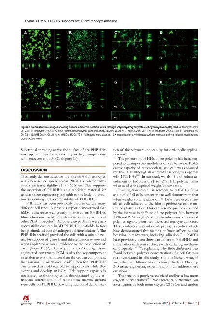
Substantial spreading across the surface <strong>of</strong> the PHBHHx<br />
was apparent after 72 h, indicating its high compatibility<br />
with tenocytes and hMSCs (Figure 3F).<br />
DISCUSSION<br />
y-z<br />
y-z<br />
This study demonstrates for the first time that tenocytes<br />
will adhere to and spread across PHBHHx polymer films<br />
with a preferred rigidity <strong>of</strong> > 420 N/m. This supports<br />
the assertion <strong>of</strong> PHBHHx as a candidate material for<br />
tendon tissue engineering and adds to the body <strong>of</strong> literature<br />
supporting the biocompatibility <strong>of</strong> PHBHHx.<br />
PHBHHx has been previously used to culture many<br />
different cell types. A previous report demonstrated that<br />
hMSC adherence was greatly improved on PHBHHx<br />
films when compared to both tissue culture plastic and<br />
other PHA molecules [6] . Adipose derived MSCs were also<br />
successfully cultured in 3D PHBHHx scaffolds before<br />
being stimulated into chondrogenic differentiation [18] . The<br />
PHBHHx scaffold provided the cells with a suitable matrix<br />
for support <strong>of</strong> growth and differentiation in vitro and<br />
when implanted in vivo as evidence by the production <strong>of</strong><br />
cartilaginous ECM, a key requirement <strong>of</strong> cartilage tissue<br />
engineered constructs. ECM is also the key component<br />
in tendon as it is this, rather than the cellular component,<br />
that sustains the mechanical load [8] . Therefore, PHBHHx<br />
can be used as a 3D scaffold to support cells while they<br />
express and develop an ECM. This support capacity is<br />
not limited to chondrocytes, as demonstrated by the osteogenic<br />
differentiation <strong>of</strong> rabbit bone marrow derived<br />
stem cells on PHBHHx providing additional demonstra-<br />
B<br />
x-y<br />
x-z<br />
F<br />
x-y<br />
x-z<br />
WJSC|www.wjgnet.com<br />
y-z<br />
y-z<br />
C<br />
x-y<br />
x-z<br />
G<br />
y-z<br />
tion <strong>of</strong> the polymers applicability for orthopedic application<br />
use [3] .<br />
The proportion <strong>of</strong> HHx in the polymer has been proposed<br />
as an important modulator <strong>of</strong> cell behavior. Proliferative<br />
capacity <strong>of</strong> rat smooth muscle cells was enhanced<br />
by 20% HHx although attachment at seeding was optimal<br />
with 12% HHx [19] . In our study we also found robust attachment<br />
<strong>of</strong> hMSC and rT to 12% HHx polymer films<br />
when used at the optimal weight/volume ratio.<br />
Investigation into rT attachment to PHBHHx films<br />
as a total <strong>of</strong> all cells present in the well demonstrates that<br />
when weight/volume ratios <strong>of</strong> ≥ 1.6% were used, virtually<br />
all cells adhered to the film in preference to the untreated<br />
plastic surface. This can in some part be explained<br />
by the increase in stiffness <strong>of</strong> the polymer film between<br />
1.6% and 2.0% weight/volume. In other words, increased<br />
polymer rigidity promoted increased tenocyte adhesion.<br />
This reinforces a number <strong>of</strong> previous studies which<br />
have demonstrated that material stiffness affects cellular<br />
behavior in many ways, including adhesion [20-22] . hMSCs<br />
have previously been shown to adhere to PHBHHx and<br />
many other different surfaces with differing mechanical<br />
properties [3,23,24] , explaining why little difference was<br />
found between polymer concentrations. As cell fate was<br />
not investigated in this study, it is not known what, if<br />
any, effect on differentiation potency this had. Ongoing<br />
3-D tissue engineering experimentation will address these<br />
questions.<br />
The tendon is poorly vascularized and has a low mean<br />
oxygen concentration [25] . We therefore performed our<br />
investigation in both room oxygen (21% O2) and tendon<br />
98 September 26, 2012|Volume 4|Issue 9|<br />
D<br />
x-y y-z<br />
Figure 3 Representative images showing surface and cross section views through poly(3-hydroxybutyrate-co-3-hydroxyhexanoate) films. A: tenocytes 21%<br />
O2, 24 h; B: tenocytes 21% O2, 72 h; C: Human mesenchymal stem cells (hMSCs) 21% O2, 24 h; D: hMSCs 21% O2, 72 h; E: Tenocytes 2% O2, 24 h; F: Tenocytes 2%<br />
O2, 72 h; G: hMSCs 2% O2, 24 h; H: hMSCs 2% O2, 72 h. All images were taken at 10 × magnification. x-y indicates surface view, x-z and y-z indicate reconstructed<br />
cross section views.<br />
x-y<br />
x-z<br />
y-z<br />
x-z<br />
H<br />
x-y<br />
x-z<br />
y-z


