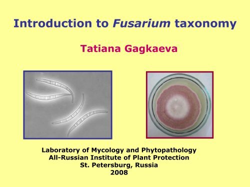
Introduction to Fusarium taxonomy
Introduction to Fusarium taxonomy
Introduction to Fusarium taxonomy

Create successful ePaper yourself
Turn your PDF publications into a flip-book with our unique Google optimized e-Paper software.
Teleomorphs of <strong>Fusarium</strong>Gibberella – the majority of <strong>Fusarium</strong> speciesHaema<strong>to</strong>nectria – <strong>Fusarium</strong> solani complexAlbonectria – <strong>Fusarium</strong> decemcellulareTeleomorph morphology usually is not sufficientlyfor identification purposes.Gibberella zeae
Key characters for identifying <strong>Fusarium</strong> speciesMacroconidiaSizelarge versus smallNumber of septarange and averageShapeelongate versus squatdegree of curvature
Key characters for identifying <strong>Fusarium</strong> speciesDorsal sideVentral sideDorsal sideVentral sideMacroconidiaApical cellsBasal cells
Apical cells shapes:A1.Basal cells shapes:B1.A2.A3.B2.B3.A4.B4.A1. BluntA2. PapillateA3.TaperingA4. HookedB1. Foot-shapedB2. Elongated foot-shapedB3. Distinctly notchedB4. Barely notched
Key characters for identifying <strong>Fusarium</strong> speciesMicroconidiaShape and SizeNumber of cellsFalse heads or/and chains
Key characters for identifying <strong>Fusarium</strong> speciesConidiophores are special structures of hyphae whichfinished in fertile cells - phialides and provide conidiawith nutrients.- unbranched, sparsely or densely branched.Conidogenous cellsMonophialides – having only one opening per cellPolyphialides – having more than one opening per cell (but notnecessarily for all conidiogenous cells)
Key characters for identifying <strong>Fusarium</strong> speciesMonophialidesPolyphialides
Length of the Conidiogenous cellLong (F.solani)Short (F.poae)
Key characters for identifying <strong>Fusarium</strong> speciesMesoconidiaSpindle-shaped conidia that are typically produced in F.sporotrichioides, F. chlamydosporum, F. semitectum, F.camp<strong>to</strong>ceras and in some isolates of F. avenaceum and F.subglutinans.These spores are not produced in sporodochia, only in the aerialmycelium.F. sporotrichioides
Key characters for identifying <strong>Fusarium</strong> speciesChlamydosporesThe presence of chlamydospores is the diagnostic character.Their absents is not informative character.They can take long period of time <strong>to</strong> form (up <strong>to</strong> 6 weeks).Formed singly, doubles, in clumps or in chains.May be found in aerial mycelia or embedded in the agar.
Key characters for identifying <strong>Fusarium</strong> speciesColony morphologyAbundance and color of aerial myceliumPigmentationGrowth rates, especially at 25°C and 30°C
ReverseSurface
Beginning the identification processThe majority of isolates are not difficult <strong>to</strong>identify if you use:•Controlled temperature and light conditions•Standard media•Cultures initiated from single spores
Growth ConditionsRemember, try <strong>to</strong> use the conditions outlined inthe identification guide you are following!Temperature – generally 25°C or fluctuating25/20°C day/night cycle.LightGood light conditions will help <strong>to</strong> stimulate conidiaformation.Artificial daylight (cool white tubes) on a 12 hrlight:12 hr dark cycle necessary <strong>to</strong> maximizeproduction of sporodochia.Black light (near-UF, emission ca. 310-360 nm) maybe essential for teleomorph development.
Standard media for growth of <strong>Fusarium</strong> fungiMedia for <strong>Fusarium</strong> isolation:Pota<strong>to</strong> dextrose agar (PDA)Czapek-Dox agarThe media are supplemented with antibiotics and fungicides after theagar is melted and the temperature is not higher than 55 degrees C.
Antibiotic solutionsAntibiotic S<strong>to</strong>ck solution, g Workingconcentration,ml/L of mediumStrep<strong>to</strong>mycin 5 /100ml H 2O 20 G-EffectiveagainsbacteriaNeomycin 1/ 100ml H 2O 12 G+Chlotetracyclinehydrochloride0.5/ 100ml H 2O 10 G- and G+Chloramphenicol 0.5/10 ml ethanol (96%) 1 G- and G+Fungicides prevent growth of saprophytesPCNB (Pentachloronitrobenzene): 750mg/L. PCNB is usuallyadded as 1 g of Terrachlor, which contained 75% PCNB (w/w).Dichloran: 200 mg in 100 ml ethanol (96%) (1 ml is added <strong>to</strong>1000 ml agar before au<strong>to</strong>claving).Propiconazole: 0.375 mg /1L after the agar is melted and thetemperature is below 55 degrees C.The linear growth of fungi may be suppressed by Tri<strong>to</strong>n X-100,s<strong>to</strong>ck solution 2% is au<strong>to</strong>claved and used at the rate of 10 ml/Lof medium.
Weak nutrient mediaCarnation Leaf-Piece Agar (CLA). CLA is a natural substratemedium prepared by aseptically placing sterile carnation leaf pieces,3-5 mm, in<strong>to</strong> a Petry dish and adding sterile 2% water agar (5-6 leafpieces is dishes). Water agar (WA) consist of 20 g agar in 1 L ofdistilled H 2 O.Spezieiller Nährs<strong>to</strong>ffarmer Agar (SNA). In 1 L of distilled H 2 O:KH 2 PO4 1 g; KNO 3 1 g; MgSO 4 x 7H 2 O 0.5 g; KCl 0.5 g; Glucose 0.2 g; Sucrose 0.2 g; Agar 20 g.Placing 1-2 pieces of sterile filter paper (approximately 1cm 2 ) on the agarsurface after the medium hag gelled can increase sporulation.Media promotes sporulation and good conidiogeneous cell development.Media transparent, so cultures can be viewed directly with microscope (x100).CLA- surface CLA- reverse SNA- surface
Criteria for identificationPDA medium:Colony morphologyPigmentation – hyphae and in agarGrowth ratesCLA and SNA media:Presence/absence of conidiaShape and formation of conidiaNature of the conidiogenous cellsPresence/absence of chlamydospores
Single spore isolationCan be used for macroconidia, microconidia and ascospores.If you want <strong>to</strong> be sure in identification, use singlespore cultures!Never use cultures before single sporing in the further studies(genetics, molecular, pathogenicity, etc.)! More than onespecies/strain of fungi or <strong>Fusarium</strong> from the same substrate.
Pro<strong>to</strong>col:Dilution Plate• Prepare sterile water in a vial• Transfer sporodochial conidia from culture with a fine needlein a water.• Check the concentration and dilute (5-10 conidia/drop) undera low power (10x) microscope.• A few drops of the suspension spread over 2% dry and thinWA plates.• Leave plates for 6-15 hr.• Check plates for germinating conidium along under a lowpower (10x).• Mark with a fine marker.• Pick small squares of agar around a germinating conidiumusing a fine sterile needle and transfer <strong>to</strong> maintenance media.
* If the culture is not form conidia then pick a hyphal tipseparated from others by spreading on WA plate and transfer<strong>to</strong> maintenance media.
<strong>Fusarium</strong> genusMain species:F. graminearum (Gibberella zeae) complexF. culmorumSection Discolor<strong>Fusarium</strong> head blight
Section DiscolorColony morphology- fast growing (5 <strong>to</strong> 7 cm >growth in 7 days)- red/pink pigmentsMacroconidia- relatively robust and thick-walled- typically formed in sporodochia- less often on hyphaeMicroconidia- absent or rareConidiophores- monophialidesChlamydospores- present in conidia and hyphae- single, chains, clumps- inconsistently produced
Section DiscolorColony appearanceF. graminearumsurfaceF. culmorumreversesurfacereverse
Section DiscolorMacroconidia<strong>Fusarium</strong>Size Cells Surface Septate Sporodochialength ratio* apical basal ventral dorsalF. graminearum 40-60 11 tapered footshapedstraightsmoothlyarched5-6 paleorangeF. culmorum 25-35 6 blunt,roundedfootshape-notchedstraight curved 3-4 brick red<strong>to</strong> brown* approximate ratio = length/ width of macroconidiaF. graminearum F. culmorum
Section RoseumMain species:F. avenaceum (Gibberella avenacea)Cosmopolitan, especially temperate zones.Wide range of plants:seedling disease, crown & root rot.Colony morphology- highly variable- surface: floccose, grey rose, yellow, white.- growth rate varies from moderate <strong>to</strong> rapidPigment - grey rose <strong>to</strong> burgundy, brownishConidiophores- branched and unbranched monophialidesSporodochia: Usually bright orangeChlamydospores - Absent
Section RoseumF. avenaceumMacroconidia formed on hyphae and in sporodochiaMacroconidia in sporodochia– long, slender, thin-walled, usually 3-5 septate– falcate <strong>to</strong> almost straight "needle-like"Apical cell – elongate, bentBasal cell – notched or foot-shapedMacroconidia &Mesoconidia in aerial mycelium– often abundant– fusiform, comma-shaped, 0-3 septate
Section SporotrichiellaMicroconidia pyriform, globular <strong>to</strong> spindle-shaped, or lemon-shaped.Main species:F. sporotrichioides F. poaeF. tricinctum(Gibberella tricincta)F. langsethiae
Section SporotrichiellaF. sporotrichioidesCosmopolitan.Occurs on a wide variety of plants & soil.Colony morphology- rapid growth, abundant floccose mycelium- sporodochia may occur in older culturesSurface: white, pinkish <strong>to</strong> brownish redPigment: rose <strong>to</strong> burgundyMacroconidia: abundant- on hyphae and sporodochia- falcate, 3-5 septateApical cell: curved, pointedBasal cell: notched or foot-shapedMicroconidia: abundant- oval <strong>to</strong> pyriform (pear-shaped), spindle-shaped- 0-1 septate, often with papillaConidiophores- monophialides & polyphialidesChlamydospores - Abudant
Section SporotrichiellaF. poaeCosmopolitanMore common on gramineous hostsColony morphology- rapid growth, abundant floccose mycelium- sporodochia rareSurface: white, pinkish whitePigment: rose <strong>to</strong> burgundyMacroconidia: generally rare- on hyphae and sporodochia- falcate, 3-5 septateApical cell: short, curved, pointedBasal cell: foot-shaped or notchedMicroconidia: abundant- globose <strong>to</strong> pyriform, often with papilla- 0-1 septate,- produced in clusters, "bunch of grapes"Conidiophores- unbranched & branched monophialidesChlamydospores - AbsentSome isolates has fruity odor
Section SporotrichiellaF. langsethiaeUp <strong>to</strong> now found only in EuropeDetected in small cereals grainColony morphology- slow growth, nearly lack of aerial myceliumSurface: white <strong>to</strong> peach, pinkish white, powderPigment: peach <strong>to</strong> violetSporodochia absentMicro morphology similar <strong>to</strong> F. poaeMacroconidia: absentMicroconidia: abundant- globose <strong>to</strong> pyriform, often with papilla- 0-1 septate,- produced in clusters, "bunch of grapes"Conidiophores- unbranched & branched monophialidesChlamydospores - AbsentNo odor
Section SporotrichiellaF. tricinctumCosmopolitan, more common in temperate areasColony morphology- moderate rapid growth,- mycelium as compact cushin- sporodochia may occur in older culturesSurface: red <strong>to</strong> vinaceous or purplePigment: red <strong>to</strong> vinaceous or purpleMacroconidia:- usually form in sporodochia- falcate <strong>to</strong> crescent-shaped, slender- 3-5 septateApical cell: curved, pointedBasal cell: notched or foot-shaped, narrow andpointedMicroconidia: abundant- lemon-shaped <strong>to</strong> pyriform, ellipsoidal- 0-1 septateConidiophores:monophialides, richly branched, slenderChlamydospores: formed singly or in chains
Section GibbosumMain species:F. equiseti (Gibberella intricans)F. acuminatum (Gibberella acuminata)Weak pathogens of wide range of plants, in soil.MacroconidiaUsually strongly curved on both ventral and dorsal sides of conidiastriking foot-shaped basal cellOften quite largeNo microconidiaChlamydospores presentPigment: red or brown
Section GibbosumF. equisetiCulture morphology:- fast growing- abundant myceliumSurface: white <strong>to</strong> beige or brownPigment: pale <strong>to</strong> dark brown with brown flecksMacroconidia- various sized - occasionally very long- 5-septate, with strong curvatureApical cell: tapering cellBasal cell: foot shapedChlamydospores abundant in hyphae, brownReverseSurface
Section GibbosumF. acuminatumCulture morphology:- slow-middle growing- floccose myceliumSurface: peach-beigePigment: carmine redSporodochia blight orangeMacroconidia- slender, sickle-shaped- 3-5-septateApical cell: tapering cellBasal cell: pedicellate shapedChlamydospores in hyphaeSurfaceReverse
Section LiseolaGibberella fujikuroi species complexF. proliferatumThe tester strains of G.fujikuroi complexWorldwide distributed on a wide variety of plantsColony morphology- rapid growth, abundant floccose mycelium- sporodochia may occurSurface: white, becoming purple-violet with agePigment: varies in violet intensityFertile perithecia G.fujikuroi after makingcrosses with the tester strain F.proliferatumReverse
Section LiseolaGibberella fujikuroi species complexF. proliferatumMacroconidia: usually abundant- on hyphae and sporodochia- slender, thin-walled, relatively straight, 3-5septateApical cell: curvedBasal cell: poorly developedMicroconidia: abundant- oval with flattened base, pyriform also mayoccur- 0 septate- in short chains and in false headsConidiophores- monophialides & polyphialidesChlamydospores - absent
Section ElegansF.oxysporum species complexWorldwide distributed on a wide variety of plants. Animportant vascular wilt and root rot pathogens.Many isolates are host specific and differentiated as formaespeciales (f.sp.) – infraspecific subdivision.Colony morphology varies widely!!!-may be floccose, sparse, abundant- sporodochia may occur, pale orangeSurface: white, becoming purple-violet with agePigment: unpigmented <strong>to</strong> dark violetMacroconidia: usually abundant- on hyphae and sporodochia- short <strong>to</strong> medium lenght- straight, <strong>to</strong> slightly curved- thin walled, usually 3- septateApical cell: tapered and curvedBasal cell: foot shaped <strong>to</strong> pointedMicroconidia: abundant- oval, elliptical- 0 septate- in false headsConidiophores- Short monophialidesChlamydospores– usually formed abundantly and quickly-singly or in pairsSurface