Burkholderia mallei and B. pseudomallei - Microbiology - American ...
Burkholderia mallei and B. pseudomallei - Microbiology - American ...
Burkholderia mallei and B. pseudomallei - Microbiology - American ...

Create successful ePaper yourself
Turn your PDF publications into a flip-book with our unique Google optimized e-Paper software.
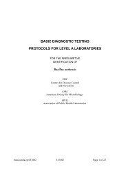
28 February 2008SENTINEL LABORATORY GUIDELINESFORSUSPECTED AGENTS OF BIOTERRORISM<strong>Burkholderia</strong> <strong>mallei</strong> <strong>and</strong> B. pseudo<strong>mallei</strong><strong>American</strong> Society for <strong>Microbiology</strong>1
Credits: <strong>Burkholderia</strong> <strong>mallei</strong> <strong>and</strong> B. pseudo<strong>mallei</strong>Subject Matter Experts, ASM:Peter H. Gilligan, Ph.D.University of North CarolinaHospitals/Clinical <strong>Microbiology</strong> & Immunology LabsChapel Hill, NCMary K. York, Ph.D.MKY <strong>Microbiology</strong> ConsultantsWalnut Creek, CAASM Sentinel Laboratory Guideline Working GroupVickie Baselski, Ph.D.University of Tennessee at MemphisMemphis, TNRoberta B. Carey, Ph.D.Karen Krisher, Ph.D., D (ABMM)Clinical <strong>Microbiology</strong> InstituteWilsonville, ORJudith Lovchik, Ph.D.Indiana State Department of HealthIndianapolis, INLarry Gray, Ph.D.TriHealth Laboratories <strong>and</strong>University of Cincinnati College of Medicine, <strong>and</strong>Clinical <strong>Microbiology</strong> Laboratory Consultants, LLCCincinnati, OHRosemary Humes, MS, MT (ASCP) SMAssociation of Public Health LaboratoriesSilver Spring, MDChris N. Mangal, MPHAssociation of Public Health LaboratoriesSilver Spring, MDDaniel S. Shapiro, M.D.Lahey ClinicBurlington, MA2
Susan E. Sharp, Ph.D.Kaiser PermanentePortl<strong>and</strong>, ORAlice Weissfeld, Ph.D.<strong>Microbiology</strong> Specialists, Inc.Houston, TXDavid Welch, Ph.D.Laboratory Corporation of AmericaDallas, TXCoordinating Editor:James W. Snyder, Ph.D.University of LouisvilleLouisville, KY3
Table of Contents: <strong>Burkholderia</strong> <strong>mallei</strong> <strong>and</strong> B. pseudo<strong>mallei</strong>I. General InformationA. Description of OrganismsB. HistoryC. Geographic DistributionD. Clinical PresentationE. Treatment <strong>and</strong> ProtectionII. ProcedureA. GeneralB. PrecautionsC. SpecimenD. MaterialsE. Quality ControlF. Stains <strong>and</strong> SmearsG. CulturesH. Interpretation <strong>and</strong> ReportingIII. References4
I. GENERAL INFORMATIONA. Description of organisms<strong>Burkholderia</strong> <strong>mallei</strong> is a nonmotile, aerobic gram-negative coccobacillus, whichmay or may not be oxidase positive or grow on MacConkey agar. <strong>Burkholderia</strong>pseudo<strong>mallei</strong> is an oxidase-positive, aerobic gram-negative bacillus that is straightor slightly curved. The organism will grow on most st<strong>and</strong>ard laboratory media, suchas sheep blood <strong>and</strong> chocolate <strong>and</strong> MacConkey agars, <strong>and</strong> it produces a characteristicmusty odor (13). The recent sequencing of the genomes of these two organismssuggest that B. <strong>mallei</strong> is a recently evolved clone of B. pseudo<strong>mallei</strong>. B. <strong>mallei</strong> hasa smaller genome which makes it much less environmentally adaptable (14).B. History<strong>Burkholderia</strong> <strong>mallei</strong> is the etiologic agent of gl<strong>and</strong>ers, a febrile illness typically seenin equines: i.e., horses, mules, <strong>and</strong> donkeys (2). During World War I, the Germanmilitary used this agent as a biological weapon against horses <strong>and</strong> mules, theprimary form of transportation during that conflict (4). Naturally occurring humaninfection is likely due to exposure to an infected animal. The last naturally acquiredhuman case of gl<strong>and</strong>ers in the United States was seen in 1945 (2). A case oflaboratory-acquired gl<strong>and</strong>ers occurred in 2000 (2).<strong>Burkholderia</strong> pseudo<strong>mallei</strong> is an environmental organism found in soil <strong>and</strong> water<strong>and</strong> is most likely obtained naturally by direct contact with, or aerosols from,environmental sources. For many years, this bacterial species as well as B. <strong>mallei</strong>were classified as a member of the genus Pseudomonas, but in 1992, they werereclassified into the genus <strong>Burkholderia</strong> (28). B. pseudo<strong>mallei</strong> was first reported ascausing human infections in 1911 by Whitmore from individuals living in Rangoon,Burma (now Yangon, Myanmar) (27) <strong>and</strong> in earlier medical literature was called“Whitmore’s disease.” These patients were described as septicemic withwidespread abscesses in the lungs, liver, spleen, <strong>and</strong> kidneys. <strong>Burkholderia</strong> was oneof the first organisms reported as a cause of infection in intravenous drug users (27).Because the organism was thought to cause a gl<strong>and</strong>ers-like illness in humans, it wascalled “pseudo<strong>mallei</strong>” by Stanton <strong>and</strong> Fletcher (8). The gl<strong>and</strong>ers-like disease inhumans due to B. pseudo<strong>mallei</strong> is now referred to in the medical literature as“melioidosis,” from the Greek word “melis,” which was the term for distemper indonkeys (19). It was found to cause disease in soldiers from both Australia <strong>and</strong> theUnited States during the Vietnam War <strong>and</strong> has been referred to as the “Vietnamtime bomb” because the disease, much like tuberculosis, can reactivate afterremaining latent for decades (8).Two organisms which are very similar to B. pseudo<strong>mallei</strong> phenotypically haverecently been described in the literature (9,10). <strong>Burkholderia</strong> thail<strong>and</strong>ensis is anenvironmental organism found in rice paddy water <strong>and</strong> soil in Thail<strong>and</strong>. It has beenshown to be of comparatively low virulence in animal models <strong>and</strong> has beeninfrequently reported to be a cause of human disease (9,10). In the clinicallaboratory, it is most easily differentiated from B. pseudo<strong>mallei</strong> on the basis of its5
ability to assimilated arabinose <strong>and</strong> test for which B. pseudo<strong>mallei</strong> is negative. Thesecond organism is <strong>Burkholderia</strong> oklahomensis. It has been reported from twocases in the US <strong>and</strong> it has been found to be essentially avirulent in animal models(9). These two organisms will not be differentiated from B. pseudo<strong>mallei</strong> by thealgorithm supplied in this protocol. However given the rarity of isolation of thesetwo organisms in clinic settings, it is unlikely that they will be encountered incritically ill individuals.C. Geographic distributionB. <strong>mallei</strong> was eradicated from the United States <strong>and</strong> Western Europe due to aprogram of compulsory slaughter of infected or seropositive horses or otheranimals. Equines) are the primary reservoir of the rare cases of gl<strong>and</strong>ers still seen inEastern Europe, the Middle East, Asia, <strong>and</strong> Africa (2).Melioidosis is a disease endemic in the tropical regions of the world, with themajority of cases in the medical literature being reported from rice-growing regionsof Southeast Asia <strong>and</strong> the tropical, northern regions of Australia. The organism hasbeen detected in very high concentrations in water found in rice paddies in bothVietnam <strong>and</strong> Thail<strong>and</strong> (13). There are data to suggest that this organism is alsoendemic in both the Philippines <strong>and</strong> the Indian subcontinent (8). There is littleknown about the prevalence of this organism in tropical regions of Africa <strong>and</strong> theAmericas although cases have been recently documented in both Brazil <strong>and</strong>Honduras (3,18). This paucity of knowledge may be due to the inability torecognize this organism in part due to poor performance of commercialidentification systems (11,17). Infections with this organism in the United States<strong>and</strong> Western Europe are almost certainly imported from regions of endemicity, <strong>and</strong>imported cases are well documented in the medical literature (13). Laboratoryacquiredinfections have also been reported (8). The recognition of multiple cases ofmelioidosis in North America or Western Europe in patients without an appropriatetravel history requires a thorough investigation of the possibility of a bioterrorismattack.D. Clinical presentation1. Gl<strong>and</strong>ers. Gl<strong>and</strong>ers can present as either a cutaneous or systemic disease (2).The incubation period is typically 1 to 14 days.Cutaneous. Patients with the cutaneous infection will have nodules withaccompanying localized lymphadenitis.Systemic. The systemic illness usually manifests itself either as broncho- or lobarpneumonia. Bacteremia may also occur, resulting in lesions being seen in the liver<strong>and</strong> spleen. Infection in humans with B. <strong>mallei</strong> is often fatal without antimicrobialtreatment.2. Melioidosis. The illness can manifest as an asymptomatic, acute, subacute, orchronic process.6
amoxicillin-clavulanate or a combination of doxycycline <strong>and</strong> trimethoprimsulfamethoxazolefor 3 to 6 months is recommended (15). Because resistance hasdeveloped with ceftazidime therapy, combination therapy is usually recommended forinitial treatment. There are no data published on prophylactic agents for B.pseudo<strong>mallei</strong>, although it is likely that the agents used for oral eradication therapywould be useful prophylactically. Of note, B. pseudo<strong>mallei</strong> organisms are frequentlyresistant to aminoglycosides, first <strong>and</strong> second generation cephalosporins, <strong>and</strong>fluoroquinolone antimicrobials in vitro, <strong>and</strong> so drugs in these classes, would likely notbe good prophylactic agents.3. There is no vaccine currently available for either B. <strong>mallei</strong> or B. pseudo<strong>mallei</strong>.II. PROCEDURESA. GeneralThe procedures described below are to be used to rule out the presence of B. <strong>mallei</strong><strong>and</strong> B. pseudo<strong>mallei</strong> in clinical specimens or as isolates. These procedures will notdifferentiate B. pseudo<strong>mallei</strong> from B. thail<strong>and</strong>ensis or B. oklahomensis. Because the2 species share >99% homology at the nucleotide level, differentiation betweenthem at the molecular level is challenging (1). Biochemical differentiation can bemore productive since they have unique characteristics.B. Precautions1. Sentinel laboratories should not accept environmental or animal specimens; suchspecimens should be forwarded directly to the State Public Health Laboratory.(A list of State Public Health Laboratory contacts can be downloaded athttp://www.aphl.org/programs/emergency_preparedness/Documents/EPrep110707_SPHL_contact_list.pdf)2. Laboratory-acquired infections have been documented. All patient specimens<strong>and</strong> culture isolates should be h<strong>and</strong>led while wearing gloves <strong>and</strong> gowns in abiosafety cabinet. Plates should be taped shut when incubating.C. Specimens1. Blood or bone marrow2. Sputum or bronchoscopically obtained specimens3. Abscess material <strong>and</strong> wound swabs4. Urine5. Serum (1 ml). Both acute- <strong>and</strong> convalescent-phase (obtained 14 days after theacute-phase specimen) specimens should be collected if serologic diagnosis of B.pseudo<strong>mallei</strong> infection is being considered. Currently, no serology for B. <strong>mallei</strong>is available in the United States.D. Materials1. Blood <strong>and</strong> bone marrow cultures can be done using:a. St<strong>and</strong>ard automated blood culture system8
. Lysis centrifugation system2. Media for isolation from other clinical specimens:a. Chocolate agar (CHOC)b. Sheep blood agar (SBA)c. MacConkey agar (MAC)d. Selective agars for <strong>Burkholderia</strong> pseudo<strong>mallei</strong> including <strong>Burkholderia</strong> cepaciaselective agars (13, 25)3. Reagentsa. Gram stain reagentsb. Oxidase reagentc. Hydrogen peroxide (3%) for catalase testd. Spot indole reagent (5% p-dimethylaminobenzaldehyde or 1%paradimethylaminocinnamaldehyde in 10% [vol/vol] concentrated HCl.)e. Colistin (10 µg) or polymyxin B (300 U) diskf. Motility semisolid medium with 2,3,5-triphenyltetrazolium chloride (TTC)indicator (Remel, Inc.:Lenexa Ks) or broth for wet mount motility.g. Optional reagentsi. Arginine dihydrolase <strong>and</strong> Moeller base controlii. Triple sugar iron agar (TSI) or Kligler’s iron agar (KIA)iii. Nitrate testa. Nitrate broth with gas indicator tubeb. Nitrite reduction reagent 1 (sulfanilic acid)c. Nitrite reduction reagent 2 (dimethyl-α-naphthylamine)d. Zinc dusth. Alternately, API 20NFT, also called 20NE (BioMerieux, Durham, N.C.), orVitek 1 gram-negative identification panels (BioMerieux) can be used forpreliminary identification of <strong>Burkholderia</strong> psuedo<strong>mallei</strong> but not B. <strong>mallei</strong> withreasonable accuracy. For a review of the performance of commercial systems,see reference 24.NOTE: While only the API 20NFT <strong>and</strong> the Vitek system have been studiedextensively (16, 22), other systems that have arginine as one of the biochemical testsmay work well. B. pseudo<strong>mallei</strong> is in the database of the Microscan overnight system,but not the rapid, gram-negative rod panel, although the sensitivity <strong>and</strong> specificity ofthe overnight product have not been studied extensively. <strong>Burkholderia</strong> pseudo<strong>mallei</strong>is not in the Phoenix (BD) database <strong>and</strong> has been reported to call the isolates B.cepacia (21).9
4. Equipment <strong>and</strong> supplies1. Blood culture instrument (optional)2. 35°C (<strong>and</strong> 42°C [optional]) incubators3. Light microscope with ×100 objective <strong>and</strong> ×10 eyepiece4. Microscope slides, disposable bacteriologic inoculating loops5. Glass tubes, sterile pipettes6. Biological safety cabinet (BSC)E. Quality controlDocument all quality control (QC) for the following tests per st<strong>and</strong>ard laboratoryprocedure/protocol.F. Stains <strong>and</strong> smears. Gram stain1. Procedure. Perform Gram stain procedure/QC per st<strong>and</strong>ard laboratory protocol.2. Interpretationa. B. <strong>mallei</strong> is a small gram-negative coccobacillus.b. B. pseudo<strong>mallei</strong> is a small gram-negative rod (Fig. 1).c. The organisms may be observed in direct gram stain from respiratoryspecimens or abscess material/wounds. They can also be seen in smears ofpositive blood culture bottles.G. Cultures1. Inoculation <strong>and</strong> plating procedures. Inoculate <strong>and</strong> streak the following mediafor isolation of the respective specimen types. NOTE: St<strong>and</strong>ard media should beused according to normal laboratory procedures.a. Blood cultures. Process according to st<strong>and</strong>ard laboratory procedure.b. Respiratory specimens, abscess material/wounds. Plate directly onto SBA<strong>and</strong> MacConkey agar; a st<strong>and</strong>ard enrichment broth can be used forwound/abscess material. Ashdown medium (13) is a selective mediumspecifically designed for recovery of B. pseudo<strong>mallei</strong>. This medium is notlikely to be available in most Sentinel laboratories. If available, <strong>Burkholderia</strong>cepacia selective agars can also be used to for the isolation of B. pseudo<strong>mallei</strong>(25). It is unlikely that BCSA selective medium could be used for isolation ofB. <strong>mallei</strong> since isolates are typically not resistant to aminoglycosides, aselective agent in this medium (26).For improved isolation, a colistin disk or polymyxin B disk may be placed inthe initial inoculation area of the SBA if isolation of <strong>Burkholderia</strong> spp. isspecifically requested.10
2. Incubationa. Temperature. 35 to 37°Cb. Atmosphere. Ambient; CO 2 acceptablec. Length of incubation. Hold primary plates for a minimum of 5 days; readdaily. B. pseudo<strong>mallei</strong> will reliably grow with 5 days of incubation from bloodcultures, so extended incubation of either broth or plated blood cultures (lysiscentrifugation)is not necessary. B. <strong>mallei</strong> will not grow as rapidly as B.pseudo<strong>mallei</strong> <strong>and</strong> may require extended incubation.3. Growth <strong>and</strong> colony characteristicsAll cultures suspected of containing B. <strong>mallei</strong> or B. pseudo<strong>mallei</strong> should beh<strong>and</strong>led in a biological safety cabinet.a. Fulminant sepsis. The recovery of B. pseudo<strong>mallei</strong> from blood culturewithin the first 24 h of incubation indicates fulminant sepsis, which has a veryhigh (90%) mortality rate.b. Growth characteristics1. B. <strong>mallei</strong>i. On SBA, the organism shows smooth, gray, translucent colonies in 2 days,without pigment or distinctive odor. B. <strong>mallei</strong> will grow without anyinhibition around the colistin or polymyxin B disk.ii. Colonies may or may not be present on MacConkey agar.2. B. pseudo<strong>mallei</strong>i. On SBA, the organism often reveals small, smooth creamy colonies in thefirst 1 to 2 days, which gradually change after a few days to dry, wrinkledcolonies similar to Pseudomonas stutzeri. Colonies are neither yellow norviolet pigmented. B. pseudo<strong>mallei</strong> will grow without any inhibition aroundthe colistin or polymyxin B disk.ii. Colonies are present on both SBA <strong>and</strong> MacConkey agars.iii. B. pseudo<strong>mallei</strong> often produces a distinctive musty or earthy odor that isvery pronounced on opening a petri dish growing the microorganism oreven opening an incubator door when a positive plate is present.“Sniffing” of plates containing B. pseudo<strong>mallei</strong> is dangerous <strong>and</strong>should not be done. However, the odor will be apparent without sniffing.4. Screening testsa. The following biochemical tests can be used to rule out an isolate as B.<strong>mallei</strong>: oxidase, indole, catalase (not needed if isolate is growing onMacConkey agar), resistance to colistin or polymyxin B, <strong>and</strong> motility. Isolatessuspected to be B. <strong>mallei</strong> based on colony <strong>and</strong> Gram stain morphology shouldhave these tests performed.11
. The following biochemical tests can be used to rule out an isolate as B.pseudo<strong>mallei</strong>: oxidase, indole, <strong>and</strong> resistance to colistin or polymyxin B.Isolates suspected to be B. pseudo<strong>mallei</strong> based on colony, odor, <strong>and</strong> Gramstain morphology should have these tests performed.1. Catalase test. Perform catalase test/QC following st<strong>and</strong>ard laboratoryprocedure, if the isolate is not growing well on MacConkey agar in 48 h:B. <strong>mallei</strong> <strong>and</strong> B. pseudo<strong>mallei</strong> are catalase positive.2. Oxidase test. Perform oxidase test/QC following st<strong>and</strong>ard laboratoryprocedure: B. <strong>mallei</strong> may be oxidase positive or negative. B. pseudo<strong>mallei</strong>is oxidase positive.3. Indole test. Perform indole test/QC following st<strong>and</strong>ard laboratoryprocedure: B. <strong>mallei</strong> <strong>and</strong> B. pseudo<strong>mallei</strong> are indole negative.4. Colistin or polymyxin B resistancei. Procedurea. Streak either an SBA or Mueller-Hinton agar plate with growth, usinga swab dipped into a broth culture corresponding to a no. 0.5McFarl<strong>and</strong> turbidity st<strong>and</strong>ard.b. Place disk in the inoculated area of the plate.c. Incubate for 24 to 48 h.d. Examine for zone of inhibition around the disk.ii. Interpretation1. No zone around the disk indicates resistance to polymyxin B orcolistin. <strong>Burkholderia</strong>, Chromobacterium violaceum, <strong>and</strong> someVibrio, <strong>and</strong> Ralstonia are resistant; most Pseudomonas species aresusceptible.2. B. <strong>mallei</strong> <strong>and</strong> B. pseudo<strong>mallei</strong> are resistant to polymyxin B <strong>and</strong>colistin.3. As an alternative, growth on B. cepacia selective agars or modifiedThayer Martin may substitute for the disk test, because these mediacontain polymyxin B or colistin. However, the lack of growth onthese media should be confirmed by the disk test.12
iii. Quality control (5)Polymyxin B: 300 Uon Mueller HintonagarPseudomonas aeruginosaATCC 27853Escherichia coli ATCC 2592214-18 mm13-19 mmColistin: 10 µgon Mueller HintonagarPseudomonas aeruginosaATCC 27853Escherichia coli ATCC 2592211-17 mm11-17 mm5. Motility testa. Procedure1. The motility test should be performed if the isolate has the colonymorphology <strong>and</strong> Gram stain reaction of B. <strong>mallei</strong> <strong>and</strong> is resistant tocolistin or polymyxin B. Because of the danger of laboratoryacquiredinfection, the wet mount motility should not be performed;the tube test is recommended.2. Inoculate medium with a stab down the center of the tube, to within0.5 in. from the bottom of the tube.3. Incubate tubes at 30°C.b. Interpretation1. A diffusible red-colored growth spreading away from the stab lineindicates motility.2. B. <strong>mallei</strong> is nonmotile, <strong>and</strong> B. pseudo<strong>mallei</strong> is motile.c. Quality control1. Since motility can be difficult to demonstrate among glucosenonfermentingrods, use Pseudomonas aeruginosa ATCC 27853 asthe positive control for the test.2. Klebsiella pneumoniae ATCC 10031 is nonmotile.6. Additional screening testsIf available, TSI or KIA, arginine dihydrolase, <strong>and</strong> nitrate may beperformed to further exclude similar organisms.a. TSI or KIA slant13
1. ProcedurePerform TSI or KIA QC following st<strong>and</strong>ard laboratory procedure.2. InterpretationB. <strong>mallei</strong> <strong>and</strong> B. pseudo<strong>mallei</strong> are glucose-nonfermenting rods orcoccobacilli, <strong>and</strong> the results that will be observed will be an alkaline(red or no change) in tube butt/no H 2 S (no black color). B.pseudo<strong>mallei</strong> may or may not produce an acid (yellow) slant, due tooxidation of lactose.b. Arginine dihydrolase with Moeller’s base control1. Procedurei. Inoculate a tube of both arginine dihydrolase <strong>and</strong> Moeller’s basewith an isolated colony of the suspected isolate.ii. Overlay the contents of both tubes with sterile mineral oil orVaspar, liquid paraffin, or petroleum jelly, maintained at 56°C inliquid form.iii. Tighten caps on tubes.iv. Incubate for 1 to 2 days at 35°C.2. Interpretationi. Compare colors in the Moeller’s base control to that in the tubecontaining arginine.a. Positive result. Arginine tube is clearly purple, while Moellerbase control has not changed color or has become yellow.b. Negative result. No difference between the tube containingarginine <strong>and</strong> the tube containing base.ii. B. <strong>mallei</strong> <strong>and</strong> B. pseudo<strong>mallei</strong> are positive for argininedihydrolase.3. Quality control using glucose-nonfermenting strains:i. Positive control strain. Pseudomonas aeruginosa ATCC 27853ii. Negative control strain. Acinetobacter lwoffi ATCC 153094. This test is available in many identification systems used routinelyin laboratories.c. Nitrate reduction1. Procedurei. Inoculate the nitrate broth with 1 to 2 isolated colonies.ii. Incubate aerobically at 35 to 37°C for up to 48 h.14
iii. After 24 h of incubation, observe for turbidity <strong>and</strong> gas in theDurham tube.a. Do not add reagents if there is no visible growth in the tube.b. If gas is present <strong>and</strong> the isolate is glucose nonfermenting(alkaline butt in TSI or KIA), do not add reagents, as the test ispositive.c. If there is growth <strong>and</strong> no gas production, remove 1 ml of brothfrom tube with a sterile pipette. Place broth in a small glasstube.i. Add 1 or 2 drops of nitrite-reduction reagents 1 <strong>and</strong> 2 tobroth. If a red color forms after the addition of nitritereductionreagents 1 <strong>and</strong> 2, the organism is positive fornitrate reductase activity.ii. If no red color forms after 10 minutes, add a pinch of zincdust. If red color forms after addition of zinc dust, nitratehas not been reduced. If no color change is observed afteraddition of zinc dust, nitrate has been reduced to nitrogengas.d. If the test is negative after 24 h (i.e., a red color is observedafter addition of zinc dust), the remaining broth should bereincubated <strong>and</strong> retested at 48 h.2. Interpretationi. B. <strong>mallei</strong> typically reduces nitrate without gas <strong>and</strong> will give a redcolor with the addition of reagents 1 <strong>and</strong> 2 <strong>and</strong> is therefore nitratereductase positive without gas.ii. B. pseudo<strong>mallei</strong> typically reduces nitrate to nitrogen gas <strong>and</strong> istherefore nitrate reductase positive with gas. B. pseudo<strong>mallei</strong>often produces only one small bubble of gas.3. Quality controli. Positive control strains. Pseudomonas aeruginosa ATCC 27853reduces nitrate to nitrogen gas. Escherichia coli 25922 reducesnitrate to nitrite with no gas.ii. Negative control strain. Acinetobacter lwoffi ATCC 15309. Nored color develops until the zinc dust is added.H. Susceptibility testingDue to the danger in working with B. <strong>mallei</strong> <strong>and</strong> B. pseudo<strong>mallei</strong>, susceptibilitytesting should be performed only in laboratories with biosafety level 3 containment<strong>and</strong> personnel precautions. Routine antimicrobial susceptibility testing is notrecommended.15
I. Interpretation <strong>and</strong> reporting1. Presumptive identification criteriaa. <strong>Burkholderia</strong> <strong>mallei</strong>- see figure 3i. Gram stain reaction. Small, straight or slightly curved gram-negativecoccobacilli. Cells are arranged in pairs, parallel bundles, or the Chineseletterform.ii. Colony characteristics. Colonies are gray, translucent, <strong>and</strong> have nopigment or distinctive odor. They may or may not grow on MacConkeyagar.iii. Oxidase variable, catalase positive, indole negative, nonmotile, colistinresistant.iv. Optional: TSI-alkaline/alkaline, KIA-alkaline/alkaline, argininedihydrolase positive, <strong>and</strong> nitrate reductase positive without gas.v. Kit identification. There is only one study that describes the reliability ofcommercial systems for the identification B. <strong>mallei</strong>. Both API 20 NE <strong>and</strong>RapID NF Plus (Remel, Lenexa, KS) either incorrectly or failed toidentify 23 B. <strong>mallei</strong> isolates. (11) Therefore isolates that screen aspotentially B. <strong>mallei</strong> <strong>and</strong> are not identified by commercial systems shouldbe referred to the LRN Reference Laboratory for identification.b. <strong>Burkholderia</strong> pseudo<strong>mallei</strong> – see Figure 4i. Gram stain reaction. Small, straight, or slightly curved gram-negativerod; may demonstrate bipolar morphology in direct specimens (Fig. 1).ii. Colony characteristics. Colonies are initially cream colored. After 3 or 4days of incubation, B. pseudo<strong>mallei</strong> colonies will have a dry, wrinkledappearance on MacConkey agar (Fig. 2). The colonies will also emit astrong, musty or dirt-like odor.iii. Oxidase positive, indole negative, motile, colistin resistantiv. Optional: TSI-acid or alkaline/alkaline, KIA-acid or alkaline/alkaline,arginine dihydrolase positive, <strong>and</strong> nitrate reductase positive with gas.v. In many cases a Kit identification is performed before B.pseudo<strong>mallei</strong> is suspected (see Figure 5 for guidelines forsupplemental tests when kits are used). Initial studies suggested that theAPI 20E <strong>and</strong> 20NE systems <strong>and</strong> VITEK 1 (but NOT VITEK 2)(BioMerieux, Durham, N.C.) were reasonably reliable systems for theidentification of B. pseudo<strong>mallei</strong> (16, 22). More recent studies suggest thatthe API 20NE is not as reliable for identification of B. pseudo<strong>mallei</strong> whena greater variety of isolates were tested (11,17). In a recent study, whencolormeteric rather than fluorometric based identification system was usedin the VITEK 2, the identification of B. pseudo<strong>mallei</strong> was still not16
particularly accurate (23). These data suggest that organisms identified asB. pseudo<strong>mallei</strong> by commercial systems should be referred to an LRNReference Laboratory for confirmation. In addition glucose nonfermenterswhich screen as potential B. pseudo<strong>mallei</strong> using the abovelisted screening tests <strong>and</strong> give “no identification” by commercial systemsshould also be referred. Finally, some strains of B. pseudo<strong>mallei</strong> have beenmisidentified by the API 20E <strong>and</strong> 20NE systems as C. violaceum (16, 22).Therefore, strains that are not violet colored but are identified as C.violaceum by the API system should also be referred to a LRN Referencelaboratory for confirmation.2. Referral of presumptive B. <strong>mallei</strong> or B. pseudo<strong>mallei</strong> isolates to LRNReference laboratorya. When to refer:i. Naturally occurring cases of B. <strong>mallei</strong> are extremely rare in humans <strong>and</strong>should be referred to LRN Reference laboratories in all cases.ii. Naturally occurring cases of B. pseudo<strong>mallei</strong> may be observed in any ofthe following situations. Confirmation of the identification of theseisolates may be requested from a LRN Reference laboratory, but they areunlikely to represent a bioterrorism event. Patients who cannot beclassified into any of the following patient populations may represent abioterrorism event.1. Patients with acute infection who have a recent history of travel to theregion of endemicity. This includes Southeast Asia (in particularThail<strong>and</strong>, Vietnam, Mynamar, or Taiwan), the Philippines, theIndian subcontinent, or the northern coast of Australia. Recent UScases have also been imported from Honduras <strong>and</strong> cases have beenreported in Brazil so B. pseudo<strong>mallei</strong> may also be considered, forbiothreat reasons, as endemic in tropical regions of Central <strong>and</strong>South America.2. Recent immigrants or visitors from the region of endemicity.3. Patients with recent onset of diabetes, renal failure, orimmunosuppressed states who have traveled in the region ofendemicity mentioned above even if that travel occurred decadesbefore.4. Individuals who work with animals (such as zoo employees) thathave recently been imported from regions of endemicity.5. Individuals who work in laboratories where they may be exposed tothis organism.b. Whom to notify <strong>and</strong> when to notify them:i. Further epidemiologic investigation is needed whenever a presumptiveidentification of B. <strong>mallei</strong> or B. pseudo<strong>mallei</strong> is made.17
ii. Within the hospital setting, the infectious disease service <strong>and</strong>/or infectioncontrol department should be notified so further investigation of thepatient’s history can be made so that naturally occurring infections can beruled out.iii. The State Laboratory Director (or designate) should be notified of thepresumptive identification of B. <strong>mallei</strong> or B. pseudo<strong>mallei</strong>.iv. When referring the isolate to the LRN Reference laboratory, the ReferenceLaboratory should provide the referring laboratory with guidance <strong>and</strong>recommendations for retaining the specimen or isolate. Once theidentification is confirmed, <strong>and</strong> the isolate is confirmed as either B. <strong>mallei</strong>or B. pseudo<strong>mallei</strong>, the Sentinel Lab is required to destroy it (e.g.autoclaving). In particular the appropriate material, including bloodculture bottles, tubes <strong>and</strong> plates, <strong>and</strong> actual clinical specimens (aspirates,biopsies, sputum specimens) should be saved until the ReferenceLaboratory confirms the identification.v. Any environmental specimens collected for detection of B. <strong>mallei</strong> or B.pseudo<strong>mallei</strong> as part of an epidemiologic/criminal investigation should bereferred directly to the State Public Health Laboratory.vi. The State Public Health Laboratory Director will coordinate notificationof the Centers for Disease Control <strong>and</strong> Prevention <strong>and</strong> State <strong>and</strong> Federallaw enforcement agencies.18
Figure 1Gram stain of B. pseudo<strong>mallei</strong> in a blood culture (8)Gram stain of B. pseudo<strong>mallei</strong> from a colony on blood agar (13)19
Figure 2B. pseudo<strong>mallei</strong> colonies on MacConkey agar (13)B. pseudo<strong>mallei</strong> colonies on blood agar (13)B. pseudo<strong>mallei</strong> colonies on Ashdown medium agar (13)20
III. References1. Bauernfeind, A., C. Roller, D. Meyer, R. Jungwirth, <strong>and</strong> I. Schneider. 1998.Molecular procedure for rapid detection of <strong>Burkholderia</strong> <strong>mallei</strong> <strong>and</strong> <strong>Burkholderia</strong>pseudo<strong>mallei</strong>. J. Clin. Microbiol. 36:2737-27412. Centers for Disease Control <strong>and</strong> Prevention. 2000. Laboratory-acquired humangl<strong>and</strong>ers—Maryl<strong>and</strong>, May 2000. Morb. Mortal. Wkly. Rep. 49:532-535.3. Centers for Disease Control <strong>and</strong> Prevention. 2006. Imported meliodosis- SouthFlorida, 2005. Morb. Mortal. Wkly. Rep. 55:873-876.4. Christopher, G. W., T. J. Cieslak, J. A. Pavlin, <strong>and</strong> E. M. Eitzen, Jr. 1997. Biologicalwarfare: a historical perspective. JAMA 278:412-417.5. Clinical <strong>and</strong> Laboratory St<strong>and</strong>ards Institute. 2005. Performance st<strong>and</strong>ards for antimicrobialsusceptibility testing. Seventeenth informational supplement. CLSI document M100-S15.Clinical <strong>and</strong> Laboratory St<strong>and</strong>ards Institute, Wayne, Pa.6. Currie, B. J., D. A. Fisher, N. M. Anstey <strong>and</strong> S. P. Jacups. 2000. Melioidosis: acute<strong>and</strong> chronic disease, relapse <strong>and</strong> re-activation. Trans. R. Soc. Trop. Med. Hyg. 94:301-304.7. Currie, B. J., D. A. Fisher, D. M. Howard, J. N. Burrow, D. Lo, S. Selva-Nayagam,N. M. Anstey, S. E. Huffam, P. L. Snelling, P. J. Marks, D. P. Stephens, G. D. Lum,S. P. Jacups, <strong>and</strong> V. L. Krause. 2000. Endemic melioidosis in tropical northernAustralia: a 10-year prospective study <strong>and</strong> review of the literature. Clin. Infect. Dis.31:981-986.8. Dance, D. A. B. 1991. Melioidosis: the tip of the iceberg? Clin. Microbiol. Rev. 4:52-60.9. DeShazer, D. 2007. Virulence of clinical <strong>and</strong> environmental isolates of <strong>Burkholderia</strong>oklahomensis <strong>and</strong> <strong>Burkholderia</strong> thail<strong>and</strong>ensis in hamsters <strong>and</strong> mice. FEMS MicrobiolLett 277:64-6910. Glass M.B., A. G. Steigerwalt, J. G. Jordan, P.P. Wilkins, J.E. Gee 2006. <strong>Burkholderia</strong>oklahomensis sp. nov., a <strong>Burkholderia</strong> pseudo<strong>mallei</strong>-like species formerly known as theOklahoma strain of Pseudomonas pseudo<strong>mallei</strong>. Int J Syst Evol Microbiol. 56: 2171-217611. Glass, M. B., <strong>and</strong> T. Popovic. 2005. Preliminary Evaluation of the API 20NE <strong>and</strong>RapID NF Plus Systems for Rapid Identification of <strong>Burkholderia</strong> pseudo<strong>mallei</strong> <strong>and</strong> B.<strong>mallei</strong>. J. Clin. Microbiol. 43: 479-48312. Gilligan, P. H. 2002. Therapeutic challenges posed by bacterial bioterrorism threats.Curr. Opin. Microbiol. 5:489-495.13. Gilligan, P. H., G. Lum, P. V<strong>and</strong>amme, <strong>and</strong> S. Whittier. 2003. <strong>Burkholderia</strong>,Stenotrophomonas, Ralstonia, Brevundimonas, Comamonas, <strong>and</strong> Acidovorax. In P. R.Murray, E. J. Baron, M. A. Pfaller, F. C. Tenover, <strong>and</strong> R. H. Yolken (ed.), Manual ofclinical microbiology, 8th ed. ASM Press, Washington, DC. p. 729-748.14. Holden MT, R.W. Titball , S.J. Peacock , A.M. Cerdeño-Tárraga , T. Atkins, L.C.Crossman, T. Pitt, C. Churcher K. Mungall, S.D. Bentley, M. Sebaihia, N.R. Thomson,N. Bason, I.R. Beacham, K. Brooks, K.A. Brown, N.F. Brown, G.L. Challis I. Cherevach,T. Chillingworth, A. Cronin, B. Crossett, P. Davis, D. DeShazer, T. Feltwell, A.Fraser, Z.Hance, H. Hauser, S. Holroyd, K. Jagels, K.E. Keith, M. Maddison, S. Moule, C. Price,M.A. Quail, E. Rabbinowitsch, K. Rutherford, M. S<strong>and</strong>ers, M. Simmonds, S. Songsivilai,K. Stevens, S. Tumapa, M. Vesaratchavest, S. Whitehead, C. Yeats, B. G. Barrell, P.C.Oyston, <strong>and</strong> J. Parkhill. 2004. Genomic plasticity of the causative agent of melioidosis,<strong>Burkholderia</strong> pseudo<strong>mallei</strong>. Proc Natl Acad Sci USA 101:14240-5.21
15. Hsueh, P. R., L. J. Teng, L. N. Lee, C. J. Yu, P. C. Yang, S. W. Ho, <strong>and</strong> K. T.Luh. 2001. Melioidosis: an emerging infection in Taiwan? Emerg. Infect. Dis. 7:428-433.16. Inglis, T. J. J., D. Chiang, G. S. H. Lee, <strong>and</strong> L. Chor-Kiang. 1998. Potentialmisidentification of <strong>Burkholderia</strong> psuedo<strong>mallei</strong> by API 20NE. Pathology 30:62-64.17. Inglis, T. J. J., A. Merritt, G. Chidlow, M. Aravena-Roman, <strong>and</strong> G. Harnett. 2005.Comparison of diagnostic laboratory methods for identification of <strong>Burkholderia</strong>pseudo<strong>mallei</strong>. J. Clin. Microbiol. 43: 2201-2206.18. Inglis T. J. J., D.B. Rolim, And A. De Queiroz Sousa. 2006. Melioidosis in theAmericas. Am J Trop Med Hyg, 75: 947 - 954.19. Ip, M., L. G. Osterberg, P. Y. Chau, <strong>and</strong> T. A. Raffin. 1995. Pulmonary meliodosis.Chest 108:1420-1424.20. Jones, A. L., T. J. Beveridge, <strong>and</strong> D. E. Woods. 1996. Intracellular survival of<strong>Burkholderia</strong> pseudo<strong>mallei</strong>. Infect. Immun. 64:782-790.21. Koh, T. H., L. S Yong Ng, J. L. Foon Ho, L.-H. Sng, G. C. Y Wang, <strong>and</strong> R.ValentineTzer Pin Lin. 2003. Automated identification systems <strong>and</strong> <strong>Burkholderia</strong> pseudo<strong>mallei</strong>.J. Clin. Microbiol. 41: 1809.22. Lowe, P., C. Engler, <strong>and</strong> R. Norton. 2002. Comparison of automated <strong>and</strong> nonautomatedsystems for identification of <strong>Burkholderia</strong> pseudo<strong>mallei</strong>. J. Clin. Microbiol. 40:4625-4627.23. Lowe, P., H. Haswell, <strong>and</strong> K. Lewis. 2006. Use of various common isolation media toevaluate the new VITEK 2 colorimetric GN card for identification of <strong>Burkholderia</strong>pseudo<strong>mallei</strong>. J. Clin. Microbiol. 44: 854-856.24. O'Hara, C. M. 2005. Manual <strong>and</strong> automated instrumentation for identification ofEnterobacteriaceae <strong>and</strong> other aerobic gram-negative bacilli. Clin. Microbiol. Rev. 18:147-162.25. Peacock S. J., G. Chieng, A. C. Cheng, D. A. B. Dance, P. Amornchai, G. Wongsuvan, N.Teerawattanasook, W.Chierakul, N. P. J. Day, <strong>and</strong> V. Wuthiekanun. 2005. Comparisonof Ashdown's Medium, <strong>Burkholderia</strong> cepacia medium, <strong>and</strong> <strong>Burkholderia</strong> pseudo<strong>mallei</strong>selective agar for clinical isolation of <strong>Burkholderia</strong> pseudo<strong>mallei</strong> J. Clin. Microbiol. 43: 5359-5361.26. Thibault FM, E. Hern<strong>and</strong>ez, D.R. Vidal, M. Girardet, <strong>and</strong> J.D. Cavallo. 2004.Antibiotic susceptibility of 65 isolates of <strong>Burkholderia</strong> pseudo<strong>mallei</strong> <strong>and</strong> <strong>Burkholderia</strong> <strong>mallei</strong>to 35 antimicrobial agents. J Antimicrob Chemother. Dec;54(6):1134-8.27. Whitmore, A. 1913. An account of a gl<strong>and</strong>ers-like disease in Rangoon. J. Hyg. 13:1-34.28. Yabuuchi, E., Y. Kosako, H. Oyaizu, I. Yano, H. Hotta, Y. Hashimoto, T. Ezaki, <strong>and</strong>M. Arakawa. 1992. Proposal of <strong>Burkholderia</strong> gen. nov; <strong>and</strong> transfer of seven species ofthe Pseudomonas homology group II to the new genus, with the type species<strong>Burkholderia</strong> cepacia (Palleroni <strong>and</strong> Holmes 1981) comb. nov. Microbiol. Immunol.36:1251-1275.22
Figure 3. <strong>Burkholderia</strong> <strong>mallei</strong> FlowchartSentinel LaboratoryMorphology: Gram-negative coccobacilli or small rodGrowth: Poor growth at 24 h; better growth of gray, translucent colonies at 48 h on SBA; may ormay not grow on MAC; no distinctive odorReactions: Oxidase-variable, variable growth on MAC, indole negative, catalase positiveIndole negative, catalasepositive, no pigmentYesPolymyxin B or colistin:no zoneYesNoNoNoNot <strong>Burkholderia</strong> <strong>mallei</strong> orB. pseudo<strong>mallei</strong>Consider Acinetobacteror BrucellaNot B. <strong>mallei</strong>.May be B. pseudo<strong>mallei</strong>NonmotileYesSubmit to LRN Reference laboratory orperform additional testing.YesTSI or KIA butt: red (no change)TSI or KIA slant: redConsider fermenters<strong>and</strong> poorly staininggram-positives.NoYesReport: Possible <strong>Burkholderia</strong> <strong>mallei</strong> submitted to LRN Referencelaboratory.Additional screening test: B. <strong>mallei</strong> is arginine positive, unlike manyother <strong>Burkholderia</strong> spp. (Test can be in kit identification systems.) It isnitrate positive without gas <strong>and</strong> does not grow at 42°C in 48 h.23
Figure 4. <strong>Burkholderia</strong> pseudo<strong>mallei</strong> FlowchartSentinel LaboratoryMorphology: Gram-negative rod, small, straight or slightly curved, may demonstrate bipolarmorphology at 24 h <strong>and</strong> peripheral staining, like endospores, when cultures are older.Growth: Poor growth at 24 h, good growth of white colonies at 48 h on SBA, may developwrinkled colonies in time, nonpigmented. Often demonstrates strong characteristic musty, earthyodor.Reactions: Oxidase-positive, growth on MacConkey in 48 h, indole negativeGrowth onMacConkeyYesNoRule out other agents,such as <strong>Burkholderia</strong><strong>mallei</strong>, Brucella, <strong>and</strong>Francisella.Oxidase positive<strong>and</strong> indole negativeNoNot <strong>Burkholderia</strong>YesPolymyxin B or colistin:no zone or growth on B.cepacia selective agarsYesResults are consistent with indole-negativeVibrio spp., nonpigmentedChromobacterium violaceum, or<strong>Burkholderia</strong> spp. Submit to LRN referencelaboratory or do additional testing.YesTSI or KIA butt: red (no change)TSI or KIA slant: variableYesNoConsider C. violaceum orindole-negative Vibrio spp.Report: Possible <strong>Burkholderia</strong> pseudo<strong>mallei</strong> submitted to LRN ReferencelaboratoryAdditional screening test: B. pseudo<strong>mallei</strong> <strong>and</strong> B. <strong>mallei</strong> are arginine positive,unlike other <strong>Burkholderia</strong>. (Test can be in kit identification systems.) Both arenitrate positive, but only B. pseudo<strong>mallei</strong> produces gas. Unlike B. <strong>mallei</strong>, B.pseudo<strong>mallei</strong> grows at 42°C in 48 h <strong>and</strong> is motile. Neither species is hemolytic onblood agar, which easily differentiates them from hemolytic Pseudomonasaeruginosa.24
Figure 5. <strong>Burkholderia</strong> pseudo<strong>mallei</strong> FlowchartSentinel Laboratory doing automated or kit methodsGrowth: Poor growth at 24 h, good growth of white colonies at 48 h on SBA, growth onMacConkey in 48 hr; may develop wrinkled colonies in time, nonpigmented. Often demonstratesstrong characteristic musty, earthy odor.Reaction: arginine positive on kitOxidase-positiveIndole-negativenoConsider otherorganismsyesNon-hemolytic on BAP<strong>and</strong> non-pigmetnedYesNoConsider Pseudomonasaeruginosa, Vibrio <strong>and</strong>other non-fermentingrods.Polymyxin B or colistin:no zone or growth on B.cepacia selective agarsNoConsider other nonfermentingrods.YesResults are consistent withindole-negative Vibrio spp.,nonpigmentedChromobacterium violaceum,or <strong>Burkholderia</strong> pseudo<strong>mallei</strong>.TSI or KIA butt: red (no change)TSI or KIA slant: variableYesNoConsider C. violaceum orindole-negative Vibrio spp.Report: Possible <strong>Burkholderia</strong> pseudo<strong>mallei</strong> submitted to LRN Reference laboratory.Additional screening test: B. pseudo<strong>mallei</strong> is nitrate positive, <strong>and</strong> produces gas. B.pseudo<strong>mallei</strong> grows at 42°C in 48 h <strong>and</strong> is motile. Note: rare Achromobacter may beresistant to polymyxin B or colistin <strong>and</strong> share these positive reactions.25