Langerhans' cell histiocytosis as a cause of periportal abnormal ...
Langerhans' cell histiocytosis as a cause of periportal abnormal ...
Langerhans' cell histiocytosis as a cause of periportal abnormal ...
Create successful ePaper yourself
Turn your PDF publications into a flip-book with our unique Google optimized e-Paper software.
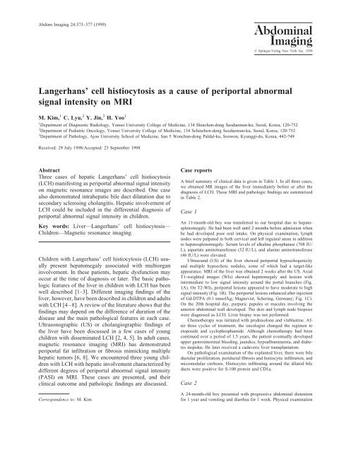
Abdom Imaging 24:373–377 (1999)AbdominalImaging© Springer-Verlag New York Inc. 1999Langerhans’ <strong>cell</strong> <strong>histiocytosis</strong> <strong>as</strong> a <strong>cause</strong> <strong>of</strong> <strong>periportal</strong> <strong>abnormal</strong>signal intensity on MRIM. Kim, 1 C. Lyu, 2 Y. Jin, 3 H. Yoo 11 Department <strong>of</strong> Diagnostic Radiology, Yonsei University College <strong>of</strong> Medicine, 134 Shinchon-dong Seodaemun-ku, Seoul, Korea, 120-7522 Department <strong>of</strong> Pediatric Oncology, Yonsei University College <strong>of</strong> Medicine, 134 Schinchon-dong Seodaemun-ku, Seoul, Korea, 120-7523 Department <strong>of</strong> Pathology, Ajou University School <strong>of</strong> Medicine, San 5 Wonchun-dong Paldal-ku, Soowon, Kyunggi-do, Korea, 442-749Received: 29 July 1998/Accepted: 23 September 1998AbstractThree c<strong>as</strong>es <strong>of</strong> hepatic Langerhans’ <strong>cell</strong> <strong>histiocytosis</strong>(LCH) manifesting <strong>as</strong> <strong>periportal</strong> <strong>abnormal</strong> signal intensityon magnetic resonance images are described. One c<strong>as</strong>ealso demonstrated intrahepatic bile duct dilatation due tosecondary sclerosing cholangitis. Hepatic involvement <strong>of</strong>LCH could be included in the differential diagnosis <strong>of</strong><strong>periportal</strong> <strong>abnormal</strong> signal intensity in children.Key words: Liver—Langerhans’ <strong>cell</strong> <strong>histiocytosis</strong>—Children—Magnetic resonance imaging.Children with Langerhans’ <strong>cell</strong> <strong>histiocytosis</strong> (LCH) usuallypresent hepatomegaly <strong>as</strong>sociated with multiorganinvolvement. In these patients, hepatic dysfunction mayoccur at the time <strong>of</strong> diagnosis or later. The b<strong>as</strong>ic pathologicfeatures <strong>of</strong> the liver in children with LCH h<strong>as</strong> beenwell described [1–3]. Different imaging findings <strong>of</strong> theliver, however, have been described in children and adultswith LCH [4–8]. A review <strong>of</strong> the literature shows that thefindings may depend on the difference <strong>of</strong> duration <strong>of</strong> thedise<strong>as</strong>e and the main pathological features in each c<strong>as</strong>e.Ultr<strong>as</strong>onographic (US) or cholangiographic findings <strong>of</strong>the liver have been discussed in a few c<strong>as</strong>es <strong>of</strong> youngchildren with disseminated LCH [2, 4, 5]. In adult c<strong>as</strong>es,magnetic resonance imaging (MRI) h<strong>as</strong> demonstrated<strong>periportal</strong> fat infiltration or fibrosis mimicking multiplehepatic tumors [6, 8]. We encountered three young childrenwith LCH with hepatic involvement characterized bydifferent degrees <strong>of</strong> <strong>periportal</strong> <strong>abnormal</strong> signal intensity(PASI) on MRI. These c<strong>as</strong>es are presented, and theirclinical outcome and pathologic findings are discussed.Correspondence to: M. KimC<strong>as</strong>e reportsA brief summary <strong>of</strong> clinical data is given in Table 1. In all three c<strong>as</strong>es,we obtained MR images <strong>of</strong> the liver immediately before or after thediagnosis <strong>of</strong> LCH. Those MRI and pathologic findings are summarizedin Table 2.C<strong>as</strong>e 1An 11-month-old boy w<strong>as</strong> transferred to our hospital due to hepatosplenomegaly.He had been well until 2 months before admission whenhe had developed poor oral intake. On physical examination, lymphnodes were palpated in both cervical and left inguinal are<strong>as</strong> in additionto hepatosplenomegaly. Serum levels <strong>of</strong> alkaline phosphat<strong>as</strong>e (708 IU/L), <strong>as</strong>partate aminotransfer<strong>as</strong>e (52 IU/L), and alanine aminotransfer<strong>as</strong>e(46 IU/L) were elevated.Ultr<strong>as</strong>ound (US) <strong>of</strong> the liver showed <strong>periportal</strong> hypoechogenicityand multiple hypoechoic nodules, some <strong>of</strong> which had a target-likeappearance. MRI <strong>of</strong> the liver w<strong>as</strong> obtained 2 weeks after the US. AxialT1-weighted images (WIs) showed hepatomegaly and lesions withintermediate to low signal intensity around the portal branches (Fig.1A). On T2-WIs, <strong>periportal</strong> lesions appeared to have moderate to highsignal intensity (Fig. 1B). The <strong>periportal</strong> lesions enhanced after injection<strong>of</strong> Gd-DTPA (0.1 mmol/kg; Magnevist, Schering, Germany; Fig. 1C).On the 20th hospital day, purpuric papules or macules involving theanterior abdominal wall developed. The skin and lymph node biopsieswere diagnosed <strong>as</strong> LCH. Liver biopsy w<strong>as</strong> not performed.Chemotherapy w<strong>as</strong> initiated with prednisolone and vinbl<strong>as</strong>tine. Afterthree cycles <strong>of</strong> treatment, the oncologist changed the regimen toetoposide and cyclophosphamide. Although chemotherapy had beencontinued over a period <strong>of</strong> 1.5 years, the patient eventually developedupper g<strong>as</strong>trointestinal bleeding, jaundice, hypoalbuminemia, and diabetesinspidus. He later received a cadaveric liver transplantation.On pathological examination <strong>of</strong> the explanted liver, there were bileductular proliferation, periductal fibrosis and histiocytic infiltration, andmicronodular cirrhosis. Histiocytes infiltrating around the dilated bileducts were positive for S-100 protein and CD1a.C<strong>as</strong>e 2A 24-month-old boy presented with progressive abdominal distentionfor 1 year and vomiting and diarrhea for 1 week. Physical examination
374 M. Kim et al.: MRI <strong>of</strong> hepatic Langerhans’ <strong>cell</strong> <strong>histiocytosis</strong>Table 1. Clinical data and outcome in three childrenC<strong>as</strong>e Age (mo) Sex Presenting symptoms Organs involved Outcome, treatment1 11 M Poor oral intake Skin, lymphnodes, liver2 24 M Abdominal distension,Scalp, livervomiting, and diarrhea3 18 M Limping gait Ilium, spine,liverHepatic dysfunction, aliver transplantationHepatic dysfunction, aliver transplantationStable, chemotherapya Hepatic dysfunction developed 1 1 ⁄2 years after an initial presentationTable 2. Summary <strong>of</strong> MR imaging and pathologic findings <strong>of</strong> the liverin three childrenC<strong>as</strong>e MR imaging Pathology1 Smooth hepatic surface,diffuse PASI2 Nodular hepatic surface,diffuse PASI, unevenlydilated intrahepatic bileducts3 Smooth hepatic surface,mild PASI in centralportal tractshowed hepatosplenomegaly and thickly crusted patches on the scalp.Laboratory examination showed an elevated serum level <strong>of</strong> alkalinephosphat<strong>as</strong>e (1075 IU/L), <strong>as</strong>partate aminotransfer<strong>as</strong>e (134 IU/L), andalanine aminotransfer<strong>as</strong>e (121 IU/L).US showed heterogeneous hepatic parenchymal echoes and <strong>periportal</strong>hypoechogenicity. MR images <strong>of</strong> the liver showed thick, <strong>abnormal</strong>PASI, intrahepatic bile duct dilatation, and nodular appearance <strong>of</strong> theenlarged liver (Fig. 2A,B). On contr<strong>as</strong>t-enhanced T1-WIs, relativelyhypointense nodular lesions were found adjacent to an <strong>abnormal</strong> <strong>periportal</strong>enhancement (Fig. 2C).In addition to the findings typical <strong>of</strong> LCH from biopsy <strong>of</strong> thescalp and liver, secondary changes <strong>of</strong> extrahepatic bile duct obstructionand cholangitis were seen. Although chemotherapy had beencontinued for 1.5 years, he developed hematemesis and melena. Hereceived a liver transplantation from a cadaveric donor. Gross andmicroscopic examination <strong>of</strong> the explanted liver showed nodularhepatic surface, severe secondary sclerosing cholangitis, and bileduct proliferations.C<strong>as</strong>e 3Biliary cirrhosis, extensive<strong>periportal</strong> fibrosisBiliary cirrhosis, extensive<strong>periportal</strong> fibrosis, secondarysclerosing cholangitisBile ductular proliferation,periductal histiocytes, andinflammatory <strong>cell</strong>sPASI; <strong>periportal</strong> <strong>abnormal</strong> signal intensity on T1, T2, and contr<strong>as</strong>tenhancedT1 weighted imagesA 18-month-old boy presented with a limping gait. Physical examinationshowed hepatomegaly. Laboratory examination showed an elevatedserum level <strong>of</strong> alkaline phosphat<strong>as</strong>e (626 IU/L), <strong>as</strong>partate aminotransfer<strong>as</strong>e(53 IU/L), and alanine aminotransfer<strong>as</strong>e (186 IU/L). An osteolyticlesion <strong>of</strong> the right iliac bone and flattening <strong>of</strong> the 8th and 9th thoracicvertebrae were found on plain radiographic examination.US <strong>of</strong> the liver showed slightly incre<strong>as</strong>ed parenchymal echoes andsmall round hypoechoic nodules in the left lobe. MR images <strong>of</strong> the livershowed PASI confined to the central portal tract (Fig. 3A,B).The diagnosis <strong>of</strong> LCH w<strong>as</strong> made from biopsy <strong>of</strong> the right ilium andthe liver. Liver biopsy showed bile ductular proliferation, periductalhistiocytic, and lymphocytic infiltration. Chemotherapy w<strong>as</strong> initiatedwith prednisolone and vinbl<strong>as</strong>tine and w<strong>as</strong> then continued with cyclophosphamideand VP-16. On follow-up MR images obtained 2 yearslater, PASI showed near complete resolution (Fig. 3C). The patient h<strong>as</strong>not developed any clinical manifestations <strong>of</strong> hepatic dysfunction for 3years after diagnosis.DiscussionIn children with LCH, hepatic involvement is common.At presentation, hepatomegaly is observed in up to 60%<strong>of</strong> patients with disseminated LCH [3], but hepatomegalydoes not always portend a poor prognosis. Hepatic dysfunctionssuch <strong>as</strong> jaundice, hypoproteinemia, and portalhypertension may be present at the time <strong>of</strong> diagnosis ormay develop over months or years [1–3]. In all threepatients, clinical manifestation <strong>of</strong> hepatic dysfunction w<strong>as</strong>not evident at the time <strong>of</strong> diagnosis.Morphologic changes <strong>of</strong> the liver in LCH have beendescribed in a number <strong>of</strong> previous studies. A reportfrom the Children’s Cancer Study Group h<strong>as</strong> describedfour patterns: (a) portal triaditis with infiltrating neutrophils,eosinophils, or mononuclear <strong>cell</strong>s; (b) bileduct proliferation with triaditis; (c) fibrohistiocyticchange; and (d) nodular parenchymal lesion with triaditis[1]. Other reports have described triaditis, <strong>periportal</strong>fibrosis, or liver cirrhosis and bile st<strong>as</strong>is and bileduct proliferation in some c<strong>as</strong>es [2, 3, 9–11]. We couldnot confirm the sequential change <strong>of</strong> hepatic pathologyin each patient, but bile ductal proliferation and periductalinflammatory <strong>cell</strong> infiltration were noted in thepretreatment liver biopsy <strong>of</strong> patient 3. Biliary cirrhosisand extensive periductal fibrosis w<strong>as</strong> seen in the explantedliver <strong>of</strong> patients 1 and 2, who had developedhepatic dysfunction. In patient 2, secondary sclerosingcholangitis w<strong>as</strong> also noted.US finding <strong>of</strong> the liver in LCH h<strong>as</strong> been described<strong>as</strong> hypoechoic nodule or <strong>periportal</strong> hyperechogenicitythat may be attributed to inflammatory <strong>cell</strong> infiltrationor fat content [4, 5, 7, 8]. US in the present c<strong>as</strong>esshowed <strong>periportal</strong> hypoechogenicity that w<strong>as</strong> band-like
M. Kim et al.: MRI <strong>of</strong> hepatic Langerhans’ <strong>cell</strong> <strong>histiocytosis</strong> 375Fig. 1. C<strong>as</strong>e 1. A T1-weighted image (500/11 TR/TE) shows <strong>periportal</strong>low signal intensity surrounding the portal veins that extends peripherallyfrom the porta hepatis. B F<strong>as</strong>t spin-echo T2-weighted image (4000/85) shows diffuse <strong>periportal</strong> high signal intensity <strong>as</strong> a <strong>periportal</strong> ring ortramline, some <strong>of</strong> which have a hyperintense nodular appearance. CContr<strong>as</strong>t-enhanced T1-weighted image (500/11) with fat suppressionshows <strong>periportal</strong> enhancement.Fig. 2.C<strong>as</strong>e 2. A T1-weighted image (650/13) shows thick, <strong>periportal</strong>low signal intensity and nodular hepatic surface. Unevenlydilated intrahepatic bile ducts or bile lakes appear to have highintensity due to stagnant bile. B F<strong>as</strong>t spin-echo T2-weighted image(4000/98) shows moderate to high signal intensity along the portaltracts. C Strong <strong>periportal</strong> enhancement is seen on contr<strong>as</strong>t-enhancedT1-weighted image (650/13). Hypointense <strong>periportal</strong> fibrosis mimickinga m<strong>as</strong>s (arrows) is more clearly defined than in the T1-weighted image.
376 M. Kim et al.: MRI <strong>of</strong> hepatic Langerhans’ <strong>cell</strong> <strong>histiocytosis</strong>or nodular. Some hypoechoic nodules looked like targets.We suspect that <strong>periportal</strong> hypoechogenicity maybe attributed to the infiltration <strong>of</strong> histiocytes and inflammatory<strong>cell</strong>s b<strong>as</strong>ed on our biopsy result, whichaccorded with the reports <strong>of</strong> Hara et al. [4] and Muwakkitet al. [5].MRI findings <strong>of</strong> the liver in LCH have been notedin two c<strong>as</strong>e reports [6, 8]. These c<strong>as</strong>es presented <strong>as</strong>m<strong>as</strong>s lesions <strong>of</strong> the liver, which had imaging characteristics<strong>of</strong> <strong>periportal</strong> fibrosis and focal fatty infiltration.In the present c<strong>as</strong>es, MRI showed PASI ratherthan a focal m<strong>as</strong>s. Matsui et al. [12] described <strong>periportal</strong><strong>abnormal</strong> intensity on MRI from patients withcholangitis, obstructive jaundice that w<strong>as</strong> due toedema, inflammatory <strong>cell</strong> infiltration, and proliferation<strong>of</strong> bile ductules. On each T1-WI and T2-WI, signalintensity <strong>of</strong> <strong>periportal</strong> lesions in our patients w<strong>as</strong> lowerand higher than that <strong>of</strong> surrounding hepatic parenchyma,respectively. Signal intensity <strong>of</strong> these lesionsw<strong>as</strong> incre<strong>as</strong>ed on contr<strong>as</strong>t-enhanced T1-WI with fatsuppression. PASI extended diffusely from the centrallarge portal tracts to peripheral small ones in patients 1and 2, who eventually developed hepatic dysfunction.In addition to the extensive PASI, intrahepatic bile ductdilatation and nodularity due to secondary biliary cirrhosiswere noted in patient 2. This finding is in contr<strong>as</strong>tto PASI confined to the central large portal tract inpatient 3 who h<strong>as</strong> not yet developed hepatic dysfunction.Pathologic examination <strong>of</strong> this patient showedbile ductular proliferation and periductal histiocyticand lymphocytic infiltration. On follow-up MRI obtained2 years later, PASI w<strong>as</strong> not seen in all sequences<strong>as</strong> compared with that <strong>of</strong> the initial study. We considerthat MRI <strong>of</strong> the liver in children with disseminatedLCH may be different according to the pathologicalchange, ranging from triaditis to <strong>periportal</strong> fibrosis andbiliary cirrhosis.In summary, we could find PASI <strong>as</strong> a common featureon initial MRI <strong>of</strong> three children with disseminated LCH.As already known, PASI itself is nonspecific. However,in conjunction with clinical and laboratory data, MRI canbe valuable in the estimation <strong>of</strong> morphologic change <strong>of</strong>the liver in children.ReferencesFig. 3. C<strong>as</strong>e 3. A T1-weighted image (550/17) with fat suppressionshows <strong>periportal</strong> low signal intensity that is confined to the central portaltracts. B Periportal enhancement is clearly seen on contr<strong>as</strong>t-enhancedT1-weighted image (600/11) with fat suppression. C T1-weighted image(500/8) obtained 2 years later shows no <strong>periportal</strong> low signal intensity.1. Heyn RM, Hamoudi A, Newton WA. Pretreatment liver biopsy in20 children with <strong>histiocytosis</strong> X: a clinicopathologic correlation.Med Pediatr Oncol 1990;18:110–1182. Squires RH, Weinberg AG, Zweiner RJ, et al. Langerhans’ <strong>cell</strong><strong>histiocytosis</strong> presenting with hepatic dysfunction. J Pediatr G<strong>as</strong>troenterolNutr 1993;16:190–1933. Leblanc A, Hadchouel M, Jehan P, et al. Obstructive jaundice inchildren with <strong>histiocytosis</strong> X. G<strong>as</strong>troenterology 1981;80:134–1394. Hara T, Mizuno Y, Ishii E, et al. Histiocytosis X presenting <strong>as</strong>multiple intrahepatic nodules. Acta Haematol Jpn 1988;51:1059–1062
M. Kim et al.: MRI <strong>of</strong> hepatic Langerhans’ <strong>cell</strong> <strong>histiocytosis</strong> 3775. Muwakkit S, Gharagozloo A, Souid AK, et al. The sonographicappearance <strong>of</strong> lesions <strong>of</strong> the spleen and pancre<strong>as</strong> in an infantwith Langerhans’ <strong>cell</strong> <strong>histiocytosis</strong>. Pediatr Radiol 1994;24:222–2236. Arakawa A, Matsukawa T, Yam<strong>as</strong>hita Y, et al. Periportal fibrosisin Langerhans’ <strong>cell</strong> <strong>histiocytosis</strong> mimicking multiple liver tumors:US, CT and MR findings. J Comput Assist Tomogr 1994;18:157–1597. Päivänsalo M, Mäkäräinen H. Liver lesions in <strong>histiocytosis</strong> X:findings on sonography and computed tomography. Br J Radiol1986;59:1123–11258. Radin DR. Langerhans <strong>cell</strong> <strong>histiocytosis</strong> <strong>of</strong> the liver: imaging findings.AJR 1992;159:63–649. Favara BE. The pathology <strong>of</strong> “<strong>histiocytosis</strong>.” Am J Pediatr HematolOncol 1981;3:45–5610. Favara BE, McCarthy RC, Meirau GW. Histiocytosis X. HumPathol 1983;14:663–67611. Thompson HH, Pitt HA, Lewin KJ, et al. Sclerosing cholangitis and<strong>histiocytosis</strong> X. Gut 1984;25:526–53012. Matsui O, Kadoya M, Tak<strong>as</strong>hima T, et al. Intrahepatic <strong>periportal</strong><strong>abnormal</strong> intensity on MR images: an indication <strong>of</strong> various hepatobiliarydise<strong>as</strong>es. Radiology 1989;171:335–338


