
You also want an ePaper? Increase the reach of your titles
YUMPU automatically turns print PDFs into web optimized ePapers that Google loves.
chapter<br />
PDHPE Application and Inquiry<br />
286<br />
16<br />
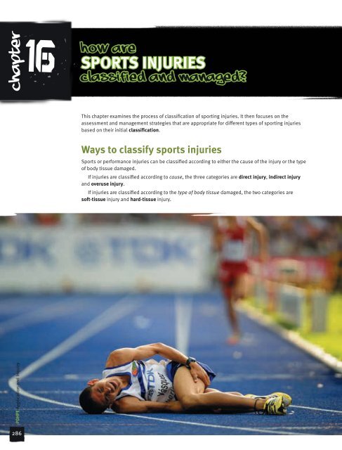
HOW ARE<br />
SPORTS INJURIES<br />
CLASSIFIED AND MANAGED?<br />
This chapter examines the process of classification of sporting <strong>injuries</strong>. It then focuses on the<br />
assessment and management strategies that are appropriate for different types of sporting <strong>injuries</strong><br />
based on their initial classification.<br />
<strong>Ways</strong> <strong>to</strong> <strong>classify</strong> <strong>sports</strong> <strong>injuries</strong><br />
Sports or performance <strong>injuries</strong> can be classified according <strong>to</strong> either the cause of the injury or the type<br />
of body tissue damaged.<br />
If <strong>injuries</strong> are classified according <strong>to</strong> cause, the three categories are direct injury, indirect injury<br />
and overuse injury.<br />
If <strong>injuries</strong> are classified according <strong>to</strong> the type of body tissue damaged, the two categories are<br />
soft-tissue injury and hard-tissue injury.
Table 16.1 Overuse <strong>injuries</strong><br />
Injury Symp<strong>to</strong>ms and signs Possible causes Management<br />
Shin soreness Tenderness<br />
Pain in shins<br />
Pain increases by running<br />
and jumping<br />
Swelling<br />
Knee pain Pain around knee<br />
Pain increased by sport,<br />
stairs, sitting, hills<br />
Swelling<br />
Discolouration<br />
Heel pain Tenderness over heel<br />
Pain increased by<br />
running, jumping<br />
Shoulder pain Pain on certain<br />
movements<br />
Reduced movement<br />
Local tenderness<br />
Elbow pain Pain in and around elbow<br />
Pain increased by certain<br />
activities, e.g. shaking,<br />
lifting, gripping<br />
Australian Coaching Council Inc.<br />
Classification according <strong>to</strong> cause<br />
Direct injury<br />
Increased activity<br />
Poor footwear<br />
Postural imbalance<br />
Muscle imbalance<br />
Increased activity<br />
Postural imbalance<br />
Poor footwear<br />
Muscle imbalance<br />
Growth spurt<br />
Tight calf muscles<br />
Growth spurt<br />
Poor footwear<br />
Increased activity,<br />
e.g. swimming<br />
Poor technique, e.g.<br />
swimming, pitching,<br />
serving<br />
Jarring<br />
Increased activity<br />
e.g. golf, tennis<br />
Muscle imbalance<br />
Poor technique<br />
Change of grip<br />
Lack of control<br />
Decrease painful activity<br />
RICER<br />
Physiotherapy<br />
Correct footwear<br />
Orthotic control<br />
Decrease activity<br />
RICER<br />
Physiotherapy<br />
Tape<br />
Correct footwear<br />
Orthotic control<br />
Decrease activity<br />
RICER<br />
Physiotherapy<br />
Stretching program<br />
Correct footwear<br />
Orthotic control<br />
Decrease activity<br />
RICER<br />
Physiotherapy<br />
Stretching program<br />
Exercises<br />
Modify activity<br />
Decrease activity<br />
RICER<br />
Physiotherapy<br />
Stretching program<br />
Elbow brace<br />
Modify technique<br />
A direct injury is caused by an external blow or force. Direct <strong>injuries</strong> can be caused by:<br />
• a collision with another person (for example, during a tackle in rugby union)<br />
• being struck with an object (for example, a cricket ball or hockey stick).<br />
Examples of <strong>injuries</strong> that result from external forces include haema<strong>to</strong>mas (‘corks’) and bruises,<br />
joint and ligament damage, dislocations and bone fractures.<br />
#<br />
RICER stands for rest, ice,<br />
compression, elevation<br />
and referral.<br />
#<br />
An orthotic control is a device<br />
placed in footwear <strong>to</strong> correct<br />
foot alignment.<br />
Chapter 16 | HOW ARE SPORTS INJURIES CLASSIFIED AND MANAGED?<br />
287
PDHPE Application and Inquiry<br />
288<br />
Figure 16.1 Direct injury: hockey stick impacting<br />
athlete’s body<br />
Indirect injury<br />
An indirect injury can occur in two ways:<br />
• The actual injury can occur some distance from the impact site. For example, falling on an outstretched<br />
hand can result in a dislocated shoulder.<br />
• The injury does not result from physical contact with an object or person, but from internal forces built<br />
up by the actions of the performer, such as may be caused by over-stretching, poor technique, fatigue<br />
and lack of fitness. Ligament sprains and muscle strains and tears are examples of these <strong>injuries</strong>.<br />
Overuse injury<br />
Figure 16.2 Indirect injury: ligament sprain/strain<br />
Overuse <strong>injuries</strong> occur when excessive and repetitive force is placed on the bones and other connective<br />
tissues of the body. Little or no pain might be experienced in the early stages of these <strong>injuries</strong> and<br />
the athlete might continue <strong>to</strong> place pressure on the injured site. This prevents the site being given the<br />
necessary time <strong>to</strong> heal. Eventually the damage accumulates, and the injured site becomes inflamed, and<br />
therefore painful.<br />
The symp<strong>to</strong>ms of overuse injury often occur when there is a change in training practices (such as<br />
increasing training frequency or intensity), and the body is unable <strong>to</strong> deal with the new stresses that are<br />
placed upon it. A large number of overuse <strong>injuries</strong> results from poorly planned training programs in which<br />
the athlete is not given appropriate<br />
time <strong>to</strong> recover between intense<br />
sessions.<br />
Other causes of overuse injury<br />
are use of poor technique and poor<br />
equipment. Athletes who practise<br />
and compete using poor technique or<br />
equipment place extra stress on their<br />
body. Examples of this include elbow<br />
injury from poor backhand technique<br />
or the use of a heavy racquet in<br />
tennis, and ankle or knee pain from<br />
an inappropriate running style or from<br />
wearing inappropriate footwear.<br />
Examples of <strong>injuries</strong> that result<br />
from repetitive forces are stress<br />
fractures (small cracks in the bone)<br />
and tendonitis (inflammation<br />
of a tendon).<br />
Figure 16.3 Stress fractures can result from a repetitive force
Classification according <strong>to</strong> tissue type<br />
Soft-tissue injury<br />
Soft-tissue <strong>injuries</strong> are the most common <strong>injuries</strong> resulting from participation in sport.<br />
They include the following:<br />
• skin <strong>injuries</strong>—abrasions, lacerations and blisters<br />
• muscle <strong>injuries</strong>—tears or strains of muscle fibres and contusions<br />
• tendon <strong>injuries</strong>—tears or strains of tendon fibres and inflammation (tendonitis)<br />
• ligament <strong>injuries</strong>—sprains and tears of ligament fibres.<br />
Soft-tissue <strong>injuries</strong> can result in internal bleeding and swelling. Prompt and effective management<br />
of this bleeding aids recovery. Soft-tissue <strong>injuries</strong> are covered in further detail on page 290.<br />
Table 16.2 Injury prevalence in AFL (missed games per club)<br />
Body area Injury type<br />
Head/neck Concussion<br />
Facial fractures<br />
Neck sprains<br />
Other head/neck <strong>injuries</strong><br />
Shoulder/arm/elbow Shoulder sprains and dislocations<br />
Acromio-clavicular joint <strong>injuries</strong><br />
Fractured clavicles<br />
Other shoulder/arm/elbow <strong>injuries</strong><br />
Forearm/wrist/hand Forearm/wrist/hand fractures<br />
Other forearm/wrist/hand <strong>injuries</strong><br />
Trunk/back Rib and chest wall <strong>injuries</strong><br />
Lumbar and thoracic spine <strong>injuries</strong><br />
Other but<strong>to</strong>ck/back/trunk <strong>injuries</strong><br />
Hip/groin/thigh Groin strains/osteitis pubis<br />
Hamstring strains<br />
Quadriceps strains<br />
Other hip/groin/thigh <strong>injuries</strong><br />
Knee Knee anterior cruciate ligament (CL)<br />
Knee medial CL or posterior CL<br />
Knee cartilage<br />
Other knee <strong>injuries</strong><br />
Shin/ankle/foot Ankle sprains or joint <strong>injuries</strong><br />
Calf strains<br />
Achilles tendon <strong>injuries</strong><br />
Fractures/stress fractures of leg or foot<br />
Other leg/foot/ankle <strong>injuries</strong><br />
0.3<br />
0.7<br />
1.1<br />
1.6<br />
6.4<br />
1.4<br />
1.8<br />
1.5<br />
2.3<br />
3.1<br />
1.9<br />
2.8<br />
1.7<br />
18.0<br />
24.3<br />
5.6<br />
5.5<br />
Other Medical illnesses/non-football <strong>injuries</strong> 4.1<br />
Missed games/club/season 147.5<br />
Adapted from 16th Annual AFL Injury Report: Season 2007<br />
Hard-tissue injury<br />
Hard-tissue <strong>injuries</strong> are those involving damage <strong>to</strong> the bones of the skele<strong>to</strong>n. They range from severe<br />
fractures and joint dislocations <strong>to</strong> bruising of the bone. A direct force can bruise a bone and cause<br />
bleeding between the outer layer of the bone and the underlying compact bone. This is common in a bone<br />
such as the tibia (shin) where there is little muscle tissue over the bone <strong>to</strong> absorb the force.<br />
Bones have a blood supply and internal bleeding can result from a fracture. In major <strong>injuries</strong>, this<br />
internal bleeding in the bone, <strong>to</strong>gether with bleeding from surrounding damaged tissue, can lead <strong>to</strong> shock<br />
and serious circula<strong>to</strong>ry complications. Hard-tissue <strong>injuries</strong> are covered in further detail on page 294.<br />
15.9<br />
6.3<br />
9.1<br />
6.0<br />
7.1<br />
3.1<br />
2.2<br />
9.5<br />
4.2<br />
#<br />
Tendons join<br />
muscle <strong>to</strong> bone<br />
while ligaments<br />
join bone <strong>to</strong> bone.<br />
#<br />
Osteitis pubis is<br />
an overuse injury<br />
<strong>to</strong> the groin region<br />
(inflammation of the<br />
pubis symphisis).<br />
Chapter 16 | HOW ARE SPORTS INJURIES CLASSIFIED AND MANAGED?<br />
289
PDHPE Application and Inquiry<br />
290<br />
Secondary injury<br />
Athletes returning <strong>to</strong> activity are also at risk of a secondary injury, which is an injury that occurs as a<br />
result of a previous injury being poorly treated or not being fully healed. Athletes risk recurrence of<br />
<strong>injuries</strong> if they commence playing before regaining full strength and range of movement.<br />
practical application<br />
Classify <strong>sports</strong> <strong>injuries</strong><br />
1 Examine Table 16.2 (page 289), then complete the following tasks.<br />
a Classify the <strong>injuries</strong> in the table in<strong>to</strong> direct and indirect<br />
<strong>injuries</strong> on the basis of their most likely cause.<br />
b Discuss how some of these <strong>injuries</strong> could be both direct<br />
and indirect.<br />
c Classify the <strong>injuries</strong> listed in the table in<strong>to</strong> soft-tissue<br />
<strong>injuries</strong> and hard-tissue <strong>injuries</strong>.<br />
d Identify the injury that resulted in the most missed<br />
matches in 2007.<br />
e Discuss the possible reasons for the identified injury<br />
resulting in the most missed matches.<br />
2 a Identify examples of direct and indirect <strong>injuries</strong> that might<br />
occur in the following <strong>sports</strong>:<br />
• field hockey<br />
• snow skiing<br />
• cricket.<br />
b Classify the <strong>injuries</strong> identified in task 2a in<strong>to</strong> soft-tissue<br />
and hard-tissue <strong>injuries</strong>.<br />
Soft-tissue <strong>injuries</strong><br />
Three common soft-tissue <strong>injuries</strong> are tears, sprains and contusions.<br />
Tears, sprains and contusions<br />
Critical inquiry<br />
1 Explain why it is necessary <strong>to</strong><br />
<strong>classify</strong> <strong>injuries</strong>.<br />
2 Investigate whether <strong>injuries</strong><br />
can be classified in other<br />
ways than those described<br />
above.<br />
Research and Review<br />
1 Describe the differences between<br />
direct and indirect <strong>injuries</strong>.<br />
2 Explain how poor technique can<br />
cause an overuse injury.<br />
3 Define soft-tissue injury and hardtissue<br />
injury.<br />
4 a Clarify what a secondary<br />
injury is.<br />
b Outline how they can be<br />
prevented.<br />
A tear is a disruption of the fibres of a muscle or tendon. This can be tiny and microscopic (often called<br />
a strain). A tear can also be more severe, and involve larger fibres of muscles and tendons. Tears (and<br />
strains) occur when a muscle or tendon is over-stretched or when a muscle contracts <strong>to</strong>o quickly. The<br />
severity of the tear can range from the microscopic level (a strain), <strong>to</strong> a small number of fibres through<br />
<strong>to</strong> a complete rupture of all muscle fibres.<br />
A sprain is a tear of ligament fibres, muscles or tendons supporting a joint. This can occur when a<br />
joint is extended beyond its normal range of movement. A sprain can involve a small number of fibres<br />
through <strong>to</strong> a complete rupture. In extreme circumstances, the fibres of the ligament, muscle or tendon<br />
can remain intact and rip from the bone.<br />
A contusion or bruise is bleeding in<strong>to</strong> the soft tissue. It is caused by a direct blow from another person,<br />
an implement or an object. A bruise can occur <strong>to</strong> any soft tissue of the body.<br />
Skin abrasions, lacerations and blisters<br />
Injuries <strong>to</strong> the skin are very common in sport. They include minor wounds, such as abrasions (grazes),<br />
blisters and small lacerations. They also include bone fractures and more serious lacerations that<br />
require suturing (stitches). Small skin abrasions, lacerations not requiring sutures and blisters are<br />
manageable conditions, and in most cases do not require referral <strong>to</strong> a doc<strong>to</strong>r.
Skin abrasions occur when the outer layer of skin is<br />
removed, usually as a result of a scraping action. The<br />
open wound can contain dirt or gravel, which should<br />
be removed. More extensive, deeper abrasions require<br />
medical attention.<br />
When the skin is lacerated (cut), the depth and<br />
location of the laceration will determine whether suturing<br />
is required. Medical attention is required if the laceration<br />
is deep enough <strong>to</strong> expose tissues, such as fat, tendons<br />
or bone. Sometimes a superficial laceration will require<br />
suturing. This can be required if the laceration is located:<br />
• over a joint (such as the knee) because flexion will<br />
continually open the wound<br />
• in a cosmetically sensitive position (for example,<br />
on the face).<br />
Deep lacerations are usually accompanied by significant<br />
bleeding.<br />
Blisters result from friction<br />
(rubbing). One layer of skin<br />
separates from another and<br />
a small pocket of fluid forms.<br />
Blisters can be caused by<br />
equipment, shoes, pressure<br />
from callus build-up, increased<br />
training loads or simply by the<br />
recommencement of training<br />
after an extended rest period.<br />
Inflamma<strong>to</strong>ry response<br />
Figure 16.4 Skin abrasions occur when the outer layer of<br />
skin is removed<br />
The initial stage of repair of body tissue is the acute inflamma<strong>to</strong>ry phase. It exists during the first 24<br />
<strong>to</strong> 72 hours after injury. The immediate response of the body <strong>to</strong> injury is <strong>to</strong> increase the flow of blood<br />
and other fluids <strong>to</strong> the injured site. If blood vessels at the site are damaged there will also be direct<br />
bleeding in<strong>to</strong> the surrounding tissue. The accumulation of fluid in the area causes an increase in<br />
tissue pressure, which produces pain.<br />
All these changes produce what we call inflammation. Inflammation consists of redness, heat,<br />
swelling, pain and loss of function. If inflammation is left unchecked and persists for a long time,<br />
formation of scar tissue will be more severe.<br />
The extent <strong>to</strong> which the formation of inflexible scar tissue can be prevented will, in part, determine<br />
the time required for rehabilitation of the injury and the degree <strong>to</strong> which normal functioning can be<br />
returned <strong>to</strong> pre-injury levels. Figure 16.5 (page 292) shows how stretching and the application of ice<br />
will limit the formation of scar tissue.<br />
Managing soft-tissue <strong>injuries</strong><br />
In order <strong>to</strong> effectively manage soft-tissue <strong>injuries</strong> the RICER procedure needs <strong>to</strong> be followed.<br />
RICER<br />
#<br />
A callus is a build-up<br />
of dead skin formed at<br />
a site where there has<br />
been frequent rubbing<br />
and pressure; for<br />
example, on the heel.<br />
The immediate management of soft-tissue <strong>injuries</strong> during the acute inflamma<strong>to</strong>ry phase is very<br />
important for successful rehabilitation after the injury. The aims of immediate treatment are <strong>to</strong>:<br />
• prevent further tissue damage<br />
• minimise swelling<br />
• ease pain<br />
• reduce the formation of scar tissue<br />
• reduce the time needed for rehabilitation.<br />
Chapter 16 | HOW ARE SPORTS INJURIES CLASSIFIED AND MANAGED?<br />
291
PDHPE Application and Inquiry<br />
292<br />
These aims are achieved through the application of the<br />
RICER procedure.<br />
• R for Rest<br />
• I for Ice<br />
• C for Compression<br />
• E for Elevation<br />
• R for Referral.<br />
Rest<br />
The injured area must remain relatively inactive for the first<br />
48–72 hours. The duration of the rest will depend on the<br />
severity of the injury.<br />
Ice<br />
IMPACT<br />
Soft tissue<br />
immediately after injury<br />
Figure 16.5 How RICER helps in the management of a soft-tissue injury<br />
The application of ice causes the blood vessels <strong>to</strong><br />
constrict, thus decreasing circulation and resulting in less<br />
inflammation at the site. Where possible, ice should be<br />
applied <strong>to</strong> the surrounding area, in addition <strong>to</strong> the direct<br />
site. Ice should be applied in a wet<br />
<strong>to</strong>wel for periods of 20–30 minutes<br />
every two hours for the first 48–72<br />
hours. Do not apply ice, or a plastic<br />
bag containing ice, directly on<strong>to</strong><br />
the skin. Care should also be taken<br />
when applying ice in the region of<br />
the eye.<br />
NO RICER<br />
Elastic bandage<br />
plus compression<br />
#<br />
Do not apply ice,<br />
or a plastic bag<br />
containing ice,<br />
directly on<strong>to</strong> the skin.<br />
Compression<br />
In addition <strong>to</strong> the application of ice, compression should be<br />
applied using a wide elastic bandage over the injured site<br />
and surrounding area. This will help <strong>to</strong> reduce the swelling<br />
by limiting fluid build-up (see Figure 16.5). It also provides<br />
support for the injured site. Care should be taken <strong>to</strong> ensure<br />
that circulation is not constricted by bandaging <strong>to</strong>o tightly.<br />
No RICER (24 hours) No RICER (3–6 weeks)<br />
RICER (24 hours)<br />
RICER plus rehab<br />
(3–6 weeks)<br />
Elevation<br />
Elevation of the injured part above the level of the heart<br />
reduces the volume and pressure of blood flow <strong>to</strong> the<br />
injured area, thus limiting inflammation. Elevation can be<br />
achieved for most <strong>injuries</strong> by supporting the injured area<br />
while the casualty is seated or lying down.<br />
Figure 16.6 Rest, ice, compression and elevation of an<br />
injured ankle
Referral<br />
Medical assessment should be sought as soon as possible <strong>to</strong> ascertain the full extent of the injury, and<br />
<strong>to</strong> commence appropriate rehabilitation.<br />
Actions <strong>to</strong> be avoided<br />
During the first 48–72 hours after an injury there are certain actions that must be avoided. These include<br />
the application of heat (for example, use of hot liniments, spas, saunas and hot baths), drinking alcohol,<br />
physical activity and massage. These actions all increase blood flow, and therefore swelling.<br />
Immediate treatment of skin <strong>injuries</strong><br />
The aims of the immediate management of skin <strong>injuries</strong> include prevention of infection for both the<br />
victim and the first aider, minimisation of blood loss and tissue damage, and promotion of healing in<br />
order <strong>to</strong> reduce recovery time.<br />
For most skin <strong>injuries</strong> the common management steps that should be followed are:<br />
1 Reduce the dangers of infection (for example, by wearing gloves).<br />
2 Control bleeding with rest, pressure and elevation.<br />
3 Assess the severity of the wound.<br />
4 Clean the wound using clean water, saline solution or a diluted antiseptic.<br />
5 Apply an antiseptic <strong>to</strong> the wound (for example, Savlon or Betadine) after ensuring that the person is<br />
not allergic <strong>to</strong> the antiseptic <strong>to</strong> be used.<br />
6 Dress the wound with a sterile pad and bandage.<br />
7 If necessary, refer the person <strong>to</strong> medical attention.<br />
Skin <strong>injuries</strong> that should be referred <strong>to</strong> medical attention include wounds that require suturing,<br />
wounds that show signs of infection or cannot be properly cleaned of foreign material and wounds <strong>to</strong><br />
the head. The reason why all head wounds should be referred <strong>to</strong> medical attention is because even<br />
minor <strong>injuries</strong> <strong>to</strong> the head might be accompanied by concussion.<br />
Critical inquiry<br />
1 a Discuss whether elevation is possible for all<br />
soft-tissue <strong>injuries</strong>.<br />
b If elevation is not possible, outline other ways<br />
that blood flow <strong>to</strong> the area can be reduced.<br />
2 If ice is not available, identify items that can be<br />
substituted.<br />
practical application<br />
Managing soft-tissue <strong>injuries</strong><br />
1 Apply the RICER procedure <strong>to</strong> the following softtissue<br />
<strong>injuries</strong>:<br />
a sprained ankle ligaments<br />
b corked thigh<br />
c sprained thumb ligaments.<br />
#<br />
Concussion is a<br />
brain injury that<br />
is not usually<br />
considered life<br />
threatening.<br />
Research and Review<br />
1 Describe the differences between a sprain and<br />
a strain.<br />
2 Explain the acute inflamma<strong>to</strong>ry response.<br />
3 Outline the aims of the immediate<br />
management of soft-tissue <strong>injuries</strong>.<br />
4 Identify the problems that can sometimes<br />
occur at the ‘ice’ and ‘compression’ stages of<br />
the RICER procedure.<br />
5 Explain what actions should be avoided after<br />
sustaining a soft-tissue injury.<br />
6 Identify the common management steps that<br />
should be followed with skin <strong>injuries</strong>.<br />
7 Clarify when a skin injury requires professional<br />
medical attention.<br />
Chapter 16 | HOW ARE SPORTS INJURIES CLASSIFIED AND MANAGED?<br />
293
PDHPE Application and Inquiry<br />
294<br />
Hard-tissue <strong>injuries</strong><br />
Types of hard-tissue <strong>injuries</strong><br />
Hard-tissue <strong>injuries</strong> include fractures and dislocations.<br />
Fractures<br />
A fracture is a break in a bone. This can result from<br />
a direct force, an indirect force or repetitive smaller<br />
impacts (as occurs in a stress fracture).<br />
If the skin over a fractured bone is intact, the fracture<br />
is described as ‘simple’ or ‘closed’. If the skin over a<br />
fracture is broken, the fracture is described as ‘open’<br />
or ‘compound’. The skin might be broken either by the<br />
force of the injury that caused the fracture or by a piece<br />
of broken bone protruding through the skin. A fracture<br />
is described as ‘complicated’ if nearby tissues and/or<br />
organs are damaged.<br />
In some cases, a simple fracture can be difficult <strong>to</strong><br />
detect. The signs and symp<strong>to</strong>ms of a fracture include:<br />
• pain at the site of the injury<br />
• inability <strong>to</strong> move the injured part<br />
• unnatural movement of the injured part<br />
• deformity of the injured part<br />
• swelling and discolouration<br />
• grating of bones.<br />
Dislocations<br />
Dislocations are <strong>injuries</strong> <strong>to</strong> joints where one bone<br />
is displaced from another. A dislocation is often<br />
accompanied by considerable damage <strong>to</strong> the surrounding<br />
connective tissue. Dislocations occur as a result of the<br />
joint being pushed past its normal range of movement.<br />
Common sites of the body where dislocations occur are<br />
the finger, shoulder and patella.<br />
Signs and symp<strong>to</strong>ms of dislocation include:<br />
• loss of movement at the joint<br />
• obvious deformity<br />
• swelling and tenderness<br />
• pain at the injured site.<br />
Table 16.3 Types of fractures<br />
Type of fracture Definition Associated fac<strong>to</strong>rs<br />
Closed The bone is fractured but there is no cut or wound<br />
at the fracture site.<br />
Open A jagged end of the fractured bone protrudes<br />
through the skin or there is a cut near the<br />
fracture site.<br />
Complicated The fractured bone damages the local tissues;<br />
i.e. the organ(s) that it protects (e.g. a lung<br />
punctured by a fractured rib).<br />
(a) (b) (c)<br />
Figure 16.7 Types of fractures: closed (a), open (b) and<br />
complicated (c)<br />
Figure 16.8 Splinting a leg fracture<br />
Bleeding remains concealed beneath the skin.<br />
Visible external bleeding occurs. Infection may<br />
enter the body and the bone through the cut.<br />
Infection will significantly delay healing and<br />
should be prevented.<br />
Seek medical assistance quickly as the damage<br />
<strong>to</strong> other structures may cause internal bleeding.
Managing hard-tissue <strong>injuries</strong><br />
Medical treatment<br />
Because hard-tissue <strong>injuries</strong> can be accompanied<br />
by significant damage <strong>to</strong> muscle, blood vessels,<br />
surrounding organs and nerves, immediate<br />
medical treatment is required. For serious hardtissue<br />
<strong>injuries</strong>, the person should not be moved,<br />
and an ambulance should be called. Immediate<br />
management in this situation is as follows:<br />
• Immobilise and support the injured site with a<br />
splint or sling.<br />
• Check for impaired circulation and other possible<br />
complications.<br />
• Arrange for transport <strong>to</strong> hospital and professional<br />
medical assessment.<br />
• Implement the RICER procedure—if it does not<br />
cause pain.<br />
Immobilisation<br />
Management of hard-tissue <strong>injuries</strong> aims <strong>to</strong><br />
minimise movement of the injured area. This is<br />
achieved by immobilising the joints above and<br />
below the injury site. If the injury site is the shaft of<br />
a long bone (for example, the femur or humerus),<br />
the injury can be supported with a sling or splint.<br />
A supporting splint should be long enough <strong>to</strong><br />
extend beyond the nearest joints of the injured site.<br />
A splint can be another limb or another part of the<br />
body or a firm, straight object.<br />
The correct application of the splint is essential.<br />
When correctly applied, a splint is secured at all<br />
these six points:<br />
• above the joint above the fracture<br />
• below the joint below the fracture<br />
• at the joint above the fracture<br />
• at the joint below the fracture<br />
• just above the fracture<br />
• just below the fracture.<br />
In some cases of fracture, a rigid splint is<br />
unnecessary. In these cases, a sling or bandaging of<br />
the injured limb <strong>to</strong> the other limb is adequate.<br />
With dislocation, immobilisation is also the<br />
immediate aim. Under no circumstances should the<br />
first aider attempt <strong>to</strong> relocate the dislocation. As<br />
a result of the dislocation there can be associated<br />
damage <strong>to</strong> the bones and <strong>to</strong> the ligaments of the<br />
joint. In most cases, an X-ray is needed before<br />
relocation. Any rushed attempt by the first-aider<br />
<strong>to</strong> relocate the dislocation might result in further<br />
damage <strong>to</strong> the joint.<br />
Figure 16.9 A sling used where the fracture does not require<br />
splinting<br />
Figure 16.10 An X-ray of a dislocated shoulder<br />
Chapter 16 | HOW ARE SPORTS INJURIES CLASSIFIED AND MANAGED?<br />
295
PDHPE Application and Inquiry<br />
296<br />
practical application<br />
Managing hard-tissue <strong>injuries</strong><br />
1 Using a variety of appropriate materials, apply a splint <strong>to</strong><br />
fractures of the:<br />
a tibia c radius<br />
b femur d finger.<br />
2 It is important <strong>to</strong> be able <strong>to</strong> discriminate between <strong>injuries</strong><br />
that require immediate medical attention and those that do<br />
not. Using the knowledge gained so far through the study of<br />
<strong>sports</strong> medicine, construct a policy entitled ‘Guidelines for<br />
Medical Referral’. This policy should list those situations where<br />
immediate medical attention for <strong>injuries</strong> should be sought.<br />
Assessment of <strong>injuries</strong><br />
It is important <strong>to</strong> follow correct assessment procedures when assisting an injured athlete.<br />
TOTAPS<br />
When attending <strong>to</strong> an injured athlete who is unconscious, the DRABCD action plan must be<br />
followed. If the athlete is conscious, the TOTAPS method of injury assessment can be used. This<br />
ordered procedure will provide information about the extent of the injury, and will indicate whether<br />
the person should be permitted <strong>to</strong> continue the game/performance or should be given professional<br />
medical help. TOTAPS stands for:<br />
• T for Talk<br />
• O for Observe<br />
• T for Touch<br />
• A for Active movement<br />
• P for Passive movement<br />
• S for Skills Test.<br />
It is important <strong>to</strong> note that the control of bleeding takes priority over TOTAPS.<br />
Talk<br />
Research and Review<br />
1 Describe the various types of<br />
fractures.<br />
2 Identify the signs and<br />
symp<strong>to</strong>ms of a fracture and<br />
of a dislocation.<br />
3 Outline what the immediate<br />
aim of management of a<br />
fracture and a dislocation<br />
is and how this can best be<br />
achieved.<br />
4 Explain why a dislocation<br />
should not be relocated.<br />
Ask the athlete questions <strong>to</strong> gather information about the cause, nature and site of the injury. For example:<br />
•<br />
•<br />
•<br />
How did the injury happen?<br />
Where does it hurt?<br />
Did you hear any snaps or cracks?<br />
•<br />
•<br />
•<br />
Do you have any ‘pins and needles’?<br />
Is the pain sharp or dull?<br />
Did you continue <strong>to</strong> play for any time?<br />
For suspected concussion, the questions should be directed at discovering the athlete’s alertness and<br />
level of consciousness.<br />
If the athlete shows signs of serious injury (that is, spinal injury, a fracture or a dislocation) the<br />
person should be immobilised and professional help should be sought immediately. The first aider<br />
might also seek information on the injury his<strong>to</strong>ry of the athlete (for example, previous <strong>injuries</strong> <strong>to</strong> the<br />
body part) and might talk <strong>to</strong> witnesses who saw the injury occur.<br />
Observe<br />
After questioning the athlete, visually examine the site of the injury. Look for deformity, swelling<br />
and redness. If the injury is <strong>to</strong> a limb, compare it with the corresponding area on the opposite<br />
limb. If there is obvious deformity, there is likely <strong>to</strong> be a fracture or serious ligament/tendon<br />
damage, and medical assistance is needed. If there is no deformity move on <strong>to</strong> the next stage of<br />
the assessment (‘<strong>to</strong>uch’).<br />
#<br />
DRABCD stands for<br />
danger, response,<br />
airway, breathing,<br />
compressions and<br />
defibrillation.
Touch<br />
If there is no obvious deformity and the athlete is not<br />
especially distressed, feel the site of the injury. Using your<br />
hands and fingers, gently <strong>to</strong>uch the site without moving it.<br />
If possible, feel the corresponding site on the other side of<br />
the body and compare the two sides. Note any differences<br />
in bone shape and skin temperature.<br />
Observe the athlete’s level of distress as you <strong>to</strong>uch the<br />
injury. If <strong>to</strong>uching the injury causes the athlete intense<br />
pain, the injury might be serious and medical diagnosis is<br />
necessary. If <strong>to</strong>uching the injury causes only slight pain,<br />
move on <strong>to</strong> the next stage of the assessment (‘active<br />
movement’).<br />
If there is evidence of a fracture or dislocation, the<br />
procedure is s<strong>to</strong>pped at this point. Specific management for<br />
a fracture should begin.<br />
Active movement<br />
Ask the athlete <strong>to</strong> attempt <strong>to</strong> move the injured part. Observe<br />
the degree of pain. Also observe the extent or range of<br />
movement that is achieved by the athlete. If possible,<br />
compare it with the other limb. As the athlete moves, feel<br />
the injured site for any clicking or grating. If the athlete<br />
cannot move the injured site, or has only minimal range<br />
of movement, the RICER procedure is used, and medical<br />
assistance is sought. If the athlete can move without<br />
intense discomfort, proceed <strong>to</strong> the next stage (‘passive<br />
movement’).<br />
Passive movement<br />
If you have reached the passive movement stage, it is likely<br />
that the injury is not serious. A decision needs <strong>to</strong> be made<br />
as <strong>to</strong> whether or not the athlete should continue <strong>to</strong> play. The<br />
‘passive movement’ stage requires the first aider <strong>to</strong> move<br />
the athlete’s injured body part and determine how much<br />
pain-free movement is possible. If the athlete cannot have<br />
the injured part manipulated through the normal range of<br />
movement without pain, the first aider should not continue.<br />
RICER treatment should be administered. If the range of<br />
movement is normal, the athlete should be asked <strong>to</strong> stand.<br />
Skills test<br />
Figure 16.11 Touch the injured site <strong>to</strong> help determine the seriousness of the injury<br />
If the athlete can stand, have the person place pressure<br />
on the injured site by performing movements similar <strong>to</strong><br />
those required in the activity <strong>to</strong> be resumed. For example,<br />
the athlete could run, hop, jump and push. If these actions<br />
can be completed, the athlete may resume the activity.<br />
For example, in the case of a <strong>to</strong>uch football player being<br />
assessed for an ankle injury, you would ask the player <strong>to</strong><br />
run forward and backward and change direction quickly as<br />
these movements are fundamental <strong>to</strong> the game.<br />
Chapter 16 | HOW ARE SPORTS INJURIES CLASSIFIED AND MANAGED?<br />
297
PDHPE Application and Inquiry<br />
298<br />
Table 16.4 A summary of the approach <strong>to</strong> an injured athlete<br />
Step Action<br />
1 Danger • Control dangers then assess injured<br />
athlete<br />
2 Life threat • Use DRABCD<br />
3 Initial injury assessment • Use STOP<br />
4 Detailed injury assessment • Use TOTAPS<br />
5 Initial management • Manage appropriately<br />
• Refer <strong>to</strong> health professional<br />
Safer Sport<br />
practical application<br />
#<br />
STOP stands for s<strong>to</strong>p,<br />
talk, observe and<br />
prevent and is a fast<br />
on-field assessment.<br />
Using TOTAPS<br />
For each scenario outlined below, apply the TOTAPS procedure <strong>to</strong> determine the nature and extent of injury.<br />
Remember that medical help is <strong>to</strong> be sought when a serious injury is suspected. Prepare a table such as the one below,<br />
and complete it.<br />
• Scenario 1: A hockey player goes in for a tackle and is involved in a heavy collision with another player.<br />
The player remains on the ground in intense pain, grasping the lower leg.<br />
•<br />
Scenario 2: At a cross-country event, a participant cannot continue and is in obvious discomfort with a leg injury.<br />
• Scenario 3: At a game of rugby union, a player remains lying on the ground after a ruck. The player is conscious<br />
and complains of pain in the spinal region.<br />
Scenario 4: You arrive at the scene of a cycling accident. The cyclist is attempting <strong>to</strong> remount the bike and<br />
• continue the race, but has severely limited movement in one arm.<br />
•<br />
Scenario 5: A 100-metre hurdler pulls up in the middle of the race and grasps his or her knee.<br />
Step Scenario 1 Scenario 2 Scenario 3 Scenario 4 Scenario 5<br />
Talk: What questions would you ask the injured<br />
person?<br />
Observe: What are some of the <strong>injuries</strong> you would<br />
be looking for?<br />
Touch: Would you <strong>to</strong>uch the injury? If so, how?<br />
Active movement: What things would you look for as<br />
the casualty moves the injury?<br />
Passive movement: Would you move the injured part?<br />
If so, how?<br />
Skills test: What skills would you require the athlete<br />
<strong>to</strong> perform?<br />
Research and Review<br />
1 Discuss the value of following the TOTAPS procedure.<br />
2 In the <strong>to</strong>uch stage, identify what the first-aider should be feeling for.<br />
3 a Compare the differences between active movement and passive movement in the TOTAPS procedure.<br />
b Discuss why passive movement follows active movement.<br />
4 At each stage of the TOTAPS procedure, describe some signs that require you <strong>to</strong> seek immediate medical treatment.
Chapter summary<br />
• Sports <strong>injuries</strong> can be classified by cause: indirect,<br />
direct or overuse. They can also be classified by<br />
tissue type: soft or hard tissue.<br />
• Soft-tissue injury is the most prevalent type of<br />
injury in sport and it occurs <strong>to</strong> muscles, ligaments,<br />
tendons and the skin. Examples include tears,<br />
sprains, strains, contusions, abrasions, lacerations<br />
and blisters.<br />
• The inflamma<strong>to</strong>ry response is the body’s initial<br />
mechanism of tissue repair and occurs in the first<br />
72 hours post injury. Blood and fluids flood the site,<br />
causing pain and inflammation.<br />
• Soft-tissue <strong>injuries</strong> are managed using the<br />
RICER (rest, ice, compression, elevation and<br />
referral) procedure.<br />
• The immediate treatment of skin <strong>injuries</strong> includes<br />
the control of bleeding, cleaning of the wound,<br />
application of antiseptic and dressing of the wound.<br />
• Hard-tissue injury involves injury <strong>to</strong> bone. Examples<br />
include fractures and dislocations.<br />
The key <strong>to</strong> the management of hard-tissue injury is<br />
• the immobilisation of the limb.<br />
• Sports <strong>injuries</strong> are assessed using the TOTAPS<br />
(talk, observe, <strong>to</strong>uch, active movement, passive<br />
movement and skills test) procedure.<br />
Revision activities<br />
1 Identify a variety of sporting <strong>injuries</strong> that can be<br />
classified as:<br />
a soft tissue<br />
b hard tissue<br />
c overuse.<br />
2 Describe practices that should be avoided after<br />
receiving a soft-tissue injury.<br />
3 Outline the procedure for the immediate<br />
management of skin <strong>injuries</strong>.<br />
4 Clarify the difference between the assessment and<br />
management of sporting injury.<br />
summary<br />
HOW ARE SPORTs INJURIES CLASSIFIED AND MANAGED?<br />
Extension activities<br />
1 Select a sport of your choice and research the most<br />
common <strong>injuries</strong> that occur within that sport. Use<br />
Publisher or a similar program <strong>to</strong> create a brochure<br />
or pamphlet in which you summarise the following:<br />
a latest injury statistics<br />
b classification of <strong>injuries</strong> sustained<br />
c primary causes of injury<br />
d preventative methods.<br />
Incorporate a variety of diagrams and graphs <strong>to</strong> visually<br />
represent the statistics you have found.<br />
2 Compile a media file that contains a variety of<br />
articles about the <strong>injuries</strong> sustained by elite<br />
athletes. For each injury identified:<br />
a Classify it according <strong>to</strong> cause and tissue type.<br />
b Outline relevant management procedures.<br />
3 Create a visual representation (such as a Vodcast)<br />
of the management procedure for:<br />
a soft-tissue injury<br />
b hard-issue injury.<br />
4 Create a series of questions using content from this<br />
chapter that could be used as part of a game show<br />
such as Jeopardy or Who Wants <strong>to</strong> be a Millionaire.<br />
Use your questions <strong>to</strong> test your classmates’<br />
knowledge. Use a game show format.<br />
Exam-style questions<br />
1 Explain how sporting <strong>injuries</strong> can be classified.<br />
(8 marks)<br />
2 Contrast the management of soft-tissue injury and<br />
hard-tissue injury. (8 marks)<br />
3 Describe the inflamma<strong>to</strong>ry response and the role it<br />
plays in injury rehabilitation. (8 marks)<br />
4 Assess each step of the TOTAPS procedure and the<br />
role it plays in the assessment of a sporting injury.<br />
(12 marks)<br />
Chapter 16 | HOW ARE SPORTS INJURIES CLASSIFIED AND MANAGED?<br />
16<br />
299