- Page 2:
Soil Biology Series Editor: Ajit Va
- Page 6:
Prof. Dr. Stéphane Declerck Univer
- Page 10:
cause the system provides a way to
- Page 14:
Contents Part I State of the Art 1
- Page 18:
Contents XI 6.2 Extraradical Myceli
- Page 22:
Contents XIII 2.3 CO2 .............
- Page 26:
Contents XV 2 Evolution of Monoxeni
- Page 30:
Contents XVII 2 Sebacinaceous Fungi
- Page 34:
Contributors Alok Adholeya Centre f
- Page 38:
Contributors XXI Giovanna Maria Gio
- Page 42:
Contributors XXIII Davide Sisti Uni
- Page 46:
Part I State of the Art
- Page 50:
4 J.A.Fortinetal. to use this appro
- Page 54:
6 J.A.Fortinetal. 4 Life Cycle of G
- Page 58:
8 J.A.Fortinetal. symbiosis involve
- Page 62:
10 J.A. Fortin et al. these fungi i
- Page 66:
12 J.A. Fortin et al. account of th
- Page 70:
14 J.A. Fortin et al. Axenic cultur
- Page 74:
2 The Monoxenic Culture of Arbuscul
- Page 78:
The Monoxenic Culture of Arbuscular
- Page 82:
The Monoxenic Culture of Arbuscular
- Page 86:
The Monoxenic Culture of Arbuscular
- Page 90:
The Monoxenic Culture of Arbuscular
- Page 94:
The Monoxenic Culture of Arbuscular
- Page 98:
The Monoxenic Culture of Arbuscular
- Page 102:
3 The Monoxenic Culture of Arbuscul
- Page 106:
The Monoxenic Culture of Arbuscular
- Page 110:
The Monoxenic Culture of Arbuscular
- Page 114:
The Monoxenic Culture of Arbuscular
- Page 118:
The Monoxenic Culture of Arbuscular
- Page 122:
The Monoxenic Culture of Arbuscular
- Page 126:
The Monoxenic Culture of Arbuscular
- Page 130:
The Monoxenic Culture of Arbuscular
- Page 134:
The Monoxenic Culture of Arbuscular
- Page 138:
4 Life Cycle of Glomus Species in M
- Page 142:
Life Cycle of Glomus Species in Mon
- Page 146:
Life Cycle of Glomus Species in Mon
- Page 150:
Life Cycle of Glomus Species in Mon
- Page 154:
Life Cycle of Glomus Species in Mon
- Page 158:
Life Cycle of Glomus Species in Mon
- Page 162:
Life Cycle of Glomus Species in Mon
- Page 166:
Life Cycle of Glomus Species in Mon
- Page 170:
Life Cycle of Glomus Species in Mon
- Page 174:
Life Cycle of Glomus Species in Mon
- Page 178:
Life Cycle of Glomus Species in Mon
- Page 182:
Life Cycle of Glomus Species in Mon
- Page 186:
74 F.A. de Souza et al. spore morph
- Page 190:
76 F.A. de Souza et al. et al. 1992
- Page 194:
78 F.A. de Souza et al. pot-culture
- Page 198:
80 F.A. de Souza et al. every growi
- Page 202:
82 F.A. de Souza et al. 5 Life Hist
- Page 206:
84 F.A. de Souza et al. Fig. 3. Sch
- Page 210:
86 F.A. de Souza et al. spores in t
- Page 214:
88 F.A. de Souza et al. References
- Page 218:
90 F.A. de Souza et al. Kang-Hyeon
- Page 222:
Part III In Vitro Development and P
- Page 226:
96 G. Nagahashi and D. Douds Jr. to
- Page 230:
98 G. Nagahashi and D. Douds Jr. of
- Page 234:
100 G. Nagahashi and D. Douds Jr. T
- Page 238:
102 G. Nagahashi and D. Douds Jr. W
- Page 242:
104 G. Nagahashi and D. Douds Jr. F
- Page 246:
106 G. Nagahashi and D. Douds Jr. e
- Page 250:
108 G. Nagahashi and D. Douds Jr. b
- Page 254:
110 G. Nagahashi and D. Douds Jr. N
- Page 258:
112 B. Bago and C. Cano species (Di
- Page 262:
114 B. Bago and C. Cano little or n
- Page 266:
116 B. Bago and C. Cano totally use
- Page 270:
118 B. Bago and C. Cano since each
- Page 274:
120 B. Bago and C. Cano and (3) per
- Page 278:
122 B. Bago and C. Cano Fig.2a-l. (
- Page 282:
124 B. Bago and C. Cano validation
- Page 286:
126 B. Bago and C. Cano Thus, caref
- Page 290:
128 B. Bago and C. Cano conditions
- Page 294:
130 B. Bago and C. Cano Fig.6a-o. M
- Page 298:
132 B. Bago and C. Cano of sporocar
- Page 302:
134 B. Bago and C. Cano Bago B, Pfe
- Page 306:
136 B. Bago and C. Cano Gerdemann J
- Page 310:
138 B. Bago and C. Cano Sanders IR
- Page 314:
140 H. Vierheilig and B. Bago signa
- Page 318:
142 H. Vierheilig and B. Bago G. pr
- Page 322:
144 H. Vierheilig and B. Bago Apart
- Page 326:
146 H. Vierheilig and B. Bago gene
- Page 330:
148 H. Vierheilig and B. Bago Cheno
- Page 334:
150 H. Vierheilig and B. Bago wild-
- Page 338:
152 H. Vierheilig and B. Bago mycel
- Page 342:
154 H. Vierheilig and B. Bago Bago
- Page 346:
156 H. Vierheilig and B. Bago Juge
- Page 350:
158 H. Vierheilig and B. Bago Tomme
- Page 354:
160 A. Grandmougin-Ferjani, J. Font
- Page 358:
162 A. Grandmougin-Ferjani, J. Font
- Page 362:
164 A. Grandmougin-Ferjani, J. Font
- Page 366:
166 A. Grandmougin-Ferjani, J. Font
- Page 370:
168 A. Grandmougin-Ferjani, J. Font
- Page 374:
170 A. Grandmougin-Ferjani, J. Font
- Page 378:
172 A. Grandmougin-Ferjani, J. Font
- Page 382:
174 A. Grandmougin-Ferjani, J. Font
- Page 386:
176 A. Grandmougin-Ferjani, J. Font
- Page 390:
178 A. Grandmougin-Ferjani, J. Font
- Page 394:
180 A. Grandmougin-Ferjani, J. Font
- Page 398:
182 Y. Desjardins, C. Hernández-Se
- Page 402:
184 Y. Desjardins, C. Hernández-Se
- Page 406:
186 Y. Desjardins, C. Hernández-Se
- Page 410:
188 Y. Desjardins, C. Hernández-Se
- Page 414:
190 Y. Desjardins, C. Hernández-Se
- Page 418:
192 Y. Desjardins, C. Hernández-Se
- Page 422:
194 Y. Desjardins, C. Hernández-Se
- Page 426:
196 Y. Desjardins, C. Hernández-Se
- Page 430:
198 Y. Desjardins, C. Hernández-Se
- Page 434:
11 1 Introduction Uptake, Assimilat
- Page 438:
Uptake, Assimilation and Translocat
- Page 442:
Uptake, Assimilation and Translocat
- Page 446:
Uptake, Assimilation and Translocat
- Page 450:
Uptake, Assimilation and Translocat
- Page 454:
Uptake, Assimilation and Translocat
- Page 458:
Uptake, Assimilation and Translocat
- Page 462:
Uptake, Assimilation and Translocat
- Page 466:
218 M. St Arnaud and A. Elsen The A
- Page 470:
220 M. St Arnaud and A. Elsen 1987,
- Page 474:
222 M. St Arnaud and A. Elsen Table
- Page 478:
224 M. St Arnaud and A. Elsen sive
- Page 482:
226 M. St Arnaud and A. Elsen palli
- Page 486:
228 M. St Arnaud and A. Elsen Bianc
- Page 490:
230 M. St Arnaud and A. Elsen Pinio
- Page 494:
Part IV Root Organ Culture of Ectom
- Page 498:
236 A.P. Coughlan and Y. Piché fun
- Page 502:
238 A.P. Coughlan and Y. Piché mal
- Page 506:
240 A.P. Coughlan and Y. Piché gen
- Page 510:
242 A.P. Coughlan and Y. Piché rat
- Page 514:
244 A.P. Coughlan and Y. Piché pro
- Page 518: 246 A.P. Coughlan and Y. Piché age
- Page 522: 248 A.P. Coughlan and Y. Piché of
- Page 526: 250 A.P. Coughlan and Y. Piché Dud
- Page 530: 252 A.P. Coughlan and Y. Piché Wen
- Page 534: 254 G. Giomaro, D. Sisti and A. Zam
- Page 538: 256 G. Giomaro, D. Sisti and A. Zam
- Page 542: 258 G. Giomaro, D. Sisti and A. Zam
- Page 546: 260 G. Giomaro, D. Sisti and A. Zam
- Page 550: 262 G. Giomaro, D. Sisti and A. Zam
- Page 554: 264 G. Giomaro, D. Sisti and A. Zam
- Page 558: 266 G. Giomaro, D. Sisti and A. Zam
- Page 562: Part V Root Organ Culture of Other
- Page 566: 272 A. Schüßler and E. Wolf
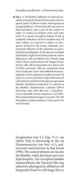
- Page 572: Geosiphon pyriformis- a Glomeromyco
- Page 576: Geosiphon pyriformis- a Glomeromyco
- Page 580: Geosiphon pyriformis- a Glomeromyco
- Page 584: Geosiphon pyriformis- a Glomeromyco
- Page 588: Geosiphon pyriformis- a Glomeromyco
- Page 592: Geosiphon pyriformis- a Glomeromyco
- Page 596: Geosiphon pyriformis- a Glomeromyco
- Page 600: Geosiphon pyriformis- a Glomeromyco
- Page 604: 292 R. Prasad et al. Scientists fro
- Page 608: 294 R. Prasad et al. members of thi
- Page 612: 296 R. Prasad et al. Table 3. Typic
- Page 616: 298 R. Prasad et al. 5 Axenic Culti
- Page 620:
300 R. Prasad et al. Fig. 2. a Axen
- Page 624:
302 R. Prasad et al. Fig.5a-c. Root
- Page 628:
304 R. Prasad et al. Fig.8a-c. Chla
- Page 632:
306 R. Prasad et al. Fig.10a, b. Ro
- Page 636:
308 R. Prasad et al. Fig.13a-d. Aut
- Page 640:
310 R. Prasad et al. Acknowledgemen
- Page 644:
312 R. Prasad et al. Institute of S
- Page 648:
17 1 Introduction Large-Scale Inocu
- Page 652:
Large-Scale Inoculum Production of
- Page 656:
Large-Scale Inoculum Production of
- Page 660:
Large-Scale Inoculum Production of
- Page 664:
Large-Scale Inoculum Production of
- Page 668:
Large-Scale Inoculum Production of
- Page 672:
Large-Scale Inoculum Production of
- Page 676:
Large-Scale Inoculum Production of
- Page 680:
Large-Scale Inoculum Production of
- Page 684:
Large-Scale Inoculum Production of
- Page 688:
Large-Scale Inoculum Production of
- Page 692:
Large-Scale Inoculum Production of
- Page 696:
Part VII Methodology
- Page 700:
342 S. Cranenbrouck et al. more tha
- Page 704:
344 S. Cranenbrouck et al. Table 1.
- Page 708:
346 S. Cranenbrouck et al. Table 1.
- Page 712:
348 S. Cranenbrouck et al. Table 2.
- Page 716:
350 S. Cranenbrouck et al. 5. Solut
- Page 720:
352 S. Cranenbrouck et al. were Lyc
- Page 724:
354 S. Cranenbrouck et al. Disinfec
- Page 728:
356 S. Cranenbrouck et al. Fig. 4.
- Page 732:
358 S. Cranenbrouck et al. Fig. 5.
- Page 736:
360 S. Cranenbrouck et al.
- Page 740:
362 S. Cranenbrouck et al. 3. Trans
- Page 744:
364 S. Cranenbrouck et al. 3. Rinse
- Page 748:
366 S. Cranenbrouck et al. 7.2 Mate
- Page 752:
368 S. Cranenbrouck et al. 8 Contin
- Page 756:
370 S. Cranenbrouck et al. 3. Citra
- Page 760:
372 S. Cranenbrouck et al. 9 Conclu
- Page 764:
374 S. Cranenbrouck et al. Legué V
- Page 768:
Subject Index Abscisic acid (ABA) 1
- Page 772:
Subject Index 379 Colony developmen
- Page 776:
Subject Index 381 - clarum 19, 34,
- Page 780:
Subject Index 383 -drying 23 - susp
- Page 784:
Subject Index 385 Primary colonizat
- Page 788:
Subject Index 387 Storage 23, 25, 3