BTS guidelines for the insertion of a chest drain - British Thoracic ...
BTS guidelines for the insertion of a chest drain - British Thoracic ...
BTS guidelines for the insertion of a chest drain - British Thoracic ...
- No tags were found...

You also want an ePaper? Increase the reach of your titles
YUMPU automatically turns print PDFs into web optimized ePapers that Google loves.
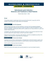
ii56Laws, Neville, Duffybe undertaken. In a study <strong>of</strong> <strong>chest</strong> tubes inserted in traumasuites using full aseptic technique, <strong>the</strong>re were no infectivecomplications in 80 cases. 38Studies <strong>of</strong> <strong>the</strong> use <strong>of</strong> antibiotic prophylaxis <strong>for</strong> <strong>chest</strong> tube<strong>insertion</strong> have been per<strong>for</strong>med but have failed to reachsignificance because <strong>of</strong> small numbers <strong>of</strong> infectious complications.However, a meta-analysis <strong>of</strong> <strong>the</strong>se studies has been per<strong>for</strong>medwhich suggested that, in <strong>the</strong> presence <strong>of</strong> <strong>chest</strong> trauma(penetrating or blunt), <strong>the</strong> use <strong>of</strong> prophylactic antibioticsreduces <strong>the</strong> absolute risk <strong>of</strong> empyema by 5.5–7.1% and <strong>of</strong> allinfectious complications by 12.1–13.4%. 39 The use <strong>of</strong> prophylacticantibiotics in trauma cases is <strong>the</strong>re<strong>for</strong>e recommended.The antibiotics used in <strong>the</strong>se studies were cephalosporins orclindamycin.The use <strong>of</strong> prophylactic antibiotics is less clear in <strong>the</strong> event<strong>of</strong> spontaneous pneumothorax or pleural effusion <strong>drain</strong>age asno studies were found which addressed <strong>the</strong>se circumstances.In one study only one infectious complication (in <strong>the</strong> <strong>chest</strong>tube track) occurred in a series <strong>of</strong> 39 spontaneous pneumothoracestreated with <strong>chest</strong> tubes. 4012 ANAESTHESIA• Local anaes<strong>the</strong>tic should be infiltrated prior to <strong>insertion</strong><strong>of</strong> <strong>the</strong> <strong>drain</strong>. [C]Local anaes<strong>the</strong>tic is infiltrated into <strong>the</strong> site <strong>of</strong> <strong>insertion</strong> <strong>of</strong> <strong>the</strong><strong>drain</strong>. A small gauge needle is used to raise a dermal blebbe<strong>for</strong>e deeper infiltration <strong>of</strong> <strong>the</strong> intercostal muscles and pleuralsurface. A spinal needle may be required in <strong>the</strong> presence <strong>of</strong>a thick <strong>chest</strong> wall.Local anaes<strong>the</strong>tic such as lignocaine (up to 3 mg/kg ) isusually infiltrated. Higher doses may result in toxic levels. Thepeak concentration <strong>of</strong> lignocaine was found to be 24 F)• Blunt dissection into <strong>the</strong> pleural space must beper<strong>for</strong>med be<strong>for</strong>e <strong>insertion</strong> <strong>of</strong> a large bore <strong>chest</strong><strong>drain</strong>. [C]13.3.1 Incision• The incision <strong>for</strong> <strong>insertion</strong> <strong>of</strong> <strong>the</strong> <strong>chest</strong> <strong>drain</strong> should besimilar to <strong>the</strong> diameter <strong>of</strong> <strong>the</strong> tube being inserted.[C]Once <strong>the</strong> anaes<strong>the</strong>tic has taken effect an incision is made. Thisshould be slightly bigger than <strong>the</strong> operator’s finger and tube.The incision should be made just above and parallel to a rib.13.3.2 Blunt dissectionMany cases <strong>of</strong> damage to essential intrathoracic structureshave been described following <strong>the</strong> use <strong>of</strong> trocars to insert largebore <strong>chest</strong> tubes. Blunt dissection <strong>of</strong> <strong>the</strong> subcutaneous tissueand muscle into <strong>the</strong> pleural cavity has <strong>the</strong>re<strong>for</strong>e becomeuniversal 43and is essential. In one retrospective study onlyfour technical complications were seen in 447 cases usingblunt dissection. 37 Using a Spencer-Wells clamp or similar, apath is made through <strong>the</strong> <strong>chest</strong> wall by opening <strong>the</strong> clamp toseparate <strong>the</strong> muscle fibres. For a large <strong>chest</strong> <strong>drain</strong>, similar insize to <strong>the</strong> finger, this track should be explored with a fingerthrough into <strong>the</strong> thoracic cavity to ensure <strong>the</strong>re are no underlyingorgans that might be damaged at tube <strong>insertion</strong>. 2–9 Thecreation <strong>of</strong> a patent track into <strong>the</strong> pleural cavity ensures thatexcessive <strong>for</strong>ce is not needed during <strong>drain</strong> <strong>insertion</strong>.13.3.3 Position <strong>of</strong> tube tip• The position <strong>of</strong> <strong>the</strong> tip <strong>of</strong> <strong>the</strong> <strong>chest</strong> tube shouldideally be aimed apically <strong>for</strong> a pneumothorax orbasally <strong>for</strong> fluid. However, any tube position can beeffective at <strong>drain</strong>ing air or fluid and an effectivelyfunctioning <strong>drain</strong> should not be repositioned solelybecause <strong>of</strong> its radiographic position. [C]In <strong>the</strong> case <strong>of</strong> a large bore tube, after gentle <strong>insertion</strong> through<strong>the</strong> <strong>chest</strong> wall <strong>the</strong> trocar positioned a few centimetres from <strong>the</strong>tube tip can af<strong>for</strong>d support <strong>of</strong> <strong>the</strong> tube and so help itspositioning without incurring organ damage. A smaller clampcan also be used to direct <strong>the</strong> tube to its desired position. 13If possible, <strong>the</strong> tip <strong>of</strong> <strong>the</strong> tube should be aimed apically to<strong>drain</strong> air and basally <strong>for</strong> fluid. However, successful <strong>drain</strong>agecan still be achieved when <strong>the</strong> <strong>drain</strong> is not placed in an idealposition, 21so effectively functioning tubes should not berepositioned simply because <strong>of</strong> a suboptimal radiographicappearance.13.3.4 Securing <strong>the</strong> <strong>drain</strong>• Large and medium bore <strong>chest</strong> <strong>drain</strong> incisions shouldbe closed by a suture appropriate <strong>for</strong> a linear incision.[C]• “Purse string” sutures must not be used. [C]Two sutures are usually inserted—<strong>the</strong> first to assist laterclosure <strong>of</strong> <strong>the</strong> wound after <strong>drain</strong> removal and <strong>the</strong> second, astay suture, to secure <strong>the</strong> <strong>drain</strong>.The wound closure suture should be inserted be<strong>for</strong>e bluntdissection. A strong suture such as “1” silk is appropriate. 621 A“mattress” suture or sutures across <strong>the</strong> incision are usuallyemployed and, whatever closure is used, <strong>the</strong> stitch must be <strong>of</strong>a type that is appropriate <strong>for</strong> a linear incision (fig 4). Complicated“purse string” sutures must not be used as <strong>the</strong>y convertwww.thoraxjnl.com
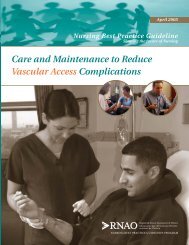
<strong>BTS</strong> <strong>guidelines</strong> <strong>for</strong> <strong>the</strong> <strong>insertion</strong> <strong>of</strong> a <strong>chest</strong> <strong>drain</strong>ii57Figure 4Example <strong>of</strong> stay and closing sutures.a linear wound into a circular one that is painful <strong>for</strong> <strong>the</strong>patient and may leave an unsightly scar. 9 A suture is not usuallyrequired <strong>for</strong> small gauge <strong>chest</strong> tubes.The <strong>drain</strong> should be secured after <strong>insertion</strong> to prevent itfalling out. Various techniques have been described, 44but asimple technique <strong>of</strong> anchoring <strong>the</strong> tube has not been <strong>the</strong> subject<strong>of</strong> a controlled trial. The chosen suture should be stout andnon absorbable to prevent breaking (e.g. “1” silk), 6and itshould include adequate skin and subcutaneous tissue toensure it is secure (fig 4).Large amounts <strong>of</strong> tape and padding to dress <strong>the</strong> site areunnecessary and concerns have been expressed that <strong>the</strong>y mayrestrict <strong>chest</strong> wall movement 6 or increase moisture collection.A transparent dressing allows <strong>the</strong> wound site to be inspectedby nursing staff <strong>for</strong> leakage or infection. An omental tag <strong>of</strong>tape has been described 2 which allows <strong>the</strong> tube to lie a littleaway from <strong>the</strong> <strong>chest</strong> wall to prevent tube kinking and tensionat <strong>the</strong> <strong>insertion</strong> site (fig 5).14 MANAGEMENT OF DRAINAGE SYSTEM14.1 Clamping <strong>drain</strong>• A bubbling <strong>chest</strong> tube should never be clamped. [C]• Drainage <strong>of</strong> a large pleural effusion should becontrolled to prevent <strong>the</strong> potential complication <strong>of</strong>re-expansion pulmonary oedema. [C]• In cases <strong>of</strong> pneumothorax, clamping <strong>of</strong> <strong>the</strong> <strong>chest</strong> tubeshould usually be avoided. [B]• If a <strong>chest</strong> tube <strong>for</strong> pneumothorax is clamped, thisshould be under <strong>the</strong> supervision <strong>of</strong> a respiratory physicianor thoracic surgeon, <strong>the</strong> patient should bemanaged in a specialist ward with experienced nursingstaff, and <strong>the</strong> patient should not leave <strong>the</strong> wardenvironment. [C]Figure 5 Omental tag to support <strong>the</strong> tube while allowing it to lie alittle away from <strong>the</strong> <strong>chest</strong> wall.• If a patient with a clamped <strong>drain</strong> becomes breathlessor develops subcutaneous emphysema, <strong>the</strong> <strong>drain</strong>must be immediately unclamped and medical advicesought. [C]There is no evidence to suggest that clamping a <strong>chest</strong> <strong>drain</strong>prior to its removal increases success or prevents recurrence <strong>of</strong>a pneumothorax and it may be hazardous. This is <strong>the</strong>re<strong>for</strong>egenerally discouraged. Clamping a <strong>chest</strong> <strong>drain</strong> in <strong>the</strong> presence<strong>of</strong> a continuing air leak may lead to <strong>the</strong> potentially fatal complication<strong>of</strong> tension pneumothorax. 369A bubbling <strong>drain</strong><strong>the</strong>re<strong>for</strong>e should never be clamped. However, many experiencedspecialist physicians support <strong>the</strong> use <strong>of</strong> <strong>the</strong> clamping <strong>of</strong>non-bubbling <strong>chest</strong> <strong>drain</strong>s inserted <strong>for</strong> pneumothorax todetect small air leaks not immediately obvious at <strong>the</strong> bedside.By clamping <strong>the</strong> <strong>chest</strong> <strong>drain</strong> <strong>for</strong> several hours, followed by a<strong>chest</strong> radiograph, a minor air leak may be detected, avoiding<strong>the</strong> need <strong>for</strong> later <strong>chest</strong> <strong>drain</strong> re<strong>insertion</strong>. In <strong>the</strong> ACCP Delphiconsensus statement 45 about half <strong>the</strong> consensus groupsupported clamping and half did not, and this seems similar to<strong>the</strong> UK spread <strong>of</strong> opinion. Drain clamping is <strong>the</strong>re<strong>for</strong>e notgenerally recommended <strong>for</strong> safety reasons, but is acceptableunder <strong>the</strong> supervision <strong>of</strong> nursing staff who are trained in <strong>the</strong>management <strong>of</strong> <strong>chest</strong> <strong>drain</strong>s and who have instructions tounclamp <strong>the</strong> <strong>chest</strong> <strong>drain</strong> in <strong>the</strong> event <strong>of</strong> any clinical deterioration.Patients with a clamped <strong>chest</strong> <strong>drain</strong> inserted <strong>for</strong>pneumothorax should not leave <strong>the</strong> specialist ward area.There have been reports <strong>of</strong> re-expansion pulmonaryoedema following rapid evacuation <strong>of</strong> large pleural effusions 4647 48as well as in association with spontaneous pneumothorax.This has been reported to be fatal in some cases (up to 20% <strong>of</strong>subjects in one series <strong>of</strong> 53 cases 49 ). In <strong>the</strong> case <strong>of</strong> spontaneouspneumothorax this is a rare complication with no cases <strong>of</strong>re-expansion pulmonary oedema reported in two large studies<strong>of</strong> 400 and 375 patients, respectively. 50 51 It is usually associatedwith delayed diagnosis and <strong>the</strong>re<strong>for</strong>e awareness <strong>of</strong> itspotential occurrence is sufficient.Milder symptoms suggestive <strong>of</strong> re-expansion oedema arecommon after large volume thoracentesis in pleural effusion,with patients experiencing discom<strong>for</strong>t and cough. It has beensuggested that <strong>the</strong> tube be clamped <strong>for</strong> 1 hour after <strong>drain</strong>ing1 litre. 52 While <strong>the</strong>re is no evidence <strong>for</strong> actual amounts, goodpractice suggests that no more than about 1.5 litres should be<strong>drain</strong>ed at one time, or <strong>drain</strong>age should be slowed to about500 ml per hour.14.2 Closed system <strong>drain</strong>age• All <strong>chest</strong> tubes should be connected to a single flow<strong>drain</strong>age system e.g. under water seal bottle or fluttervalve. [C]• Use <strong>of</strong> a flutter valve system allows earlier mobilisationand <strong>the</strong> potential <strong>for</strong> earlier discharge <strong>of</strong>patients with <strong>chest</strong> <strong>drain</strong>s.The <strong>chest</strong> tube is <strong>the</strong>n attached to a <strong>drain</strong>age system whichonly allows one direction <strong>of</strong> flow. This is usually <strong>the</strong> closedunderwater seal bottle in which a tube is placed under waterat a depth <strong>of</strong> approximately 3 cm with a side vent whichallows escape <strong>of</strong> air, or it may be connected to a suctionpump. 2–4 7 This enables <strong>the</strong> operator to see air bubble out as <strong>the</strong>lung re-expands in <strong>the</strong> case <strong>of</strong> pneumothorax or fluid evacuationrate in empyemas, pleural effusions, or haemothorax. Thecontinuation <strong>of</strong> bubbling suggests a continued visceral pleuralair leak, although it may also occur in patients on suctionwhen <strong>the</strong> <strong>drain</strong> is partly out <strong>of</strong> <strong>the</strong> thorax and one <strong>of</strong> <strong>the</strong> tubeholes is open to <strong>the</strong> air. The respiratory swing in <strong>the</strong> fluid in<strong>the</strong> <strong>chest</strong> tube is useful <strong>for</strong> assessing tube patency andconfirms <strong>the</strong> position <strong>of</strong> <strong>the</strong> tube in <strong>the</strong> pleural cavity. The disadvantages<strong>of</strong> <strong>the</strong> underwater seal system include obligatoryinpatient management, difficulty <strong>of</strong> patient mobilisation, and<strong>the</strong> risk <strong>of</strong> knocking over <strong>the</strong> bottle.www.thoraxjnl.com
ii58Laws, Neville, DuffyThe use <strong>of</strong> integral Heimlich flutter valves has beenadvocated in patients with pneumothoraces, especially as <strong>the</strong>ypermit ambulatory or even outpatient management which hasbeen associated with a 85–95% success rate. 53 54 In 176 cases <strong>of</strong>pneumothorax treated with small <strong>chest</strong> tubes and a Heimlichflutter valve <strong>the</strong>re were only eight failures (hospital admissions<strong>for</strong> problems with tube function or placement). Themean length <strong>of</strong> inpatient stay has been quoted at 5 hours witha thoracic vent and 144 hours with an underwater seal, with acost saving US$5660. 53Case reports <strong>of</strong> incorrect use (wrongdirection <strong>of</strong> flow) <strong>of</strong> such valves have been described, however,with tension pneumothorax as a result. 55 Flutter valves cannotbe used with fluid <strong>drain</strong>age as <strong>the</strong>y tend to become blocked.However, in <strong>the</strong> UK a similar short hospital stay is achieved byinitial aspiration <strong>of</strong> pneumothoraces (see <strong>guidelines</strong> on pneumothorax,page ii39).The use <strong>of</strong> a <strong>drain</strong>age bag with an incorporated flutter valveand vented outlet has been successfully usedpostoperatively. 56 57 A randomised trial <strong>of</strong> 119 cases followingelective thoracotomy compared <strong>the</strong> use <strong>of</strong> an underwater sealwith <strong>the</strong> flutter bag and found no difference in <strong>drain</strong>age volumes,requirement <strong>for</strong> suction, or complications with <strong>the</strong>added advantage <strong>of</strong> earlier mobilisation with <strong>drain</strong>age bags. 57In cases <strong>of</strong> malignant pleural effusion <strong>drain</strong>age a closedsystem using a <strong>drain</strong>age bag or aspiration via a three way taphas been described to aid palliation and outpatientmanagement. 33One report <strong>of</strong> a modified urinary collectingbag <strong>for</strong> prolonged underwater <strong>chest</strong> <strong>drain</strong>age has beendescribed <strong>for</strong> use with empyemas, bronchopulmonary fistula,and pneumothorax associated with emphysema with no complicationsin <strong>the</strong> 12 patients studied. 5814.3 Suction• When <strong>chest</strong> <strong>drain</strong> suction is required, a high volume/low pressure system should be used. [C]• When suction is required, <strong>the</strong> patient must be nursedby appropriately trained staff. [C]The use <strong>of</strong> high volume/low pressure suction pumps has beenadvocated in cases <strong>of</strong> non-resolving pneumothorax or followingchemical pleurodesis, 6 but <strong>the</strong>re is no evidence to supportits routine use in <strong>the</strong> initial treatment <strong>of</strong> spontaneouspneumothorax. 59 60 If suction is required, this may beper<strong>for</strong>med via <strong>the</strong> underwater seal at a level <strong>of</strong> 10–20 cm H 2O.A high volume pump (e.g. Vernon-Thompson) is required tocope with a large leak. A low volume pump (e.g. Robertspump) is inappropriate as it is unable to cope with <strong>the</strong> rapidflow, <strong>the</strong>reby effecting a situation similar to clamping andrisking <strong>for</strong>mation <strong>of</strong> a tension pneumothorax. A wall suctionadaptor may also be effective, although <strong>chest</strong> <strong>drain</strong>s must notbe connected directly to <strong>the</strong> high negative pressure availablefrom wall suction.In <strong>the</strong> management <strong>of</strong> pleural infection, <strong>the</strong> use <strong>of</strong> suctionis less clear. Most studies are observational and have used suctionapplied via <strong>the</strong> <strong>chest</strong> tube after flushing to prevent blockingand have reported success, but this has not been comparedwith cases without suction. This is discussed fur<strong>the</strong>r in <strong>the</strong>guideline on pleural infection (page ii18).There is no evidence that briefly disconnecting a <strong>drain</strong> fromsuction used <strong>for</strong> spontaneous pneumothorax or pleuraleffusion is disadvantageous. There<strong>for</strong>e, as long as adequateinstruction is given to patient, portering and nursing staffwith regard to keeping <strong>the</strong> underwater seal bottle below <strong>the</strong>level <strong>of</strong> <strong>the</strong> <strong>chest</strong>, it is acceptable to stop suction <strong>for</strong> short periodssuch as <strong>for</strong> radiography.14.4 Ward instructions• Patients with <strong>chest</strong> tubes should be managed onspecialist wards by staff who are trained in <strong>chest</strong><strong>drain</strong> management. [C]Audit points• The presence and use <strong>of</strong> an appropriate nursing <strong>chest</strong> <strong>drain</strong>observation chart should be noted.• The frequency <strong>of</strong> <strong>chest</strong> <strong>drain</strong> complications should berecorded.• The use <strong>of</strong> premedication and analgesics and patient painscores relating to <strong>chest</strong> <strong>drain</strong> <strong>insertion</strong> should be recorded.• The duration <strong>of</strong> <strong>chest</strong> tube <strong>drain</strong>age should be recorded.• A <strong>chest</strong> radiograph should be per<strong>for</strong>med after <strong>insertion</strong><strong>of</strong> a <strong>chest</strong> <strong>drain</strong>. [C]Patients should be managed on a ward familiar with <strong>chest</strong>tubes. Instruction to and appropriate training <strong>of</strong> <strong>the</strong> nursingstaff is imperative. If an underwater seal is used, instructionsmust be given to keep <strong>the</strong> bottle below <strong>the</strong> <strong>insertion</strong> site at alltimes, to keep it upright, and to ensure that adequate water isin <strong>the</strong> system to cover <strong>the</strong> end <strong>of</strong> <strong>the</strong> tube. 9 Daily reassessment<strong>of</strong> <strong>the</strong> amount <strong>of</strong> <strong>drain</strong>age/bubbling and <strong>the</strong> presence <strong>of</strong> respiratoryswing should be documented, preferably on a dedicated<strong>chest</strong> <strong>drain</strong> chart. Instruction with regard to <strong>chest</strong> <strong>drain</strong>clamping must be given and recorded. 61Patients should be encouraged to take responsibility <strong>for</strong><strong>the</strong>ir <strong>chest</strong> tube and <strong>drain</strong>age system. They should be taughtto keep <strong>the</strong> underwater seal bottle below <strong>the</strong> level <strong>of</strong> <strong>the</strong>ir<strong>chest</strong> and to report any problems such as pulling on <strong>the</strong> <strong>drain</strong><strong>insertion</strong> site. Educational material (e.g. leaflets) should beavailable on <strong>the</strong> ward <strong>for</strong> patients and nursing staff.A <strong>chest</strong> radiograph should be per<strong>for</strong>med to assess tubeposition, exclude complications such as pneumothorax or surgicalemphysema, and assess <strong>the</strong> success <strong>of</strong> <strong>the</strong> procedure in<strong>the</strong> volume <strong>of</strong> fluid <strong>drain</strong>age or pneumothorax resolution.Concern has previously been expressed in cases where <strong>the</strong>tube enters <strong>the</strong> lung fissure. In a study <strong>of</strong> 66 patients with<strong>chest</strong> tubes inserted <strong>for</strong> acute <strong>chest</strong> trauma, 58% <strong>of</strong> whichwere located within a pulmonary fissure, 62no difference inoutcome was seen between <strong>the</strong>se cases and those in whom <strong>the</strong>tube was located outside <strong>the</strong> fissures.14.5 Removal <strong>of</strong> <strong>the</strong> <strong>chest</strong> tube• In cases <strong>of</strong> pneumothorax, <strong>the</strong> <strong>chest</strong> tube should notbe clamped at <strong>the</strong> time <strong>of</strong> its removal. [B]In cases <strong>of</strong> pneumothorax, <strong>the</strong>re is no evidence that clampinga <strong>chest</strong> <strong>drain</strong> at <strong>the</strong> time <strong>of</strong> its removal is beneficial. 60The <strong>chest</strong> tube should be removed ei<strong>the</strong>r while <strong>the</strong> patientper<strong>for</strong>ms Valsalva’s manoeuvre or during expiration with abrisk firm movement while an assistant ties <strong>the</strong> previouslyplaced closure suture. 2–478 The timing <strong>of</strong> removal is dependenton <strong>the</strong> original reason <strong>for</strong> <strong>insertion</strong> and clinical progress (see<strong>guidelines</strong> <strong>for</strong> management <strong>of</strong> pneumothorax (page ii39),malignant pleural effusions (page ii29), and pleural infections(page ii18)).In <strong>the</strong> case <strong>of</strong> pneumothorax, <strong>the</strong> <strong>drain</strong> should not usuallybe removed until bubbling has ceased and <strong>chest</strong> radiographydemonstrates lung reinflation. 4 Clamping <strong>of</strong> <strong>the</strong> <strong>drain</strong> be<strong>for</strong>eremoval is generally unnecessary. In one study <strong>the</strong> removal <strong>of</strong><strong>chest</strong> tubes after continuous suction was compared with <strong>the</strong>removal after a period <strong>of</strong> disconnection from suction to anunderwater seal. No significant difference was seen between<strong>the</strong>se two methods with only two <strong>of</strong> 80 cases (2.5%) requiringre<strong>insertion</strong> <strong>of</strong> a <strong>chest</strong> tube. 3815 PATIENTS REQUIRING ASSISTED VENTILATIONDuring <strong>the</strong> <strong>insertion</strong> <strong>of</strong> a <strong>chest</strong> tube in a patient on a highpressure ventilator (especially with positive end expiratorypressure (PEEP), it is essential to disconnect from <strong>the</strong> ventilatorat <strong>the</strong> time <strong>of</strong> <strong>insertion</strong> to avoid <strong>the</strong> potentially seriouswww.thoraxjnl.com
<strong>BTS</strong> <strong>guidelines</strong> <strong>for</strong> <strong>the</strong> <strong>insertion</strong> <strong>of</strong> a <strong>chest</strong> <strong>drain</strong>ii59complication <strong>of</strong> lung penetration, 63 although as long as bluntdissection is carried out and no sharp instruments are used,this risk is reduced. 64ACKNOWLEDGEMENTSThe authors are grateful to Dr Richard Holmes <strong>for</strong> figs 3, 4, and 5......................Authors’ affiliationsD Laws, Department <strong>of</strong> <strong>Thoracic</strong> Medicine, Royal Bournemouth Hospital,Bournemouth BH7 7DW, UKE Neville, Respiratory Centre, St Mary’s Hospital, Portsmouth PO3 6AD,UKJ Duffy, Cardiothoracic Surgery Department, City Hospital, NottinghamNG5 1PB, UKREFERENCES1 American College <strong>of</strong> Surgeons Committee on Trauma. In:<strong>Thoracic</strong>trauma. Advanced Trauma Life Support program <strong>for</strong> physicians: instructormanual. Chicago: AmericanCollege <strong>of</strong> Surgeons, 1993. [IV]2 Miller KS, Sahn SA. Review. Chest tubes. Indications, technique,management and complications. Chest 1987;91:258–64. [IV]3 Parmar JM. How to insert a <strong>chest</strong> <strong>drain</strong>. Br J Hosp Med1989;42:231–3. [IV]4 Treasure T, Murphy JP. Pneumothorax. Surgery 1989;75:1780–6. [IV]5 Westaby S, Brayley N. <strong>Thoracic</strong> trauma – I. BMJ 1990;330:1639–44.[IV]6 Harriss DR, Graham TR. Management <strong>of</strong> intercostal <strong>drain</strong>s. Br J HospMed 1991;45:383–6. [IV]7 Iberti TJ, Stern PM. Chest tube thoracostomy. Crit Care Clin1992;8:879–95. [IV]8 Quigley R.L. Thoracentesis and <strong>chest</strong> tube <strong>drain</strong>age. Crit Care Cli1995;11:111–26. [IV]9 Tomlinson MA. Treasure T. Insertion <strong>of</strong> a <strong>chest</strong> <strong>drain</strong> : how to do it. Br JHosp Med 1997;58:248–52. [IV]10 Collop NA, Kim S, Sahn SA. Analysis <strong>of</strong> tube thoracostomy per<strong>for</strong>medby pulmonologists at a teaching hospital. Chest 1997;112:709–13. [III]11 Luketich JD, Kiss MD, Hershey J, et al. Chest tube <strong>insertion</strong>: aprospective evaluation <strong>of</strong> pain management. Clin J Pain1998;14:152–4. [IIa]12 Reinhold C, Illescas FF, Atri M, et al. The treatment <strong>of</strong> pleural effusionsand pneumothorax with ca<strong>the</strong>ters placed percutaneously under imageguidance. AJR 1989;152:1189–91. [III]13 Ward EW, Hughes TE. Sudden death following <strong>chest</strong> tube <strong>insertion</strong>: anunusual case <strong>of</strong> vagus nerve irritation. J Trauma 1994;36:258–9. [IV]14 Boland GW, Lee MJ, Silverman S, et al. Review. Interventional radiology<strong>of</strong> <strong>the</strong> pleural space. Clin Radiol 1995;50:205–14. [IV]15 Klein JS, Schultz S, Heffner JE. Intervential radiology <strong>of</strong> <strong>the</strong> <strong>chest</strong>:image-guided percutaneous <strong>drain</strong>age <strong>of</strong> pleural effusions, lung abscess,and pneumothorax. AJR 1995;164:581–8. [IV]16 Rosenberg ER. Ultrasound in <strong>the</strong> assessment <strong>of</strong> pleural densities. Chest1983;84:283–5. [IV]17 Harnsberger HR, Lee TG, Mukuno DH. Rapid, inexpensive real timedirected thoracocentesis. Radiology 1983;146:545–6. [IV]18 Holden MP. Management <strong>of</strong> intercostal <strong>drain</strong>age tubes. In: Practice <strong>of</strong>cardiothoracic surgery. Bristol: John Wright, 1982: 3. [IV]19 Aslam PA, Hughes FA. Insertion <strong>of</strong> an apical tube. Surg Gynecol Obstet1970;130:1097. [IV]20 Galvin IF, Gibbons JRP, Magout M, et al. Placement <strong>of</strong> an apical <strong>chest</strong>tube by a posterior approach. Br J Hosp Med 1990;44:330–1. [IV]21 Hyde J, Sykes T, Graham T. Reducing morbidity from <strong>chest</strong> <strong>drain</strong>s. BMJ1997;311:914–5. [IV]22 Clementsen P, Evald T, Grode G, et al. Treatment <strong>of</strong> malignant pleuraleffusion : pleurodesis using a small bore ca<strong>the</strong>ter. A prospectiverandomized study. Respir Med 1998;92:593–6. [Ib]23 Patz EF, Goodman PC, Erasmus JJ. Percutaneous <strong>drain</strong>age <strong>of</strong> pleuralcollections. J Thorac Imaging 1998;13:83–92. [IV]24 Henderson AF, Banham SW, Moran F. Re-expansion pulmonaryoedema: a potentially serious complication <strong>of</strong> delayed diagnosis <strong>of</strong>pneumothorax. BMJ 1985;29:593–4. [IV]25 Thomas RJ, Sagar SM. What size pleural tube <strong>for</strong> pleural effusions(letter)? Br J Hosp Med 1990;43:184. [IV]26 Taylor PM. Ca<strong>the</strong>ters smaller <strong>the</strong>n 24 French gauge can be used <strong>for</strong><strong>chest</strong> <strong>drain</strong>s (letter). BMJ 1997;315:186. [IV]27 Conces DJ, Tarver RD, Gray WC, et al. Treatment <strong>of</strong> pneumothoracesutilizing small caliber <strong>chest</strong> tubes. Chest 1988;94:55–7. [III]28 Parker LA, Charnock GC, Delany DJ. Small bore ca<strong>the</strong>ter <strong>drain</strong>age andsclero<strong>the</strong>rapy <strong>for</strong> malignant pleural effusions. Cancer 1989;64:1218–21. [III]29 Morrison MC, Mueller PR, Lee MJ, et al. Sclero<strong>the</strong>rapy <strong>of</strong> malignantpleural effusion through sonographically placed small-bore ca<strong>the</strong>ters. AJR1992;158:41–3. [III]30 G<strong>of</strong>f BA, Mueller PR, Muntz HG, et al. Small <strong>chest</strong> tube <strong>drain</strong>agefollowed by bleomycin sclerosis <strong>for</strong> malignant pleural effusions. ObstetGynecol 1993;81:993–6. [III]31 Seaton KG, Patz EF, Goodman PC. Palliative treatment <strong>of</strong> malignantpleural effusions: value <strong>of</strong> small-bore ca<strong>the</strong>ter thoracostomy anddoxycycline sclero<strong>the</strong>rapy. AJR 1995;164:589–91. [IIb]32 Thompson RL, Yau JC, Donnelly RF, et al. Pleurodesis with iodized talc<strong>for</strong> malignant effusions using pigtail ca<strong>the</strong>ters. Ann Pharmaco<strong>the</strong>r1998;32:739–42. [IIb]33 Van Le L, Parker LA, DeMars LR, et al. Pleural effusions: outpatientmanagement <strong>of</strong> pigtail ca<strong>the</strong>ter <strong>chest</strong> tubes. Gynecol Oncol1994;54:215–7. [IV]34 Matsumoto AH. Image-guided <strong>drain</strong>age <strong>of</strong> complicated pleural effusionsand adjunctive use <strong>of</strong> intrapleural urokinase. Chest 1995;108: 1190–1.[IV]35 Moulton JS, Benkert RE, Weisiger KH, et al. Treatment <strong>of</strong> complicatedpleural fluid collections with image guided <strong>drain</strong>age and intra cavitaryurokinase. Chest 1995;108:1252–9. [III]36 Parry GW, Morgan WE, Salama FD. Management <strong>of</strong> haemothorax. AnnR Coll Surg Engl 1996;78:325–6. [IV]37 Millikan JS, Moore EE, Steiner E, et al. Complications <strong>of</strong> tubethoracostomy <strong>for</strong> acute trauma. Am J Surg 1980;140:738–41. [III]38 Davis JW, MacKersie RC, Hoyt DB, et al. Randomised study <strong>of</strong>algorithms <strong>for</strong> discontinuing tube thoracostomy <strong>drain</strong>age. JAmCollSurg1994;179:553–7. [Ib]39 Fallon WF, Wears RL. Prophylactic antibiotics <strong>for</strong> <strong>the</strong> prevention <strong>of</strong>infectious complications including empyema following tube thoracoscopy<strong>for</strong> trauma: results <strong>of</strong> a meta-analysis. J Trauma 1992;33:110–7. [Ia]40 LeBlanc KA, Tucker WY. Prophylactic antibiotics and closed tubethoracostomy. Surg Gynecol Obstet 1985;160:259–63. [Ib]41 Wooten SA, Barbarash RA, Strange C, et al. Systemic absorption <strong>of</strong>tetracycline following intrapleural instillation. Chest 1988;94:960–3.[IIa]42 Mellor DJ. A new method <strong>of</strong> <strong>chest</strong> <strong>drain</strong> <strong>insertion</strong>. Anaes<strong>the</strong>sia1996;51:713–4. [IV]43 Haggie JA. Management <strong>of</strong> pneumothorax: <strong>chest</strong> <strong>drain</strong> trocar is unsafeand unnecessary. BMJ 1993;307:443. [IV]44 Rashid MA, Wikstrom T, Ortenwall P. A simple technique <strong>for</strong> anchoring<strong>chest</strong> tubes. Eur Respir J 1998;12:958–9. [IV]45 Baumann MH, Strange C, Heffner JE, et al. Management <strong>of</strong>spontaneous pneumothorax. An American College <strong>of</strong> Chest PhysiciansDelphi Consensus Statement. Chest 2001;119:590–602.46 Trapnell DH, Thurston JGB. Unilateral pulmonary oedema after pleuralaspiration. Lancet 1970;i:1367–9. [IV]47 Rozenman J,Yellin A, Simansky DA, et al. Re-expansion pulmonaryoedema following pneumothorax. Respir Med 1996;90:235–8. [IV]48 Henderson AK. Fur<strong>the</strong>r advice on inserting a <strong>chest</strong> <strong>drain</strong> (letter andreply). Br J Hosp Med 1990;43:82. [IV]49 Mafhood S, Hix WR, Aaron BI, et al. Re-expansion pulmonary oedema.Ann Thorac Surg 1988;45:340–5. [IV]50 Mills M, Balsch B. Spontaneous pneumothorax : a series <strong>of</strong> 400 cases.Ann Thorac Surg 1965;1:286. [IV]51 Brooks J. Open thoracotomy in <strong>the</strong> management <strong>of</strong> spontaneouspneumothorax. Ann Surg 1973;177:798. [IV]52 Hall M, Jones A. Clamping may be appropriate to prevent discom<strong>for</strong>tand reduce risk <strong>of</strong> oedema (letter). BMJ 1997;315:313. [IV]53 Roegela M, Roeggla G, Muellner M, et al. The cost <strong>of</strong> treatment <strong>of</strong>spontaneous pneumothorax with <strong>the</strong> thoracic vent compared withconventional thoracic <strong>drain</strong>age (letter). Chest 1996;110:303. [Ib]54 Ponn RB, Siverman HJ, Federico JA. Outpatient <strong>chest</strong> tube management.Ann Thorac Surg 1997;64:1437–40. [III]55 Mainini SE, Johnson FE. Tension pneumothorax complicatingsmall-caliber <strong>chest</strong> tube <strong>insertion</strong>. Chest 1990; 97:759–60. [IV]56 Mat<strong>the</strong>ws HR, Mcguigan JA. Closed <strong>chest</strong> <strong>drain</strong>age without anunderwater seal. Thorax 1988;41:804P. [IV]57 Graham ANJ, Cosgrove AP, Gibbons JRP, et al. Randomised clinicaltrial <strong>of</strong> <strong>chest</strong> <strong>drain</strong>age systems. Thorax 1992;47:461–2. [Ib]58 Bar-El Y, Leiberman Y, Yellin A. Modified urinary collecting bag <strong>for</strong>prolonged underwater <strong>chest</strong> <strong>drain</strong>age. Ann Thorac Surg1992;54:995–6. [IIb]59 Sharma TN, Agrihotri SP, Jain NK, et al . Intercostal tube thoracostomyin pneumothorax : factors influencing re-expansion <strong>of</strong> lung. Ind J ChestDis Allied Sci 1988;30:32–5. [III]60 So SY, Yu DY. Ca<strong>the</strong>ter <strong>drain</strong>age <strong>of</strong> spontaneous pneumothorax: suctionor no suction, early or late removal? Thorax 1982;37:46–8. [Ib]61 Williams T. To clamp or not to clamp. Nursing Times 1992;88:33. [IV]62 Curtin JJ, Goodman LR, Quebberman EJ, et al. Thoracostomy tubes afteracute <strong>chest</strong> injury: relationship between location in a pleural fissure andfunction. AJR1994;163:1339–42. [IIa]63 Peek GJ, Firmin RK, Arsiwala S. Chest tube <strong>insertion</strong> in <strong>the</strong> ventilatedpatient. Injury 1995;26:425–6. [IV]64 Main A. As few sharp objects as possible should be used on enteringpleural space (letter). BMJ 1998;316:68. [IV]www.thoraxjnl.com