Arabidopsis mesophyll protoplasts: a versatile cell system for
Arabidopsis mesophyll protoplasts: a versatile cell system for
Arabidopsis mesophyll protoplasts: a versatile cell system for
- No tags were found...

Create successful ePaper yourself
Turn your PDF publications into a flip-book with our unique Google optimized e-Paper software.
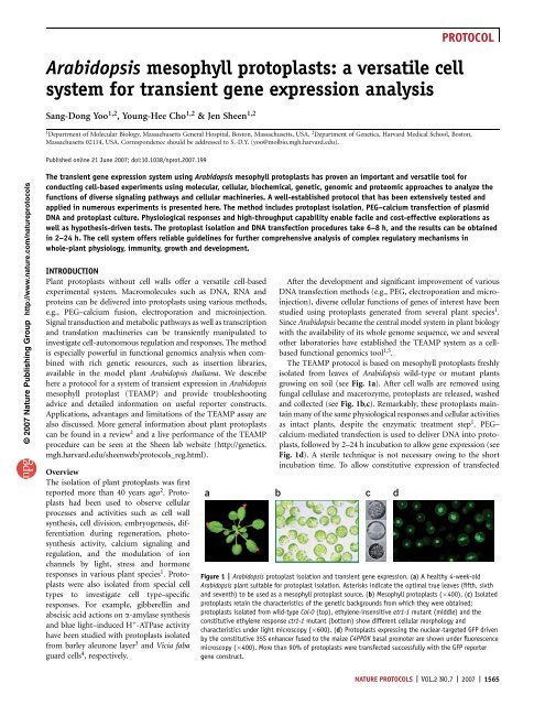
PROTOCOL© 2007 Nature Publishing Group http://www.nature.com/ natureprotocolsprotein–protein interaction has also been examined in <strong>Arabidopsis</strong><strong>mesophyll</strong> <strong>protoplasts</strong> to establish a heterodimerization map ofspecific groups of bZIP transcription factors 18 .Single-<strong>cell</strong> imaging of diverse fluorescence marker proteins,e.g., GFP and its variants, fused to functional proteins have alsobeen extensively utilized to examine protein localizations, proteindomain functions in protein targeting and protein transportmachinery functions in vesicle trafficking. For instance, sub<strong>cell</strong>ularlocalization of various proteins has been determined using the GFPtag 11,12,17–19 . Novel roles of the transmembrane domain andC-terminal 7 amino acids of <strong>Arabidopsis</strong> small outer envelopemembrane protein 7 have been identified <strong>for</strong> chloroplast targeting20 . Interesting regulatory roles of dynamin-like ADL6 (ref. 21),small GTP binding protein Rha1 (ref. 22) and actin filaments 23have been studied in vacuolar trafficking. The protoplast isolationmethod and fluorescence-assisted <strong>cell</strong> sorting technique have beenfurther applied to reveal genome-wide <strong>cell</strong> type–specific transcriptprofiles in <strong>Arabidopsis</strong> roots 24 .Advantages and limitationsUni<strong>for</strong>m and abundant <strong>mesophyll</strong> <strong>protoplasts</strong> isolated frommature <strong>Arabidopsis</strong> leaves can respond to diverse signals in aphysiological manner similar to the responses observed in leavesof whole plants. Importantly, application of the TEAMP <strong>system</strong>is not limited to processes and activities that occur in leaves.Proteins that are normally expressed in other <strong>cell</strong> types andtissues can be ectopically expressed and examined in <strong>mesophyll</strong><strong>protoplasts</strong> to learn their molecular and <strong>cell</strong>ular functions.Analysis using <strong>mesophyll</strong> <strong>protoplasts</strong> often provides in<strong>for</strong>mationof greater functional and physiological relevance to plants thandata obtained using heterologous <strong>cell</strong> <strong>system</strong>s, e.g., bacteria,yeast, insect and mammalian <strong>cell</strong>s. As most of the quantitativeand physiological responses can be observed and measuredwithin 2–10 h after DNA transfection, the experiments donot require sterile techniques and complex culture medium,which represents a tremendous saving in time and costs. Thereis no other biological <strong>system</strong> <strong>for</strong> gene expression analysis(including bacterial, yeast and mammalian <strong>cell</strong> culture) thatcan be handled with the same minimal laboratory requirement.The requirement <strong>for</strong> a relatively small number of <strong>protoplasts</strong> isanother advantage of the TEAMP <strong>system</strong> owing to its highefficiency and sensitivity. In some other protoplast protocols,10–1,000 times more <strong>protoplasts</strong> (10 5 –10 7 ) are recommended<strong>for</strong> each DNA transfection experiment 25 .On the other hand, the conserved functions of moleculardomains and regulatory mechanisms among bacteria, yeasts, animalsand plants allow us to utilize molecular switches, originallyidentified in other organisms, to control gene and protein expressionlevels in plant <strong>mesophyll</strong> <strong>protoplasts</strong>. For instance, themammalian steroid nuclear receptor has been used to provide aligand inducible gene expression <strong>system</strong> in <strong>mesophyll</strong> <strong>protoplasts</strong> 12 .As depicted in Figure 2c, an artificial transcription activator (ATA)is constructed with a bacterial DNA-binding domain (LexA-DB), aviral transcription activation domain (VP16) and a mammalian(rat) glucocorticoid receptor (GR). The ATA is driven underthe constitutive 35S enhancer fused to the maize C4PPDK basalpromoter 6 . Upon dexamethasone (DEX) treatment, the ATA(LexA-VP16-GR) is released from the cytoplasm HSP90 complexand moves into the nucleus to induce target gene transcription 26 .In the nucleus, the ATA controls the transcription of a DNAconstruct that is driven by eight copies of the bacterial LexAoperator (8xLexA-OP) and the minimum 35S promoter (seeFig. 2c). In the example presented (in which the LUC reportergene is included in the 8xLexA-OP-driven construct; see Fig. 2d),LUC expression is induced upon DEX treatment and shows linearexpression kinetics. The expression level of LUC is also controlledin a ligand dosage–dependent manner, indicating that the expressionlevel of desired genes and proteins can be manipulatedusing this inducible <strong>system</strong>.Mesophyll <strong>protoplasts</strong> can be isolated from fresh leaves ofwild-type and most <strong>Arabidopsis</strong> mutant plants and used immediately<strong>for</strong> experiments without the time-consuming step of <strong>cell</strong>culture establishment. Wild-type <strong>mesophyll</strong> <strong>protoplasts</strong> maintainmost leaf features and responses, which are often lost inundifferentiated suspension culture <strong>cell</strong>s. The mutant <strong>protoplasts</strong>possess the <strong>cell</strong>ular nature that is characteristic of the originalmutant plants. For example, <strong>mesophyll</strong> <strong>protoplasts</strong> isolated fromthe ethylene-insensitive etr1-1 and the constitutive ethyleneresponsectr1-1 mutants show <strong>cell</strong>ular morphology and characteristicsdistinct from those in the wild type (see Fig. 1c). Thereare many successful examples of using the TEAMP assay withvarious mutant plants, including the analysis of ARF7 functionsin auxin signaling 11 , the antagonistic interaction between HXK1-dependent glucose and ethylene signaling 12 , the receptor kinaseFLS2 function in flg22 signaling 15 and roles of various planthormone signaling pathways in programmed <strong>cell</strong> death inducedby the fungal toxin fumonisin B1 (ref. 27). Conceptually, theuse of <strong>protoplasts</strong> generated from mutant plants in TEAMPassays to screen <strong>for</strong> new regulators is similar to per<strong>for</strong>mingmutant suppressor or enhancer screens, which are commonlyapplied in the genetic analysis of specific signaling pathways. Ingeneral, it is necessary empirically to optimize the protoplastisolation and DNA transfection conditions <strong>for</strong> different <strong>Arabidopsis</strong>accessions and mutants. Once conditions are optimized,the use of genetically diverse <strong>mesophyll</strong> <strong>protoplasts</strong> makes theTEAMP <strong>system</strong> even more powerful in functional genomicsresearch.Unlike plant and animal <strong>cell</strong> culture lines, <strong>mesophyll</strong> <strong>protoplasts</strong>do not divide in the simple incubation buffer described in thisprotocol. Thus, this TEAMP protocol is not suitable <strong>for</strong> the study of<strong>cell</strong> cycle regulation. To use <strong>protoplasts</strong> to study <strong>cell</strong> division anddedifferentiation or other long-term plant responses, specializedplant growth conditions and sterile culture media will be required.Such protoplast regeneration protocols have been developed overthe past two decades as well 28–30 .For successful experiments, a high level of protoplast transfectionefficiency (greater than 50%) is very important to obtainreliable and reproducible data using the TEAMP <strong>system</strong>. Werecommend the use of an improved GFP vital marker donatedto the ABRC to visually monitor protoplast transfectionefficiency 19 (see Fig. 1d). Careful practice, keen observation andaccumulated experience contribute to the acquisition of meaningfulresults. All experiments need to be repeated multiple times toensure consistency and to avoid pitfalls and false positive results.Even though the protocol appears to be simple and straight<strong>for</strong>ward,the importance of practice and empirical optimization of eachexperiment <strong>for</strong> the individual researcher’s purpose cannot be overemphasized.NATURE PROTOCOLS | VOL.2 NO.7 | 2007 | 1567
PROTOCOL© 2007 Nature Publishing Group http://www.nature.com/ natureprotocolsMATERIALSREAGENTSGeneric razor blade (single-edged blade; VWR Scientific, cat. no. 55411-055). 0.2 M 4-morpholineethanesulfonic acid (MES, pH 5.7; Sigma,. Petri dish (100 25 mm 2 <strong>for</strong> 10 ml enzyme solution; VWR Scientific,cat. no. M8250), sterilize using a 0.45-mm filtercat. no. 25389-000)0.8 M mannitol (Sigma, cat. no.M4125), sterilize using a 0.45-mm filter . Nylon mesh (75 mm, laboratory sifters, Carolina Biological Supplies,1 M CaCl2 (Sigma, cat. no. C7902), sterilize using a 0.45-mm filtercat. no. 65-2222N)2 M KCl (Sigma, cat. no. P3911), sterilize using a 0.45-mm filter . Improved Neubauer 0.1-mm-deep Reichert hemacytometer (Hausser2 M MgCl2 (Sigma, cat. no. M9272), sterilize using a 0.45-mm filterScientific, cat. no. 1483)b-Mercaptoethanol (Sigma, cat. no. M6250)30-ml round-bottomed tube (Sarstedt, cat. no. 55.517)10% (wt/vol) BSA (Sigma, cat. no. A-6793), sterilize using a 0.45-mm filter . 2-ml round-bottomed natural microcentrifuge tube (SealRite; USA Scientific,Cellulase R10 (Yakult Pharmaceutical Ind. Co., Ltd., Japan)cat. no. 1620-2700). Macerozyme R10 (Yakult Pharmaceutical Ind. Co., Ltd., Japan) . 6-well culture dish (Falcon, cat. no. 3046)m CRITICAL We have obtained the best results with this enzyme provider. REAGENT SETUP. PEG4000 (Fluka, cat. no. 81240) m CRITICAL We found that the PEG source Enzyme solution Prepare 20 mM MES (pH 5.7) containing 1.5% (wt/vol)is very critical to achieve high transfection efficiency.<strong>cell</strong>ulase R10, 0.4% (wt/vol) macerozyme R10, 0.4 M mannitol and 20 mM KCl.1 M Tris–phosphate (pH 7.8), sterilize using a 0.45-mm filterWarm the solution at 55 1C <strong>for</strong> 10 min to inactivate DNAse and proteases and. 100 mM trans-1,2-diaminocyclo-hexane-N,N,N¢,N¢-tetraacetic acidenhance enzyme solubility. Cool it to room temperature (25 1C) and add 10 mM(DACTAA; Sigma, cat. no. D-1383)CaCl 2 , 1–5 mM b-mercaptoethanol (optional) and 0.1% BSA. m CRITICAL50% (vol/vol) glycerol (Fisher, cat. no. 15892), sterilize using a 0.45-mm filter Addition of 1–5 mM b-mercaptoethanol is optional, and its use should be20% (vol/vol) Triton X-100 (Sigma, cat. no. T-8787)determined according to the experimental purpose. m CRITICAL Be<strong>for</strong>e the1 M DTT (Sigma, cat. no. D-9779)enzyme powder is added, the MES solution is preheated at 70 1C <strong>for</strong> 3–5 min.LUC assay <strong>system</strong> (Promega, cat. no. E1501)The final enzyme solution should be clear light brown. Filter the final enzyme. 1 M Tris–HCl (pH 8.0) (US Biological, cat. no. T8650), sterilize using a solution through a 0.45-mm syringe filter device into a Petri dish (100 25 mm 20.45-mm filter<strong>for</strong> 10 ml enzyme solution). m CRITICAL The enzyme solution should be. 0.1 M 4-methylumbelliferyl glucuronide (MUG; Gold BioTechnology, Inc., prepared fresh.cat. no. MUG-1G)WI solution Prepare 4 mM MES (pH 5.7) containing 0.5 M mannitol and0.2 M Na2 CO 3 (Sigma, cat. no. S7795)20 mM KCl. The prepared WI solution can be stored at room temperature1 M methylumbelliferone (MU; Fluka, cat. no. 69580)(22–25 1C).Metro-Mix 360 (Sun Gro Horticulture, Inc.)W5 solution Prepare 2 mM MES (pH 5.7) containing 154 mM NaCl, 125 mMJiffy7 (Jiffy Products Ltd., Canada)CaCl 2 and 5 mM KCl. The prepared W5 solution can be stored at room<strong>Arabidopsis</strong> accessions: Col-0 and Ler (ABRC)temperature.Enzyme solution (see REAGENT SETUP)MMG solution Prepare 4 mM MES (pH 5.7) containing 0.4 M mannitol andWashing and incubation (WI) solution (see REAGENT SETUP)15 mM MgCl 2 . The prepared MMG solution can be stored at room temperature.W5 solution (see REAGENT SETUP)PEG–calcium transfection solution Prepare 20–40% (wt/vol) PEG4000 inMMG solution (see REAGENT SETUP)ddH 2 O containing 0.2 M mannitol and 100 mM CaCl 2 . m CRITICAL PreparePEG–calcium transfection solution (see REAGENT SETUP)PEG solution at least 1 h be<strong>for</strong>e transfection to completely dissolve PEG. TheProtoplast lysis buffer (see REAGENT SETUP)PEG solution can be stored at room temperature and used within 5 d. However,MUG substrate mix <strong>for</strong> GUS assay (see REAGENT SETUP)freshly prepared PEG solution gives relatively better protoplast transfection. Ampicillinefficiency. Do not autoclave PEG solution.EQUIPMENTProtoplast lysis buffer Prepare 2.5 mM Tris–phosphate (pH 7.8) containingFluorescence microscope (Leica DM5000; Leica)1 mM DTT, 2 mM DACTAA, 10% (vol/vol) glycerol and 1% (vol/vol) TritonLuminometer (Monolite3010; Pharmigen)X-100. The lysis buffer should be prepared fresh.Fluorometer (TK100; Hoefer)Bench-top centrifuge (IEC Centra CL2; International Equipment Company). 0.45-mm syringe sterilization filter (Whatman, cat. no. 6870-2504)MUG substrate mix <strong>for</strong> GUS assay Prepare 10 mM Tris–HCl (pH 8)containing 1 mM MUG and 2 mM MgCl 2 . The prepared GUS assay substratecan be stored at –20 1C.PROCEDUREPlant growth TIMING 3–4 weeks1| Grow <strong>Arabidopsis</strong> plants on either Metro-Mix 360 or Jiffy7 soil in a greenhouse or an environment-controlled chamber witha relatively short photoperiod (10–13 h light at 23 1C/11–14 h dark at 20 1C) under low light (50–75 mE m –2 s –1 ) and 40–65%relative humidity.m CRITICAL STEP Col-0 and Ler have been extensively tested in our lab. In general, <strong>Arabidopsis</strong> plants are very sensitive to all kindsof environmental changes (e.g., drought, flooding, extreme temperature and constant mechanical perturbation). Try to maintain aconstant environment as much as possible.Protoplast isolation TIMING 4–5 h2| Choose well-expanded leaves from 3–4-week-old plants (usually true leaf numbers five to seven) be<strong>for</strong>e flowering(see Fig. 1a).m CRITICAL STEP The selection of healthy leaves at the proper developmental stage is a very important factor in protoplastexperiments. Protoplasts prepared from leaves recovered from stress conditions (e.g., drought, flooding, extreme temperature andconstant mechanical perturbation) may look similar to those from healthy leaves. However, we have often experienced lowtransfection efficiency with the <strong>protoplasts</strong> from stressed leaves. High stress–induced <strong>cell</strong>ular status can also be a problem in thestudy of stress, defense and hormonal signaling pathways.3| Cut 0.5–1-mm leaf strips from the middle part of a leaf using a fresh sharp razor blade without tissue crushing at thecutting site. A good preparation yields approximately 10 7 <strong>protoplasts</strong> per gram fresh weight (approximately 100–150 leaves1568 | VOL.2 NO.7 | 2007 | NATURE PROTOCOLS
PROTOCOLdigested in 40–60 ml of enzyme solution). For routine experiments, 10–20 leaves digested in 5–10 ml enzyme solution will give0.5–1 10 6 <strong>protoplasts</strong>, enough <strong>for</strong> more than 25–100 samples (1–2 10 4 <strong>protoplasts</strong> per sample).m CRITICAL STEP Change the blade after cutting four to five leaves. We routinely cut leaves on a piece of clean white paper(8" 11") on top of the solid and clean laboratory bench, which provides <strong>for</strong> good support and easy inspection of wounded/crushedtissue (juicy and dark green stain).4| Transfer leaf strips quickly and gently into the prepared enzyme solution (10–20 leaves in 5–10 ml) by dipping both sidesof the strips (completely submerged) using a pair of flat-tip <strong>for</strong>ceps.m CRITICAL STEP Immediate dipping and submerging of leaf strips is very critical <strong>for</strong> protoplast yield. When leaf strips are dried outon the paper during cutting, the enzyme solution cannot penetrate and protoplast yield is decreased significantly.5| Vacuum infiltrate leaf strips <strong>for</strong> 30 min in the dark using a desiccator.© 2007 Nature Publishing Group http://www.nature.com/ natureprotocols6| Continue the digestion, without shaking, in the dark <strong>for</strong> at least 3 h at room temperature. The enzyme solution should turngreen after a gentle swirling motion, which indicates the release of <strong>protoplasts</strong>.m CRITICAL STEP Digestion time depends on the experimental goals, desirable responses and materials used, and it needs to beoptimized empirically. After 3 h digestion, most <strong>protoplasts</strong> are released from leaf strips in case of Col-0. However, the protoplastisolation efficiency differs significantly <strong>for</strong> different ecotypes and genotypes. The digesting time has to be optimized <strong>for</strong> eachecotype and genotype. Prolonged incubation of leaves (16–18 h) in the dark is stressful and might eliminate physiological responsesof leaf <strong>cell</strong>s. However, the stress might be important <strong>for</strong> the dedifferentiation and regeneration processes recommended in otherprotoplast protocols.7| Check <strong>for</strong> the release of <strong>protoplasts</strong> in the solution under the microscope; the size of <strong>Arabidopsis</strong> <strong>mesophyll</strong> <strong>protoplasts</strong> isapproximately 30–50 mm.m CRITICAL STEP It is not necessary to release all the <strong>protoplasts</strong> from leaf strips. Be gentle with <strong>protoplasts</strong>, but you can handlethem with regular pipettes and pipette tips.8| Dilute the enzyme/protoplast solution with an equal volume of W5 solution be<strong>for</strong>e filtration to remove undigested leaf tissues.9| Wash a clean 75-mm nylon mesh with water to remove ethanol (the mesh is normally kept in 95% ethanol) then removeexcess water be<strong>for</strong>e protoplast filtration. Filter the enzyme solution containing <strong>protoplasts</strong> after wetting the 75-mm nylon meshwith W5 solution.10| Centrifuge the flow-through at 100g to pellet the <strong>protoplasts</strong> in a 30-ml round-bottomed tube <strong>for</strong> 1–2 min. Remove asmuch supernatant as possible and re-suspend the protoplast pellet by gentle swirling.m CRITICAL STEP A higher speed (200g) of centrifugation may help to increase protoplast recovery.? TROUBLESHOOTING11| Re-suspend <strong>protoplasts</strong> at 2 10 5 ml –1 in W5 solution after counting <strong>cell</strong>s under the microscope (100) using ahemacytometer. Rest the <strong>protoplasts</strong> by keeping on ice <strong>for</strong> 30 min.m CRITICAL STEP If cold response is desired, keep <strong>protoplasts</strong> at room temperature. Although the <strong>protoplasts</strong> can be kept on ice <strong>for</strong>at least 24 h, freshly prepared <strong>protoplasts</strong> should be used <strong>for</strong> the study of gene expression regulation, signal transduction andprotein trafficking, processing and localization.12| Protoplasts should begin to settle at the bottom of the tube by gravity after 15 min. Remove the W5 solution as much aspossible without touching the protoplast pellet. Re-suspend <strong>protoplasts</strong> at 2 10 5 ml –1 in MMG solution kept at room temperature.DNA-PEG–calcium transfection TIMING Less than 1 h <strong>for</strong> 20 samples13| Add 10 ml DNA (10–20 mg of plasmid DNA of 5–10 kb in size) to a 2-ml microfuge tube.m CRITICAL STEP To study the role of a candidate regulator, three plasmids expressing a regulatory effector, a specific reporter anda transfection control reporter are used at the ratio of 5:4:1. However, it is highly recommended to optimize the plasmid ratiodepending on each reporter’s sensitivity. For example, a highly sensitive reporter needs only half the amount. Whenever the plasmidamount is changed, the same total plasmid DNA amount is compensated with a control plasmid that does not express any effectoror reporter. The plasmid DNA has to be purified appropriately to support high transfection efficiency. Additional in<strong>for</strong>mation onthe preparation of plasmid DNA using the economical CsCl gradient is provided on the Sheen lab website (http://genetics.mgh.harvard.edu/sheenweb/protocols.html).14| Add 100 ml <strong>protoplasts</strong> (2 10 4 <strong>protoplasts</strong>) and mix gently.15| Add 110 ml of PEG solution, and then mix completely by gently tapping the tube (handle six to ten samples <strong>for</strong> eachtransfection).NATURE PROTOCOLS | VOL.2 NO.7 | 2007 | 1569
PROTOCOL© 2007 Nature Publishing Group http://www.nature.com/ natureprotocols11. Wang, S., Tiwari, S.B., Hagen, G. & Guilfoyle, T.J. Auxin response factor7 restoresthe expression of auxin-responsive genes in mutant <strong>Arabidopsis</strong> leaf <strong>mesophyll</strong><strong>protoplasts</strong>. Plant Cell 17, 1979–1993 (2005).12. Yanagisawa, S., Yoo, S.-D. & Sheen, J. Differential regulation of EIN3stability by glucose and ethylene signalling in plants. Nature 425, 521–525(2003).13. Hwang, I. & Sheen, J. Two-component circuitry in <strong>Arabidopsis</strong> cytokinin signaltransduction. Nature 413, 383–389 (2001).14. Mueller, B. & Sheen, J. <strong>Arabidopsis</strong> cytokinin signalling pathway. Sci. STKE 2007(Connections Map Pathways, in the press), http://stke.sciencemag.org/cgi/cm/stkmcm;CMP_10021 (2007).15. Asai, T. et al. MAP kinase signalling cascade in <strong>Arabidopsis</strong> innate immunity.Nature 415, 977–983 (2002).16. He, P. et al. Specific bacterial suppressors of MAMP signalling upstream ofMAPKKK in <strong>Arabidopsis</strong> innate immunity. Cell 125, 563–575 (2006).17. Cho, Y.-H., Yoo, S.-D. & Sheen, J. Regulatory functions of nuclear hexokinase1complex in glucose signalling. Cell 127, 579–589 (2006).18. Ehlert, A. et al. Two-hybrid protein-protein interaction analysis in <strong>Arabidopsis</strong><strong>protoplasts</strong>: establishment of a heterodimerization map of group C and group SbZIP transcription factors. Plant J. 46, 890–900 (2006).19. Chiu, W. et al. Engineered GFP as a vital reporter in plants. Curr. Biol. 6, 325–330(1996).20. Lee, Y., Kim, D., Kim, Y. & Hwang, I. Identification of a signal that distinguishesbetween the chloroplast outer envelope membrane and the endomembrane<strong>system</strong> in vivo. Plant Cell 13, 2175–2190 (2001).21. Jin, J.B et al. A new dynamin-like protein, ADL6, is involved in traffickingfrom the trans-Golgi network to the central vacuole in <strong>Arabidopsis</strong>. Plant Cell 13,1511–1526 (2001).22. Sohn, E.J. et al. Rha1, an <strong>Arabidopsis</strong> Rab5 homolog, plays a critical role in thevacuolar trafficking of soluble cargo proteins. Plant Cell 15, 1057–1070 (2003).23. Kim, H., Park, M., Kim, S. & Hwang, I. Actin filaments play a critical role invacuolar trafficking at the Golgi complex in plant <strong>cell</strong>s. Plant Cell 17, 888–902(2005).24. Birnbaum, K. et al. A gene expression map of the <strong>Arabidopsis</strong> root. Science 302,1956–1960 (2003).25. Boudsocq, M., Barbier-Brygoo, H. & Lauriere, C. Identification of nine sucrosenonfermenting 1-related protein kinases 2 activated by hyperosmotic and salinestresses in <strong>Arabidopsis</strong> thaliana. J. Biol. Chem. 279, 41758–41766 (2004).26. Picard, D., Salser, S.J. & Yamamoto, K.R. A movable and regulable inactivationfunction within the steroid binding domain of the glucocorticoid receptor. Cell54, 1073–1080 (1988).27. Asai, T. et al. Fumonisin B1-induced <strong>cell</strong> death in <strong>Arabidopsis</strong> <strong>protoplasts</strong> requiresjasmonate-, ethylene-, and salicylate-dependent signaling pathways. Plant Cell12, 1823–1835 (2000).28. Wenck, A.R. & Marton, L. Large-scale protoplast isolation and regeneration of<strong>Arabidopsis</strong> thaliana. Biotechniques 18, 640–643 (1995).29. Damm, B., Schmidt, R. & Willmitzer, L. Efficient trans<strong>for</strong>mation of <strong>Arabidopsis</strong>thaliana using direct gene transfer to <strong>protoplasts</strong>. Mol. Gen. Genet. 217, 6–12 (1989).30. Masson, J. & Paszkowski, J. Culture response of <strong>Arabidopsis</strong> thaliana <strong>protoplasts</strong>is determined by growth conditions of donor plants. Plant J. 2, 829–833 (1992).1572 | VOL.2 NO.7 | 2007 | NATURE PROTOCOLS