primal pictures launches 3d anatomy software for the dental profession
primal pictures launches 3d anatomy software for the dental profession
primal pictures launches 3d anatomy software for the dental profession
- No tags were found...

Create successful ePaper yourself
Turn your PDF publications into a flip-book with our unique Google optimized e-Paper software.
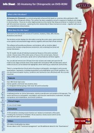
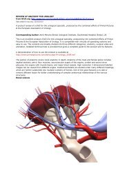
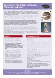
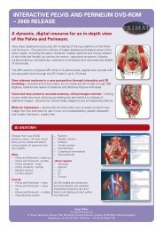
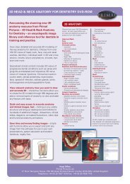
PRIMAL PICTURES LAUNCHES 3D ANATOMY SOFTWARE FOR THEDENTAL PROFESSIONPrimal Pictures is delighted to announce <strong>the</strong> recent release of 3D Head & Neck Anatomy <strong>for</strong> Dentistry.This stunning new <strong>anatomy</strong> resource has been developed using <strong>the</strong> expertise of <strong>dental</strong> specialists and isdesigned to meet <strong>the</strong> needs of <strong>dental</strong> students, professors and practitioners worldwide.“The graphics are remarkably clear,” comments Professor Patricia A. Reynolds, Director of FlexibleLearning, King’s College London Dental Institute. “The DVD helps make <strong>anatomy</strong> much easier tovisualise and includes notes on clinical relevance. It provides an important 3D teaching and learning aid.”3D Head & Neck <strong>for</strong> Dentistry provides clear, detailed and accurate 3D modelling of more than 100 viewsof key <strong>anatomy</strong> from <strong>the</strong> head to individual teeth. It offers a comprehensive image library that can be used<strong>for</strong> reference, to enhance consultations, presentations, teaching and study.“I think it is fantastic, really impressive,” comments Dr Natasha Berridge a qualified dentist andFoundation Year Two doctor who is currently working in critical care on her general surgery rotation. “Ithink it is a superb tool <strong>for</strong> any <strong>dental</strong> student. Head and neck <strong>anatomy</strong> can be really difficult to learn butPrimal’s <strong>software</strong> allows you to visualise structures in 3D perfectly and is as close to live dissection as youcould possibly get.”Views include oral and nasal cavities, dentition, individual teeth in 3D and cross section, nerves, larynx andpharynx, sinuses, <strong>the</strong> eye and brain. “It is great as a reference point <strong>for</strong> medical students who are lookingto extend <strong>the</strong>ir knowledge of specialist areas such as maxillofacial or oral <strong>anatomy</strong> - especially if <strong>the</strong>y areworking in an acute clinical setting, such as Accident and Emergency or during placement on a head andneck firm – an additional bonus is that it provides a great means to support patient education in a clinicalsetting,” adds Dr Berridge.The DVD also features specialised clinical content, including 3D views of progressive <strong>dental</strong> conditionssuch as caries and gingivitis, as well as interactive 3D nerve views of intraoral injections. The clinical textsection covers a range of subjects from <strong>dental</strong> anaes<strong>the</strong>sia and <strong>the</strong> spread of infection to embryogenesisand pterygopalatine fossa.“We were really keen to ensure that <strong>the</strong> DVD was truly relevant to dentists and so we spent a lot of timelooking at <strong>the</strong> potential problems that dentists face, such as how infection spreads in <strong>the</strong> head and neck,”says Dr Scott Rice, clinical fellow in oral and maxillofacial surgery at Barts and The London NHS Trust.“We also wanted to focus on <strong>the</strong> use of local anaes<strong>the</strong>sia because it is an everyday job requirement <strong>for</strong> anydentist.”“When students learn <strong>anatomy</strong>, having clinical relevance gives <strong>the</strong>m something to hook onto – <strong>anatomy</strong>can seem very dry when <strong>the</strong>re is little context and <strong>the</strong>re<strong>for</strong>e, more difficult to learn,” explains ProfessorReynolds. “It is <strong>the</strong>re<strong>for</strong>e very useful to be able to see what you might cut when you per<strong>for</strong>m surgery or
what tissues you are in when looking <strong>for</strong> a particular pathology. So putting <strong>the</strong> <strong>anatomy</strong> into a clinicalcontext brings it alive and enhances <strong>the</strong> learning experience.”Users can link slices of <strong>the</strong> model with MRI scan data in <strong>the</strong> axial, sagittal and coronal planes <strong>for</strong>comparison and study purposes and animations showing functional <strong>anatomy</strong> of <strong>the</strong> TemporomandibularJoint and facial and neck muscle function help users to visualise key movements and functionality.“The most useful aspect of <strong>the</strong> DVD <strong>for</strong> me is this ability to link <strong>anatomy</strong> and MRI or CT images,”comments Dr Rice. “As a postgraduate working in clinics, I am analysing scans on a daily basis and this isa really useful tool <strong>for</strong> training students in <strong>the</strong> accurate interpretation of scan data.”The DVD allows users to view <strong>anatomy</strong> from whatever angle <strong>the</strong>y wish in clear, anatomically accurate 3Dimages which can be exported into o<strong>the</strong>r <strong>for</strong>mats <strong>for</strong> use in presentations and dissertations. The fullyinteractive <strong>software</strong> provides detailed labelling and explanatory text, clinical notes and links to o<strong>the</strong>rrelevant content within <strong>the</strong> <strong>software</strong>.Dr Rice recommends <strong>the</strong> DVD <strong>for</strong> anyone who is studying Dentistry or a post-graduate <strong>dental</strong>qualification such as <strong>the</strong> MClinDent: “The fact that this is a DVD and not a book is what makes is soattractive. Although it is not <strong>the</strong> same as cadaver dissection by any stretch of <strong>the</strong> imagination, Primalallows you to follow your area of interest as far as you want to go. There is no prescribed route as <strong>the</strong>re isin a standard <strong>anatomy</strong> text book and so you can choose what to zoom in on.“What’s more, if you make a mess of it, unlike a cadaver, you can just click reset, go back to <strong>the</strong> beginningand start again!”“We are delighted with <strong>the</strong> feedback that we have received already on this product,” comments PrimalPictures Managing Director, Peter Allan. “We wanted to give <strong>the</strong> <strong>dental</strong> <strong>profession</strong> all of <strong>the</strong> benefits ofour interactive <strong>software</strong> models but we needed to provide specialist content in order to make it relevant.Our authors have worked extremely hard with us to develop this DVD and we are very proud of <strong>the</strong> result.We hope that it will make as much difference to <strong>the</strong> teaching and learning experience in dentistry as ouro<strong>the</strong>r products have made to <strong>the</strong> medical <strong>profession</strong>.”Additional comments from Professor Patricia Reynolds“The 3D visuals in <strong>the</strong> DVD were very good so we knew that if we could enhance it fur<strong>the</strong>r with clinicalrelevance, it could really come alive,” comments Professor Reynolds. “When students learn <strong>anatomy</strong>,clinical relevance gives <strong>the</strong>m something to hook onto – <strong>anatomy</strong> can seem very dry when <strong>the</strong>re is littlecontext and <strong>the</strong>re<strong>for</strong>e, more difficult to learn. It’s great if you have 3D spatial ability and can visualise andmemorise anatomical <strong>pictures</strong>, but if it isn’t related to anything in clinical practice, you will find it muchharder to understand why you need to know <strong>the</strong> details.“It is <strong>the</strong>re<strong>for</strong>e very useful to be able to see what you might cut when you per<strong>for</strong>m surgery or what tissuesyou are in when looking <strong>for</strong> a particular pathology. For instance, if you are looking <strong>for</strong> a calculus in agland, you will need to understand <strong>the</strong> relationships of <strong>the</strong> <strong>anatomy</strong> around it. You need to know <strong>the</strong><strong>anatomy</strong> well enough to ensure that you do not cut or damage something that you should not. So putting<strong>the</strong> <strong>anatomy</strong> into a clinical context brings it alive and enhances <strong>the</strong> learning experience.“Dental students may have less area to cover in <strong>anatomy</strong> compared to medical students but it is just asimportant that <strong>the</strong>y know where <strong>the</strong>y are putting <strong>the</strong>ir needles or knives, what pathologies may relate tothis part of <strong>the</strong> body or what <strong>anatomy</strong> is involved in a fractured jaw so that when <strong>the</strong>y do begin to practice<strong>the</strong>y can put <strong>the</strong>ir studies into action with confidence. Cadavers are playing a smaller role in <strong>anatomy</strong>
teaching <strong>the</strong>se days <strong>for</strong> a number of reasons, but <strong>the</strong>re are now alternative models to learn from and Primalprovides a tool that enables <strong>the</strong> user to appreciate 3D relationships.”