Definiens in Medical Imaging
Definiens in Medical Imaging
Definiens in Medical Imaging

You also want an ePaper? Increase the reach of your titles
YUMPU automatically turns print PDFs into web optimized ePapers that Google loves.
6<br />
<strong>Def<strong>in</strong>iens</strong> <strong>in</strong> <strong>Medical</strong> Imag<strong>in</strong>g<br />
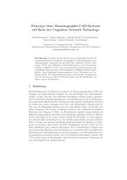
Example: Case-based retrieval <strong>in</strong> mammography<br />
<strong>Def<strong>in</strong>iens</strong> developed a prototype which extracts breast, nipples, calcifi cations and<br />
masses from x-ray mammograms. The statistical results can be used for a similarity<br />
search <strong>in</strong> a case database.<br />
The image data is analyzed via the follow<strong>in</strong>g steps:<br />
1. Accurate segmentation of object of <strong>in</strong>terest<br />
2. Detection and classification of masses and calcifications and creation of typical<br />
f<strong>in</strong>gerpr<strong>in</strong>t <strong>in</strong>clud<strong>in</strong>g lesion morphology<br />
3. Record<strong>in</strong>g the results <strong>in</strong> a table<br />
4. Comparison of the actual case with similar cases <strong>in</strong> the table<br />
5. Presentation of similar cases<br />
The Results<br />
The prototype was applied to 28 cancer cases. In 6 cases, clustered calcifications were<br />
the only <strong>in</strong>dications of cancer. In 8 cases malignant masses <strong>in</strong>dicated cancer. The<br />
rema<strong>in</strong><strong>in</strong>g 14 cases had calcifications with<strong>in</strong> masses.<br />
The <strong>Def<strong>in</strong>iens</strong>‘ approach detected at least one cancer region <strong>in</strong> 92.9 % of the cancer<br />
cases.Where there were malignant calcification clusters, the algorithm detected at least<br />
one cluster <strong>in</strong> all cases. The detection rate for cancer cases that conta<strong>in</strong>ed malignant<br />
masses was 90.9 %.<br />
In a multi-modal approach, images from different image modalities are analyzed<br />
<strong>in</strong> parallel. The correspond<strong>in</strong>g prototype can l<strong>in</strong>k tumor objects to correspond<strong>in</strong>g<br />
objects <strong>in</strong> images of the same or a different modality. Representative statistical results,<br />
for example on tumor morphology can be exported to support an expert’s diagnosis.<br />
Conclusions<br />
The results on different data modalities perform<strong>in</strong>g Computer Aided Detection of<br />
breast lesions look very promis<strong>in</strong>g. The technology will be the basis for the creation<br />
of new generation of CAD Systems, not only for the breast, but also for other organs by<br />
us<strong>in</strong>g different patient-relevant data <strong>in</strong> parallel.<br />
Figure 5 Screenshot of l<strong>in</strong>ked image <strong>in</strong>formation, with<br />
a1) US-, a2) x-ray- and a3) MR-mammography, analyzed<br />
by us<strong>in</strong>g a CNL prototype, which consists of a process<br />
hierarchy b1) and a class hierarchy b2). Tumors are l<strong>in</strong>ked<br />
to each other (outl<strong>in</strong>ed <strong>in</strong> red)<br />
Acknowledgment<br />
This work was supported by the Bayerische For schungsstiftung<br />
with<strong>in</strong> the scope of the project Mammo-iCAD.