Automated CT Based Liver Volume Assessment - Definiens
Automated CT Based Liver Volume Assessment - Definiens
Automated CT Based Liver Volume Assessment - Definiens

Create successful ePaper yourself
Turn your PDF publications into a flip-book with our unique Google optimized e-Paper software.
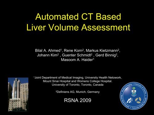
<strong>Automated</strong> <strong>CT</strong> <strong>Based</strong><br />
<strong>Liver</strong> <strong>Volume</strong> <strong>Assessment</strong><br />
Bilal A. Ahmed 1 , Rene Korn 2 , Markus Kietzmann 2 ,<br />
Johann Kim 2 , Guenter Schmidt 2 , Gerd Binnig 2 ,<br />
Masoom A. Haider 1<br />
1 Joint Department of Medical Imaging, University Health Netowork,<br />
Mount Sinai Hospital and Womens College Hospital.<br />
University of Toronto, Toronto, Canada<br />
2 <strong>Definiens</strong> AG, Munich, Germany<br />
RSNA 2009
Background<br />
• <strong>Liver</strong> volume is an important measure of functional<br />
capacity<br />
• Much prior work has been done in normal livers without<br />
metastases<br />
• It is routinely used for surgical planning for liver<br />
resections<br />
• Manual volumetry is time-consuming, tedious and<br />
generally not reproducible<br />
• Therefore, there has been a growing interest in the<br />
development of fast and accurate segmentation methods<br />
Medical Imaging – Princess Margaret Hospital – University Health Network – Mount Sinai Hospital – University of Toronto
Purpose<br />
To evaluate the accuracy of liver volume<br />
measured by a fully-automated<br />
segmentation software that quantifies liver<br />
volume in patients with metastatic liver<br />
disease using contextual information from<br />
adjacent structures, including lungs, the<br />
spine, ribs and gallbladder<br />
Medical Imaging – Princess Margaret Hospital – University Health Network – Mount Sinai Hospital – University of Toronto
Methods<br />
• 30 consecutive <strong>CT</strong> scans from distinct patients with liver<br />
metastases were obtained. Patients had tumor burdens form<br />
minimal to heavily occupying much of the liver<br />
– 320 slice Toshiba <strong>CT</strong> scanner<br />
– Study details:<br />
• 1mm collimation<br />
• Portal venous phase contrast-enhanced scans of the abdomen<br />
• All scans were manually segmented to define reference standard<br />
for liver boundaries (including metastatic lesions) using in-<br />
house software<br />
– Parts of the inferior vena cava and portal vein were included within<br />
the liver boundary when enclosed by liver parenchyma in order to<br />
make the liver contour smooth<br />
Medical Imaging – Princess Margaret Hospital – University Health Network – Mount Sinai Hospital – University of Toronto
Methods<br />
• Five representative cases were selected as a<br />
training set for the automatic segmentation<br />
algorithm<br />
• The remaining 25 cases were used to test the<br />
refined algorithm<br />
• Resultant automatic segmentations generated were<br />
compared to the manually generated reference<br />
segmentations and deviations were measured<br />
Medical Imaging – Princess Margaret Hospital – University Health Network – Mount Sinai Hospital – University of Toronto
Automatic Segmentation Algorithm<br />
• Fully automatic segmentation algorithm developed in the Cognition Network<br />
Language (CNL) with <strong>Definiens</strong> Developer XD 1.0<br />
• Automatic pre-processing using rule-based segmentation<br />
– Defines a set of rules which is used to successively extract different<br />
structures from the test images<br />
– Order of segmentation/classification is: background and body � lungs<br />
and other intra-body air � fat, muscle and bones � spine and ribs<br />
– Incorporates knowledge about intensity distributions, neighborhood<br />
relations and object geometry<br />
• Automatic selection of a seed based on geometry, position and size<br />
relationship<br />
– Order of segmentation/classification is: liver seed � gall bladder<br />
• Automatic seed growing<br />
– Growing process is halted by previously detected contextual structures<br />
– Order of segmentation/classification is: liver seed � liver<br />
Medical Imaging – Princess Margaret Hospital – University Health Network – Mount Sinai Hospital – University of Toronto
Automatic Segmentation Algorithm<br />
Automatic Preprocessing<br />
Automatic <strong>Liver</strong> Seed Selection<br />
Algorithm: <strong>Liver</strong> Refinement/Growing<br />
Original Image updating<br />
Medical Imaging – Princess Margaret Hospital – University Health Network – Mount Sinai Hospital – University of Toronto
Automatic Segmentation Algorithm<br />
Preprocessing<br />
1. Background is separated from the body<br />
2. Right and left lungs are identified as context objects<br />
3. <strong>Based</strong> on size and intensity (HU), additional context objects are<br />
generated (e.g. fat, muscle, other organs, bones)<br />
4. Within the bone objects, spine and ribs are identified as additional<br />
context objects based on ROI (using lungs) and shape<br />
5. An additional layer of 3D edge information is calculated<br />
Note: Context objects do not have to be 100% perfectly segmented to guide the analysis.<br />
Medical Imaging – Princess Margaret Hospital – University Health Network – Mount Sinai Hospital – University of Toronto
<strong>Based</strong> on<br />
intensity and<br />
size the<br />
background is<br />
separated from<br />
the body<br />
Algorithm: Preprocessing<br />
Medical Imaging – Princess Margaret Hospital – University Health Network – Mount Sinai Hospital – University of Toronto
Algorithm: Preprocessing<br />
<strong>Based</strong> on<br />
intensity,<br />
position and<br />
size right and<br />
left lungs are<br />
identified as<br />
context objects<br />
Medical Imaging – Princess Margaret Hospital – University Health Network – Mount Sinai Hospital – University of Toronto
Algorithm: Preprocessing<br />
<strong>Based</strong> on size<br />
and intensity<br />
(HU),<br />
additional<br />
context objects<br />
are generated<br />
(e.g. fat,<br />
muscle and<br />
bones)<br />
Medical Imaging – Princess Margaret Hospital – University Health Network – Mount Sinai Hospital – University of Toronto
Algorithm: Preprocessing<br />
Within the<br />
bone objects,<br />
spine and ribs<br />
are identified<br />
as additional<br />
context objects<br />
based on ROI<br />
(using lungs)<br />
and shape<br />
Medical Imaging – Princess Margaret Hospital – University Health Network – Mount Sinai Hospital – University of Toronto
Algorithm: <strong>Liver</strong> Seed<br />
<strong>Liver</strong> Seed<br />
1. Exploiting the 3D edge layer, potential liver seeds within the<br />
muscle context objects are identified<br />
2. <strong>Based</strong> on size, a single liver seed is automatically selected<br />
3. <strong>Based</strong> on right lung and liver seed, the gallbladder context<br />
object is segmented<br />
Medical Imaging – Princess Margaret Hospital – University Health Network – Mount Sinai Hospital – University of Toronto
Potential<br />
liver seeds<br />
are identified<br />
within the<br />
muscle<br />
context<br />
objects<br />
Algorithm: <strong>Liver</strong> Seed<br />
Medical Imaging – Princess Margaret Hospital – University Health Network – Mount Sinai Hospital – University of Toronto
<strong>Based</strong> on<br />
size, a single<br />
liver seed is<br />
automatically<br />
selected<br />
Algorithm: <strong>Liver</strong> Seed<br />
Medical Imaging – Princess Margaret Hospital – University Health Network – Mount Sinai Hospital – University of Toronto
<strong>Based</strong> on<br />
muscle, right<br />
lung, liver<br />
seed and<br />
size, the<br />
gallbladder<br />
context<br />
object is<br />
identified<br />
Algorithm: <strong>Liver</strong> Seed<br />
Medical Imaging – Princess Margaret Hospital – University Health Network – Mount Sinai Hospital – University of Toronto
Algorithm: <strong>Liver</strong> refinement/Growing<br />
<strong>Liver</strong> Refinement/Growing<br />
1. The liver seed is split into an upper and lower part<br />
2. First, the lower liver seed is grown (gallbladder, ribs,<br />
edge)<br />
3. Second, the upper liver is grown (ribs, lung wings, edge)<br />
Update Original Image<br />
Results are transferred back to original image size and refined<br />
(prior to this, all work was done based on a downscaled image by a factor of 0.5)<br />
Medical Imaging – Princess Margaret Hospital – University Health Network – Mount Sinai Hospital – University of Toronto
Algorithm: <strong>Liver</strong> Refinement/Growing<br />
The liver<br />
seed is split<br />
into an upper<br />
and lower<br />
part<br />
Medical Imaging – Princess Margaret Hospital – University Health Network – Mount Sinai Hospital – University of Toronto
Algorithm: <strong>Liver</strong> Refinement/Growing<br />
First, the<br />
lower liver<br />
seed is<br />
grown<br />
(gallbladder,<br />
ribs, edge)<br />
Medical Imaging – Princess Margaret Hospital – University Health Network – Mount Sinai Hospital – University of Toronto
Algorithm: <strong>Liver</strong> Refinement/Growing<br />
Second, the<br />
upper liver<br />
seed is<br />
grown (ribs,<br />
lung wings,<br />
edge)<br />
Medical Imaging – Princess Margaret Hospital – University Health Network – Mount Sinai Hospital – University of Toronto
Algorithm: Update Original Image<br />
Results are<br />
transferred to<br />
original<br />
image size<br />
(100%) and<br />
refined<br />
further<br />
Medical Imaging – Princess Margaret Hospital – University Health Network – Mount Sinai Hospital – University of Toronto
Results: Measurements<br />
• Two measurements were used to evaluate segmentation accuracy:<br />
– Minimum overlap<br />
• A, B Sets of voxels from tracing and classification data respectively<br />
– Correlation Coefficient<br />
• tp = number of true positive voxels<br />
• fp = number of false positive voxels<br />
• tn = number of true negative voxels<br />
• fn = number of false negative voxels<br />
Medical Imaging – Princess Margaret Hospital – University Health Network – Mount Sinai Hospital – University of Toronto
Results: Sample Segmentation I<br />
Statistics<br />
• CC 0.86<br />
• MO 86%<br />
• 9:47 min<br />
Medical Imaging – Princess Margaret Hospital – University Health Network – Mount Sinai Hospital – University of Toronto
Results: Sample Segmentation II<br />
Statistics<br />
• CC 0.94<br />
• MO 89%<br />
• 6:29 min<br />
(best case)<br />
Medical Imaging – Princess Margaret Hospital – University Health Network – Mount Sinai Hospital – University of Toronto
Results: Summary<br />
• Correlation coefficient between the measured and<br />
estimated volumes: 0.864 ± 0.04 (0.777 – 0.935)<br />
• Mean volumetric overlap: 77% ± 6 (64 – 88%)<br />
• Mean liver volume measured by the automatic<br />
segmentation algorithm: 1820 cm³ ± 788 (710 – 4565 cm³)<br />
• Mean liver volume measured by manual segmentation:<br />
1806 cm³ ± 685 (934 – 3460 cm³)<br />
• Mean total time for the automatic segmentation: 4.8<br />
minutes ± 1.2 on E6750 @2.66GHz Intel Core 2 Duo<br />
CPU with 3.25 GB RAM<br />
Medical Imaging – Princess Margaret Hospital – University Health Network – Mount Sinai Hospital – University of Toronto
Discussion<br />
• Initial evaluation of fully automated algorithm using<br />
contextual information from surrounding structures<br />
shows promising results<br />
• Small training set (5 cases) and large test set (25 cases)<br />
is challenging test of performance<br />
• Unique advantage of this approach is a universal toolkit<br />
that can be used to develop highly specific algorithms<br />
detecting all kinds of objects<br />
– Has wide array of applications in image segmentation<br />
• Further evaluation will include specific segmentation of<br />
liver metastases<br />
Medical Imaging – Princess Margaret Hospital – University Health Network – Mount Sinai Hospital – University of Toronto
Conclusion<br />
• Fully-automated segmentation software that<br />
quantifies liver volumes using contextual<br />
information from adjacent structures<br />
exhibits promising accuracies in acceptable<br />
time frames in patients with varying degrees<br />
of metastatic disease<br />
Medical Imaging – Princess Margaret Hospital – University Health Network – Mount Sinai Hospital – University of Toronto
References<br />
• Heimann T, van Ginneken B, Styner MA, et al. Comparison and evaluation of methods<br />
for liver segmentation from <strong>CT</strong> datasets. IEEE Trans Med Imaging. 2009<br />
Aug;28(8):1251-65.<br />
• Schmidt G, Athelogou MA, Schonmeyer R, Korn R, and Binnig G. Cognition network<br />
technology for a fully automated 3D segmentation of liver. Proc MICCAI Workshop on<br />
3D Segmentation in the Clinic: a Grand Challenge. 2007; pp. 125–133.<br />
• Prasad SR, Jhaveri KS, Saini S, Hahn PF, Halpern EF, Sumner JE. <strong>CT</strong> tumor<br />
measurement for therapeutic response assessment: comparison of unidimensional,<br />
bidimensional, and volumetric techniques initial observations. Radiology 2002;<br />
225:416-419.<br />
• Lamecker H, Lange T, Seebass M, Eulenstein S, Westerhoff M, Hege HC. Automatic<br />
segmentation of the liver for preoperative planning of resections. Stud Health Technol<br />
Inform 2003; 94:171-173.<br />
• Selver MA, Kocaoglu A, Demir GK, Dogan H, Dicle O, Guzelis C. Patient oriented and<br />
robust automatic liver segmentation for pre-evaluation of liver transplantation. Comput<br />
Biol Med 2008; 38:765-784.<br />
• Athelogou, M. et al.: <strong>Definiens</strong> Cognition Network Technology – a novel multimodal<br />
image analysis technique for automatic identification and quantification of biological<br />
image contents. In Shorte, S.L., Frischknecht, F., eds.: Imaging Cellular and Molecular<br />
Biological Functions. Springer-Verlag, Berlin Heidelberg (2007) 407-421.<br />
Medical Imaging – Princess Margaret Hospital – University Health Network – Mount Sinai Hospital – University of Toronto