Automated CT Based Liver Volume Assessment - Definiens
Automated CT Based Liver Volume Assessment - Definiens
Automated CT Based Liver Volume Assessment - Definiens
Create successful ePaper yourself
Turn your PDF publications into a flip-book with our unique Google optimized e-Paper software.
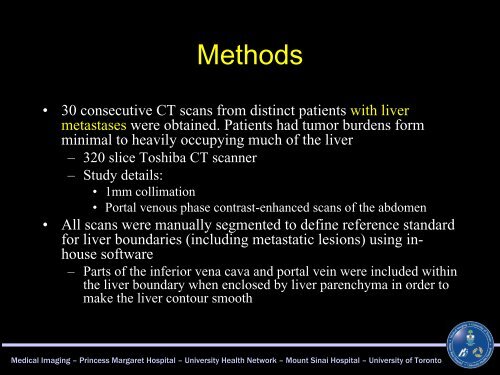
Methods<br />
• 30 consecutive <strong>CT</strong> scans from distinct patients with liver<br />
metastases were obtained. Patients had tumor burdens form<br />
minimal to heavily occupying much of the liver<br />
– 320 slice Toshiba <strong>CT</strong> scanner<br />
– Study details:<br />
• 1mm collimation<br />
• Portal venous phase contrast-enhanced scans of the abdomen<br />
• All scans were manually segmented to define reference standard<br />
for liver boundaries (including metastatic lesions) using in-<br />
house software<br />
– Parts of the inferior vena cava and portal vein were included within<br />
the liver boundary when enclosed by liver parenchyma in order to<br />
make the liver contour smooth<br />
Medical Imaging – Princess Margaret Hospital – University Health Network – Mount Sinai Hospital – University of Toronto


