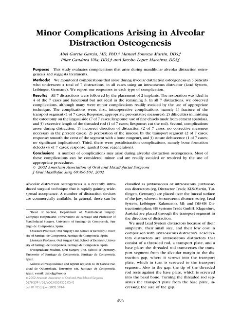
Minor Complications Arising in Alveolar Distraction Osteogenesis
Minor Complications Arising in Alveolar Distraction Osteogenesis
Minor Complications Arising in Alveolar Distraction Osteogenesis
Create successful ePaper yourself
Turn your PDF publications into a flip-book with our unique Google optimized e-Paper software.
J Oral Maxillofac Surg<br />
60:496-501, 2002<br />
<strong>M<strong>in</strong>or</strong> <strong>Complications</strong> <strong>Aris<strong>in</strong>g</strong> <strong>in</strong> <strong>Alveolar</strong><br />
<strong>Distraction</strong> <strong>Osteogenesis</strong><br />
Abel Garcia Garcia, MD, PhD,* Manuel Somoza Mart<strong>in</strong>, DDS,†<br />
Pilar Gandara Vila, DDS,‡ and Jacobo Lopez Maceiras, DDS§<br />
Purpose: This study evaluates complications that arise dur<strong>in</strong>g mandibular alveolar distraction osteogenesis<br />
and suggests treatments.<br />
Methods: We monitored complications that arose dur<strong>in</strong>g alveolar distraction osteogenesis <strong>in</strong> 5 patients<br />
who underwent a total of 7 distractions, <strong>in</strong> all cases us<strong>in</strong>g an <strong>in</strong>traosseous distractor (Lead System,<br />
Leib<strong>in</strong>ger, Germany). We report our responses to each type of complication.<br />
Results: All 7 distractions were followed by the placement of 2 implants. The restoration was ideal <strong>in</strong><br />
4 of the 7 cases and functional but not ideal <strong>in</strong> the rema<strong>in</strong><strong>in</strong>g 3. In all 7 distractions, we observed<br />
complications, although many were m<strong>in</strong>or complications readily avoided by the use of appropriate<br />
technique. The complications were, first, <strong>in</strong>traoperative complications, namely 1) fracture of the<br />
transport segment (1 of 7 cases; Response: appropriate preventative measures), 2) difficulties <strong>in</strong> f<strong>in</strong>ish<strong>in</strong>g<br />
the osteotomy on the l<strong>in</strong>gual side (7 of 7 cases; Response: use of f<strong>in</strong>e chisels made from cement spatulas),<br />
and 3) excessive length of the threaded rod (1 of 7 cases; Response: cut the rod). Second, complications<br />
arose dur<strong>in</strong>g distraction: 1) <strong>in</strong>correct direction of distraction (2 of 7 cases; no corrective measures<br />
necessary <strong>in</strong> the present cases), 2) perforation of the mucosa by the transport segment (2 of 7 cases;<br />
response: smooth the crest of the segment with a bone rongeur), and 3) suture dehiscence (1 of 7 cases;<br />
no significant implications). Third, there were postdistraction complications, namely bone formation<br />
defects (4 of 7 cases; response: guided bone regeneration).<br />
Conclusion: A number of complications may arise dur<strong>in</strong>g alveolar distraction osteogenesis. Most of<br />
these complications can be considered m<strong>in</strong>or and are readily avoided or resolved by the use of<br />
appropriate procedures.<br />
© 2002 American Association of Oral and Maxillofacial Surgeons<br />
J Oral Maxillofac Surg 60:496-501, 2002<br />
<strong>Alveolar</strong> distraction osteogenesis is a recently <strong>in</strong>troduced<br />
surgical technique that is rapidly ga<strong>in</strong><strong>in</strong>g widespread<br />
acceptance. A number of distraction devices<br />
are commercially available. In general, these can be<br />
*Head of Section, Department of Maxillofacial Surgery,<br />
Complejo Hospitalario Universitario de Santiago and Professor of<br />
Maxillofacial Surgery, University of Santiago de Compostela, Santiago<br />
de Compostela, Spa<strong>in</strong>.<br />
†Assistant Professor, Oral Surgery Unit, School of Dentistry, University<br />
of Santiago de Compostela, Santiago de Compostela, Spa<strong>in</strong>.<br />
‡Assistant Professor, Oral Surgery Unit, School of Dentistry, University<br />
of Santiago de Compostela, Santiago de Compostela, Spa<strong>in</strong>.<br />
§Postgraduate Student, Oral Surgery Unit, School of Dentistry,<br />
University of Santiago de Compostela, Santiago de Compostela,<br />
Spa<strong>in</strong>.<br />
Address correspondence and repr<strong>in</strong>t requests to Dr Garcia: Facultad<br />
de Odontologia, Entrerrios s/n, Santiago de Compostela,<br />
Spa<strong>in</strong>; e-mail: ciabelgg@usc.es<br />
© 2002 American Association of Oral and Maxillofacial Surgeons<br />
0278-2391/02/6005-0004$35.00/0<br />
doi:10.1053/joms.2002.31844<br />
496<br />
classified as juxtaosseous or <strong>in</strong>traosseous. Juxtaosseous<br />
distractors (eg, Distractor Track; KLS/Mart<strong>in</strong>, Tuttl<strong>in</strong>gen,<br />
Germany) are placed over the buccal surface<br />
of the jaw, whereas <strong>in</strong>traosseous distractors (eg, Lead<br />
System, Leib<strong>in</strong>ger, Kalamazoo, MI; and DIS-SIS <strong>Distraction</strong>implant;<br />
SIS Systems Trade GmbH, Klagenfurt,<br />
Austria) are placed through the transport segment <strong>in</strong><br />
the direction of distraction.<br />
We used Lead System distractors because of their<br />
simplicity, their small size, and their low cost <strong>in</strong><br />
comparison with juxtaosseous distractors. Lead System<br />
distractors are <strong>in</strong>traosseous distractors that<br />
consist of a threaded rod, a transport plate, and a<br />
base plate: the threaded rod transverses the transport<br />
segment from the alveolar marg<strong>in</strong> to the distraction<br />
gap, where it screws <strong>in</strong>to the transport<br />
plate, which <strong>in</strong> turn is screwed to the transport<br />
segment. Also <strong>in</strong> the gap, the tip of the threaded<br />
rod rests aga<strong>in</strong>st the base plate, which is screwed<br />
<strong>in</strong>to the basal bone. Turn<strong>in</strong>g the threaded rod separates<br />
the transport plate from the base plate, <strong>in</strong>creas<strong>in</strong>g<br />
the size of the gap. 1
GARCIA ET AL 497<br />
Here we report a study of complications aris<strong>in</strong>g <strong>in</strong><br />
5 patients dur<strong>in</strong>g osteotomy and subsequent distraction<br />
with a Lead System distractor, and we propose<br />
treatments for each type of complication.<br />
Materials and Methods<br />
SAMPLE<br />
We studied 5 patients who had undergone a total of<br />
7 mandibular alveolar distractions. In all cases, the<br />
distraction was performed us<strong>in</strong>g a Lead System distractor.<br />
Six of the 7 distractions were performed <strong>in</strong><br />
the posterior mandible, and 1 was performed <strong>in</strong> the<br />
<strong>in</strong>cisive-can<strong>in</strong>e region.<br />
SURGICAL TECHNIQUE<br />
All patients were treated while under local anesthesia.<br />
A crestal <strong>in</strong>cision was made along the alveolar<br />
ridge, and a vestibular mucoperiosteal flap was raised,<br />
ma<strong>in</strong>ta<strong>in</strong><strong>in</strong>g the attachment of the l<strong>in</strong>gual mucoperiosteum<br />
to the transport segment. The transport segment<br />
was cut to an <strong>in</strong>verted trapezoidal shape, so as<br />
not to <strong>in</strong>terfere with mobility dur<strong>in</strong>g distraction. Osteotomy<br />
was performed with rotary <strong>in</strong>struments (sidecutt<strong>in</strong>g<br />
burrs, discs, and reciprocat<strong>in</strong>g saws) and chisels.<br />
The transport segment was totally mobilized<br />
although rema<strong>in</strong>ed attached to the l<strong>in</strong>gual mucoperiosteum.<br />
The distractor (ie, threaded rod, transport<br />
plate, and base plate) was assembled and positioned<br />
accord<strong>in</strong>g to the procedure of Ch<strong>in</strong>1 (Fig 1).<br />
Once the distractor had been positioned, and without<br />
sutur<strong>in</strong>g the mucoperiosteal flap, the transport<br />
segment was immediately raised (ie, with<strong>in</strong> the same<br />
surgical session) to a height of 5 mm to confirm<br />
adequate mobility and appropriate direction of movement<br />
and absence of <strong>in</strong>terference between the transport<br />
segment and the basal bone. The transport segment<br />
was then returned to its orig<strong>in</strong>al position.<br />
<strong>Distraction</strong> was commenced 7 days later at a rate of<br />
0.5 mm every 12 hours for 5 days. After 12 weeks, the<br />
distractor was removed, and the implants were<br />
placed. At 14 weeks after implant placement, the<br />
prosthetic restoration was commenced and subjected<br />
to load. Restorations were subsequently classified as<br />
ideal, functional but not ideal, or nonfunctional.<br />
The complications that arose are considered <strong>in</strong> 3<br />
groups: <strong>in</strong>traoperative complications, complications<br />
aris<strong>in</strong>g dur<strong>in</strong>g distraction, and postdistraction complications.<br />
Results<br />
In all cases, bone formed <strong>in</strong> the regeneration chamber,<br />
and the transport segment was stable. Likewise,<br />
<strong>in</strong> all cases, 2 implants were subsequently successfully<br />
FIGURE 1. Photograph show<strong>in</strong>g placement of the Lead System distractor.<br />
A crestal <strong>in</strong>cision is made along the alveolar ridge, and a<br />
vestibular mucoperiosteal flap is raised, ma<strong>in</strong>ta<strong>in</strong><strong>in</strong>g the attachment of<br />
the l<strong>in</strong>gual mucoperiosteum (B) to the transport segment (A) (C, basal<br />
bone). The distractor is then placed as shown: D, threaded rod; E,<br />
transport plate; and F, base plate.<br />
placed <strong>in</strong> the distracted region (8 ITI � 4.1 mm, 12.0<br />
mm PLUS; Straumann, Waldenburg, Switzerland and 4<br />
Frialoc D4/L13; Friadent, Mannheim, Germany). In all<br />
cases, a prosthetic restoration was subsequently performed.<br />
The restoration was ideal <strong>in</strong> 4 of the 7 cases<br />
and functional but not ideal <strong>in</strong> the rema<strong>in</strong><strong>in</strong>g 3 cases.<br />
<strong>Complications</strong> are summarized <strong>in</strong> Table 1.<br />
INTRAOPERATIVE COMPLICATIONS<br />
Fracture of the Transport Segment<br />
This type of complication occurred <strong>in</strong> 1 case, due<br />
to an attempt to free the transport segment us<strong>in</strong>g a<br />
chisel. As a result, a small fragment of cortical bone<br />
was lost. Subsequent bone formation <strong>in</strong> the region of<br />
the lost fragment was deficient.<br />
Difficulties <strong>in</strong> Complet<strong>in</strong>g the Osteotomy on the<br />
L<strong>in</strong>gual Side<br />
In all 7 cases, difficulties were encountered <strong>in</strong> complet<strong>in</strong>g<br />
the osteotomy on the l<strong>in</strong>gual side, which we<br />
had to access from the labial vestibular side. To do<br />
this, we constructed f<strong>in</strong>e chisels from cement spatulas<br />
(Fig 2), which we carefully <strong>in</strong>troduced from the vestibular<br />
side, check<strong>in</strong>g their exit from the l<strong>in</strong>gual side<br />
with a f<strong>in</strong>ger so as to avoid damage to the l<strong>in</strong>gual<br />
mucoperiosteum or the floor of the mouth.<br />
Excessive Length of the Threaded Rod<br />
In 1 case, the threaded rod may have <strong>in</strong>terfered<br />
with occlusion. This complication can be predicted
498 MINOR COMPLICATIONS AND ALVEOLAR DISTRACTION<br />
Table 1. COMPLICATIONS OF THE ALVEOLAR DISTRACTION<br />
Patient<br />
No. <strong>Distraction</strong> Location<br />
1 Left posterior<br />
mandibular region<br />
2 Left posterior<br />
mandibular region<br />
3 Incisive-can<strong>in</strong>e<br />
mandibular region<br />
4 Left posterior<br />
mandibular region<br />
Right posterior<br />
mandibular region<br />
5 Left posterior<br />
mandibular region<br />
Right posterior<br />
mandibular region<br />
with the aid of articulator-mounted casts and is readily<br />
resolved by cutt<strong>in</strong>g the rod to the appropriate length<br />
before placement.<br />
COMPLICATIONS DURING DISTRACTION<br />
Incorrect Direction of <strong>Distraction</strong><br />
This complication occurred twice, due to l<strong>in</strong>gual<br />
deviation of the threaded rod. As a consequence,<br />
excessive bone formed <strong>in</strong> the l<strong>in</strong>gual direction. In<br />
both cases, however, sufficient bone was obta<strong>in</strong>ed to<br />
fit 12-mm implants.<br />
Perforation of the Mucosa by the Transport<br />
Segment<br />
This complication occurred twice, due to the sharp<br />
edges of the transport segment. In 1 case, a l<strong>in</strong>gual<br />
<strong>Complications</strong> Implants<br />
Intraoperative Dur<strong>in</strong>g <strong>Distraction</strong> Postdistraction Placed Restoration<br />
Excessive length of the<br />
threaded rod<br />
Fracture of the transport<br />
segment<br />
Difficulties <strong>in</strong> complet<strong>in</strong>g<br />
the osteotomy on the<br />
l<strong>in</strong>gual side<br />
Difficulties <strong>in</strong> complet<strong>in</strong>g<br />
the osteotomy on the<br />
l<strong>in</strong>gual side<br />
Difficulties <strong>in</strong> complet<strong>in</strong>g<br />
the osteotomy on the<br />
l<strong>in</strong>gual side<br />
Difficulties <strong>in</strong> complet<strong>in</strong>g<br />
the osteotomy on the<br />
l<strong>in</strong>gual side<br />
Difficulties <strong>in</strong> complet<strong>in</strong>g<br />
the osteotomy on the<br />
l<strong>in</strong>gual side<br />
Difficulties <strong>in</strong> complet<strong>in</strong>g<br />
the osteotomy on the<br />
l<strong>in</strong>gual side<br />
Difficulties <strong>in</strong> complet<strong>in</strong>g<br />
the osteotomy on the<br />
l<strong>in</strong>gual side<br />
FIGURE 2. Osteotomes constructed from cement spatulas.<br />
Incorrect<br />
direction of<br />
distraction<br />
Perforation of the<br />
mucosa by the<br />
transport<br />
segment<br />
Suture<br />
dehiscence<br />
Perforation of the<br />
mucosa by the<br />
transport<br />
segment<br />
Incorrect<br />
direction of<br />
distraction<br />
Bone formation<br />
defects<br />
Bone formation<br />
defect<br />
Bone formation<br />
defect<br />
Bone formation<br />
defect<br />
ulcer arose. Treatment <strong>in</strong>volves elim<strong>in</strong>ation of the<br />
sharp edge with use of a burr or a rongeur. In both<br />
cases, the mucosa grew over the bone, without any<br />
need to <strong>in</strong>terrupt the distraction.<br />
Suture Dehiscence<br />
This occurred <strong>in</strong> 1 case, lead<strong>in</strong>g to some exposure<br />
of the transport segment. There was no need to <strong>in</strong>terrupt<br />
distraction, and the mucosa subsequently<br />
grew completely over the bone.<br />
POSTDISTRACTION COMPLICATIONS<br />
2 ITI Functional<br />
but not<br />
ideal<br />
2 ITI Ideal<br />
2 ITI Ideal<br />
2 ITI Ideal<br />
2 ITI Ideal<br />
2 Frialoc Functional<br />
but not<br />
ideal<br />
2 Frialoc Functional<br />
but not<br />
ideal<br />
Bone Formation Defects<br />
<strong>Complications</strong> of this type arose <strong>in</strong> 4 cases. In 3<br />
cases, bone formation was not uniform, giv<strong>in</strong>g rise to<br />
bone formation defects. In all cases, these defects led<br />
to <strong>in</strong>complete coverage of the implant (dehiscence or<br />
fenestration). In 1 case (noted <strong>in</strong> section on fracture<br />
of the transport segment), the defect was a dehiscence<br />
defect due to loss of a fragment of the transport<br />
segment dur<strong>in</strong>g osteotomy. In the other 2 cases, the<br />
defects were fenestration defects. Treatment was<br />
bone regeneration us<strong>in</strong>g Bio-Oss and Bio-Gide reabsorbable<br />
membranes (Geistlich Pharma AG, Wolhusen,<br />
Switzerland).
GARCIA ET AL 499<br />
Discussion<br />
S<strong>in</strong>ce the 19th century, there have been numerous<br />
attempts to develop techniques to extend the long<br />
bones. 2,3 <strong>Distraction</strong> osteogenesis of the long bones<br />
was pioneered by Ilizarov. 4-6 More recently, distraction<br />
techniques have been applied to the facial bones<br />
and soft tissues, 7-13 <strong>in</strong>clud<strong>in</strong>g use <strong>in</strong> the treatment of<br />
<strong>in</strong>adequate height of the alveolar ridge. 14-19<br />
<strong>Alveolar</strong> distraction osteogenesis promises to have<br />
very useful applications <strong>in</strong> the field of implantology,<br />
particularly <strong>in</strong> cases of mandibular alveolar hypoplasia.<br />
In such cases, the lack of sufficient bone height<br />
between the alveolar canal and the alveolar rim means<br />
that the implant must be short; at the same time, the<br />
reduced height of the rim means that the crown must<br />
be long. By <strong>in</strong>creas<strong>in</strong>g the height of the alveolar rim,<br />
alveolar distraction osteogenesis overcomes both<br />
problems.<br />
An understand<strong>in</strong>g of the potential complications of<br />
a given surgical technique, and of appropriate treatments,<br />
is fundamental for correct implementation of<br />
that technique. In the present study, we therefore<br />
evaluated complications aris<strong>in</strong>g <strong>in</strong> 7 alveolar distractions,<br />
all performed with Lead System distractors (Table<br />
2).<br />
Fracture of the transport segment dur<strong>in</strong>g osteotomy<br />
is a complication that can be avoided only by<br />
preventative measures. The thickness of the transport<br />
segment should be sufficient to withstand the osteotomy<br />
maneuvers. Care should be taken <strong>in</strong> manipulation,<br />
and no attempt should be made to move the<br />
segment until the osteotomy is complete.<br />
With rotary <strong>in</strong>struments, it is easy to perform the<br />
osteotomy on the vestibular side, but the osteotomy<br />
on the l<strong>in</strong>gual side is more difficult. To overcome this<br />
complication, we designed and constructed chisels<br />
from cement spatulas, allow<strong>in</strong>g us to complete the<br />
osteotomy of the l<strong>in</strong>gual cortex while respect<strong>in</strong>g the<br />
<strong>in</strong>tegrity of the mucoperiosteum.<br />
Excessive length of the threaded rod, h<strong>in</strong>der<strong>in</strong>g<br />
proper occlusion, is a significant problem, because<br />
the distractor must rema<strong>in</strong> <strong>in</strong> the patient’s mouth for<br />
at least 14 weeks. However, presurgical plann<strong>in</strong>g<br />
with articulator-mounted casts should enable the<br />
problem to be predicted. In the case of Lead System<br />
distractors, the problem is readily solved, because the<br />
threaded rod can be cut without affect<strong>in</strong>g its function.<br />
Inappropriate direction of distraction may be due<br />
to any of several factors. Generally, the distractor will<br />
tend to lean to the l<strong>in</strong>gual side. The hole drilled for<br />
<strong>in</strong>sertion of the threaded rod may be angled <strong>in</strong>correctly.<br />
Either the base plate or the transport plate may<br />
not fit correctly. The force exerted by the l<strong>in</strong>gual<br />
periosteum attached to the transport segment may<br />
lead to <strong>in</strong>appropriate direction of distraction if not<br />
taken <strong>in</strong>to account (Fig 3). To solve this complication,<br />
Table 2. TREATMENT AND CONSEQUENCES OF THE COMPLICATIONS OF ALVEOLAR DISTRACTION<br />
<strong>Complications</strong> Treatment Consequences<br />
Intraoperative<br />
Fracture of the transport segment Appropriate preventative measures Absence of bone formation<br />
Difficulties <strong>in</strong> complet<strong>in</strong>g the osteotomy on<br />
the l<strong>in</strong>gual side<br />
Use of appropriate <strong>in</strong>struments Extended surgery time<br />
Excessive length of the threaded rod<br />
Dur<strong>in</strong>g distraction<br />
Cut the rod If not corrected, <strong>in</strong>terference<br />
with occlusion<br />
Incorrect direction of distraction Care <strong>in</strong> position<strong>in</strong>g the distractor at the Bone formation <strong>in</strong> the wrong<br />
correct angle<br />
Take <strong>in</strong>to account the effect of the l<strong>in</strong>gual<br />
mucoperiosteum<br />
Use of orthodontic devices (Ch<strong>in</strong><br />
direction<br />
1 )<br />
Perforation of the mucosa by the transport Smooth the extremes of the segment with L<strong>in</strong>gual ulcer<br />
segment<br />
a burr or rongeur<br />
Suture dehiscence<br />
Breakage or loss of the distractor (Millesi-<br />
Schobel et al,<br />
No action usually required; closure by<br />
second <strong>in</strong>tention<br />
No sequelae observed<br />
21 Gaggl et al22 )<br />
Post distraction<br />
Bone formation defects Guided bone regeneration<br />
Application of a titanium membrane dur<strong>in</strong>g<br />
the osteotomy (Klug et al<br />
Gaps <strong>in</strong> the bone around the<br />
implant<br />
20 Other<br />
Dysesthesia of the mental nerve (Klug et<br />
al,<br />
)<br />
20 Gaggl et al, 22 Noc<strong>in</strong>i et al23 )
500 MINOR COMPLICATIONS AND ALVEOLAR DISTRACTION<br />
FIGURE 3. Diagram show<strong>in</strong>g the tension exerted by the l<strong>in</strong>gual<br />
mucoperiosteum (C) on the transport segment, lead<strong>in</strong>g if not corrected<br />
to distraction direction (B) as opposed to the desired distraction direction<br />
(A).<br />
Ch<strong>in</strong> 1 proposed the use of an orthodontic appliance<br />
to guide the threaded rod. However, this requires<br />
that the edentulous space should have teeth at both<br />
extremes, which was not the case <strong>in</strong> either of the<br />
subjects who had this complication <strong>in</strong> the present<br />
study. When the distractor is assembled and positioned,<br />
it is thus important to bear <strong>in</strong> m<strong>in</strong>d the forces<br />
exerted by the l<strong>in</strong>gual mucoperiosteum attached to<br />
the segment and to angle the threaded rod slightly<br />
outward to compensate for this force dur<strong>in</strong>g distraction.<br />
Perforation of the mucosa by the sharp edges of the<br />
transport segment is typically observed as the distraction<br />
proceeds. Elim<strong>in</strong>ation of the sharp edges allows<br />
rapid growth of the mucosa over the bone, <strong>in</strong> contrast<br />
to the situation expected with a free bone graft. This<br />
complication does not require <strong>in</strong>terruption of the<br />
distraction procedure.<br />
Suture dehiscence was observed <strong>in</strong> a s<strong>in</strong>gle case<br />
and did not constitute a problem; <strong>in</strong>deed, it was not<br />
even necessary to <strong>in</strong>terrupt the distraction procedure.<br />
The transport segment is vascularized, so epithelium<br />
grows normally over it. Normal epithelium growth<br />
does not occur with free bone grafts. This complication<br />
may lead to loss of the graft.<br />
Bone formation defects were observed <strong>in</strong> 3 cases.<br />
In 1 case, the defect was a dehiscence defect due to<br />
the loss of a fragment of bone from the transport<br />
segment dur<strong>in</strong>g the osteotomy. In the other 2 cases<br />
(both fenestration defects), there was no evident explanation<br />
for the defect observed. All of these defects<br />
were successfully treated with Bio-Oss covered with a<br />
Bio-Gide membrane. Klug et al 20 proposed that complications<br />
of this type can be avoided by fitt<strong>in</strong>g a<br />
titanium membrane over the defect immediately after<br />
osteotomy, to avoid <strong>in</strong>vasion by connective tissue.<br />
However, this technique may give rise to further complications,<br />
such as exposure of the titanium membrane.<br />
In no case did we observe complications that <strong>in</strong>volved<br />
the <strong>in</strong>ferior dental nerve, which are to be<br />
expected <strong>in</strong> particular dur<strong>in</strong>g osteotomy. Such complications<br />
are especially likely if the mandible is<br />
highly atrophic.<br />
Millesi-Schobel et al 21 reported a case of fracture<br />
of the distractor dur<strong>in</strong>g mandibular alveolar distraction<br />
<strong>in</strong> a patient <strong>in</strong> whom a juxtaosseous-type distractor<br />
was used. In reports presented at the XVth<br />
Congress of the European Association for Cranio-<br />
Maxillofacial Surgery <strong>in</strong> Ed<strong>in</strong>burgh <strong>in</strong> September 5<br />
through 9, 2000, Klug et al, 20 Gaggl et al, 22 and<br />
Noc<strong>in</strong>i et al, 23 mention other complications, such<br />
as dysesthesia of the mental nerve and mandibular<br />
fracture. In addition, Gaggl et al, 22 reported loss<br />
of the implant <strong>in</strong> a patient <strong>in</strong> whom a DIS-SIS distraction<br />
implant (SIS Systems Trade GmbH) was<br />
used. We have not observed any of these complications.<br />
In view of our results and our review of the<br />
literature, it may appear that complications are unacceptably<br />
frequent <strong>in</strong> this procedure. However, it<br />
should be stressed that the complications that arose<br />
can all be considered m<strong>in</strong>or and <strong>in</strong> all cases had<br />
simple solutions; <strong>in</strong> no case did the complications<br />
cause the technique to fail or reduce the rate of<br />
distraction.<br />
References<br />
1. Ch<strong>in</strong> M: <strong>Distraction</strong> osteogenesis for dental implants. Atlas Oral<br />
Maxillofac Cl<strong>in</strong> North Am 7:41, 1999<br />
2. Bertram CH, Nieländer KH, Konig DP: Pioniere der Extremitätenverlängerung.<br />
Chirurg 70:1374, 1999<br />
3. Codivilla A: On the means of lengthen<strong>in</strong>g <strong>in</strong> the lower limbs,<br />
the muscles and tissues which are shortened through deformity.<br />
Am J Orthop Surg 2:353, 1905<br />
4. Green SA: Ilizarov method. Cl<strong>in</strong> Orthop 280:2, 1992<br />
5. Ilizarov GA: The tension-stress effect on the genesis and growth<br />
of tissues, part I: The <strong>in</strong>fluence of stability of fixation and soft<br />
tissues preservation. Cl<strong>in</strong> Orthop 238:249, 1989<br />
6. Ilizarov GA: The tension-stress effect on the genesis and growth<br />
of tissues, part II: The <strong>in</strong>fluence of the rate of frequency of<br />
distraction. Cl<strong>in</strong> Orthop 239:263, 1989<br />
7. Snyder CC, Lev<strong>in</strong>e GA, Swanson HM, et al: Mandibular lengthen<strong>in</strong>g<br />
by gradual distraction: A prelim<strong>in</strong>ary report. Plast Reconstr<br />
Surg 51:506, 1973<br />
8. Michieli S, Miotti B: Lengthen<strong>in</strong>g of mandibular body by<br />
gradual surgical-orthodontic distraction. J Oral Surg 35:187,<br />
1977<br />
9. Karp NS, Thorne CHM, McCarthy JG, et al: Bone lengthen<strong>in</strong>g <strong>in</strong><br />
the craniofacial skeleton. Ann Plast Surg 24:231, 1990<br />
10. Karp NS, McCarthy JG, Schreiber JS, et al: Membranous bone<br />
lengthen<strong>in</strong>g: A serial histological study. Ann Plast Surg 29:2,<br />
1992<br />
11. McCarthy JG, Schreider J, Karp N, et al: Lengthen<strong>in</strong>g the<br />
human mandible by gradual distraction. Plast Reconstr Surg<br />
89:1, 1992<br />
12. Mol<strong>in</strong>a F, Ortiz Monasterio F: Mandibular elongation and remodel<strong>in</strong>g<br />
by distraction: A farewell to major osteotomies. Plast<br />
Reconstr Surg 96:825, 1995
GARCIA ET AL 501<br />
13. Ch<strong>in</strong> M, Toth BA: <strong>Distraction</strong> osteogenesis <strong>in</strong> maxillofacial<br />
surgery us<strong>in</strong>g <strong>in</strong>ternal devices: Review of five cases. J Oral<br />
Maxillofac Surg 54:45, 1996<br />
14. Block MS, Chang A, Crawford C: Mandibular alveolar ridge<br />
augmentation <strong>in</strong> the dog us<strong>in</strong>g distraction osteogenesis. J Oral<br />
Maxillofac Surg 54:309, 1996<br />
15. Kle<strong>in</strong> C, Papageorge M, Kovacs A, et al: Erste Erfahrungen<br />
mit e<strong>in</strong>em neuen Distraktionsimplantatsystem zur Kieferkammaugmentation.<br />
Mund Kiefer Gesichtschir 3:S74, 1999<br />
(suppl 1)<br />
16. Oda T, Sawaki Y, Ueda M: <strong>Alveolar</strong> ridge augmentation<br />
by distraction osteogenesis us<strong>in</strong>g titanium implants: An<br />
experimental study. Int J Oral Maxillofac Surg 28:151,<br />
1999<br />
17. Gaggl A, Schultes G, Karcher H: <strong>Distraction</strong> implants: A new<br />
operative technique for alveolar ridge augmentation. J Craniomaxillofac<br />
Surg 27:214, 1999<br />
18. Hidd<strong>in</strong>g J, Lazar F, Zöller JE: Erste Ergebnisse bei der vertikalen<br />
Distraktionsosteogenese des atrophischen <strong>Alveolar</strong>kamms.<br />
Mund Kiefer Gesichtschir 3:S79, 1999 (suppl 1)<br />
19. Ch<strong>in</strong> M: <strong>Distraction</strong> osteogenesis <strong>in</strong> maxillofacial surgery, <strong>in</strong><br />
Lynch SE, Genco RJ, Marx RE (eds): Tissue Eng<strong>in</strong>eer<strong>in</strong>g. Chicago,<br />
IL, Qu<strong>in</strong>tessence Books, 1999, pp 147-159<br />
20. Klug C, Millesi G, Millesi W, et al: Vertical callus distraction for<br />
mandibular augmentation: L-Shaped osteotomy and GBR by<br />
titanium membranes. J Craniomaxillofac Surg 28:55, 2000<br />
(suppl 3)<br />
21. Millesi-Schobel GA, Millesi W, Glaser C, et al: The L-shaped osteotomy<br />
for vertical callus distraction <strong>in</strong> the molar region of the<br />
mandible: A technical note. J Craniomaxillofac Surg 28:176, 2000<br />
22. Gaggl A, Schultes G, Kärcher H: <strong>Distraction</strong> implants <strong>in</strong> alveolar<br />
ridge aumentation: A 2-year follow-up. J Craniomaxillofac Surg<br />
28:101, 2000 (suppl 3)<br />
23. Noc<strong>in</strong>i PF, Wanger<strong>in</strong> K, Cortelazzi R, et al: <strong>Distraction</strong> osteogenesis<br />
<strong>in</strong> preprosthetic surgery. J Craniomaxillofac Surg 28:<br />
100, 2000 (suppl 3)


