CONTRIBUTIONS TO THE ANATOMY OF ASPLENIUM RUTA ...
CONTRIBUTIONS TO THE ANATOMY OF ASPLENIUM RUTA ...
CONTRIBUTIONS TO THE ANATOMY OF ASPLENIUM RUTA ...
- No tags were found...

You also want an ePaper? Increase the reach of your titles
YUMPU automatically turns print PDFs into web optimized ePapers that Google loves.
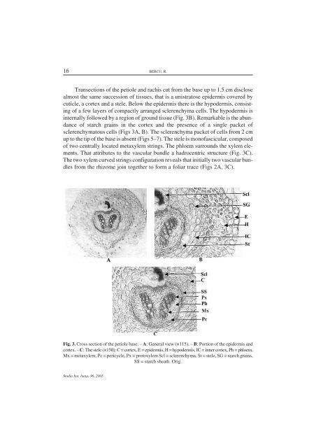
16 BERCU, R.Transections of the petiole and rachis cut from the base up to 1.5 cm disclosealmost the same succession of tissues, that is a unistratose epidermis covered bycuticle, a cortex and a stele. Below the epidermis there is the hypodermis, consistingof a few layers of compactly arranged sclerenchyma cells. The hypodermis isinternally followed by a region of ground tissue (Fig. 3B). Remarkable is the abundanceof starch grains in the cortex and the presence of a single packet ofsclerenchymatous cells (Figs 3A, B). The sclerenchyma packet of cells from 2 cmup to the tip of the base is absent (Figs 5–7). The stele is monofascicular, composedof two centrally located metaxylem strings. The phloem surrounds the xylem elements.That attributes to the vascular bundle a hadrocentric structure (Fig. 3C).The two xylem curved strings configuration reveals that initially two vascular bundlesfrom the rhizome join together to form a foliar trace (Figs 2A, 3C).Fig. 3. Cross section of the petiole base. – A: General view (×115). – B: Portion of the epidermis andcortex. – C: The stele (×150): C = cortex, E = epidermis, H = hypodermis, IC = inner cortex, Ph = phloem,Mx = metaxylem, Pc = pericycle, Px = protoxylem Scl = sclerenchyma, St = stele, SG = starch grains,SS = starch sheath. Orig.Studia bot. hung. 36, 2005