CONTRIBUTIONS TO THE ANATOMY OF ASPLENIUM RUTA ...
CONTRIBUTIONS TO THE ANATOMY OF ASPLENIUM RUTA ...
CONTRIBUTIONS TO THE ANATOMY OF ASPLENIUM RUTA ...
- No tags were found...

You also want an ePaper? Increase the reach of your titles
YUMPU automatically turns print PDFs into web optimized ePapers that Google loves.
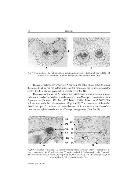
18 BERCU, R.Fig. 7. Cross section of the rachis tip (6 cm from the petiole base). – A: General view (×115). – B:Portion of the stele with a pinnulae trace (×266): Pt = pinnulae trace. Orig.The cross section, performed at 1.5 cm from the petiole base, exhibits almostthe same structure but the xylem strings of the monostele are joined, towards thecentre, by their abaxial protoxylem vessels (Figs 4A, B).The cross section cut at 2 cm from the petiole base shows a monofascicularstele, composed of metaxylem vessels arranged in an X-shape, characteristic to theAspleniaceae (OGURA 1972, BIR 1957, BERCU 1998a, BERCU et al. 2000). Thephloem surrounds the xylem elements (Figs 4A, B). The transection of the rachisfrom 2 cm up to 6 cm (from the petiole base) exhibits the same succession of tissuesbut the xylem vessels are in a T-shape arrangement (Figs 5A, B).Fig. 8. Cross section of pinnulae. – A: Portion of the mesophyll and midrib (×205). – B: Portion of thelower epidermis (×250): Cl = chloroplasts, EC = epidermal cell, LE = lower epidermis, O = ostiole,PT = parenchyma tissue, S = stoma, Sp = sporangium, SC = subsidiary cell, ST = spongy tissue, UE =upper epidermis, VB = vascular bundle. Orig.Studia bot. hung. 36, 2005