huntingdon castle mound, cambridgeshire ... - English Heritage
huntingdon castle mound, cambridgeshire ... - English Heritage
huntingdon castle mound, cambridgeshire ... - English Heritage
- No tags were found...

Create successful ePaper yourself
Turn your PDF publications into a flip-book with our unique Google optimized e-Paper software.
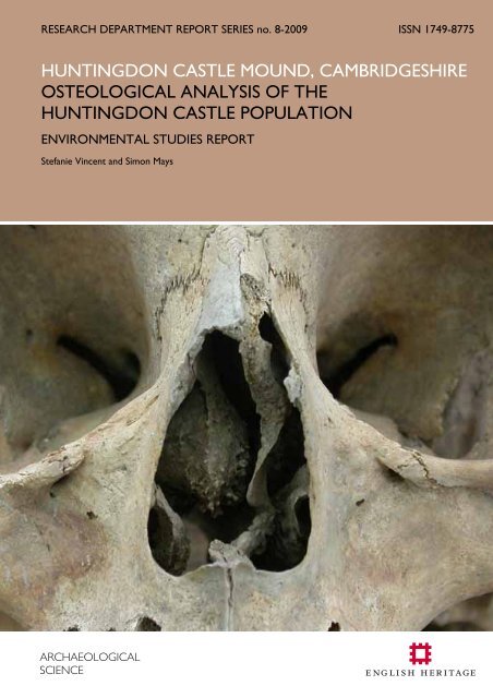
RESEARCH DEPARTMENT REPORT SERIES no. 8-2009 ISSN 1749-8775HUNTINGDON CASTLE MOUND, CAMBRIDGESHIREOSTEOLOGICAL ANALYSIS OF THEHUNTINGDON CASTLE POPULATIONENVIRONMENTAL STUDIES REPORTStefanie Vincent and Simon MaysARCHAEOLOGICALSCIENCE
Research Department Report Series 8-2009Simon MaysFort Cumberland, Fort Cumberland RoadEastney, Portsmouth. PO4 9LDOsteological Analysis of the Huntingdon Castle Population.S. Vincent and S. MaysNGR: TM3969580356© <strong>English</strong> <strong>Heritage</strong>ISSN 1749-8775The Research Department Report Series incorporates reports from all the specialist teams withinthe <strong>English</strong> <strong>Heritage</strong> Research Department: Archaeological Science; Archaeological Archives;Historic Interiors Research and Conservation; Archaeological Projects; Aerial Survey andInvestigation; Archaeological Survey and Investigation; Architectural Investigation; Imaging, Graphicsand Survey, and the Survey of London. It replaces the former Centre for Archaeology ReportsSeries, the Archaeological Investigation Report Series and the Architectural Investigation ReportSeries.Many of these are interim reports which make available the results of specialist investigations inadvance of full publication. They are not usually subject to external refereeing, and theirconclusions may sometimes have to be modified in the light of information not available at thetime of the investigation. Where no final project report is available, readers are advised to consultthe author before citing these reports in any publication. Opinions expressed in ResearchDepartment reports are those of the author(s) and are not necessarily those of <strong>English</strong> <strong>Heritage</strong>.Requests for further hard copies, after the initial print run, can be made by emailing:Res.reports@english-heritage.org.ukor by writing to:<strong>English</strong> <strong>Heritage</strong>, Fort Cumberland, Fort Cumberland Road, Eastney, Portsmouth PO4 9LDPlease note that a charge will be made to cover printing and postage.© ENGLISH HERITAGE 8 - 2009
SUMMARYFifty-five inhumations from Huntingdon Castle Mound, Cambridgeshire were examined.The majority of the burials are Anglo-Saxon in date while the remainder are postmedieval.20 males, 16 females, 6 unsexed adults and 13 juveniles were examined anddemographic, metric, non-metric and pathological data is presented. A case of possibletreponemal disease dating to 1010-1170 was identified.ARCHIVE LOCATIONFort Cumberland, Fort Cumberland Road, Eastney, Portsmouth. PO4 9LDDATE OF RESEARCHResearch was undertaken in 2008-2009.CONTACT DETAILSFort Cumberland, Fort Cumberland Road, Eastney, Portsmouth. PO4 9LD.Simon Mays; 02392 856779; Simon.Mays@english-heritage.org.uk.© ENGLISH HERITAGE 8 - 2009
CONTENTSIntroduction to Huntingdon Castle Mound 1Dating 3Bone preservation and skeletal completeness. 4Demographic composition of the Huntingdon Castle site. 6Demography. 6Metric variation 8Stature 8Cranial measurements 8Non-metric variation 10Oral Pathology. 13Caries. 13Ante-mortem tooth loss. 14Calculus 15Linear Enamel Hypoplasia 16Joint disease. 18Osteoarthritis. 18Degenerative disc disease. 20Diffuse Idiopathic Skeletal Hyperostosis (DISH). 20Hallux Valgus. 21Trauma. 22Fractures. 22Schmorl’s nodes. 24Supra-acetabular cysts. 24Osteochondritis Dissicans. 25Traumatic Myositis Ossificans. 25Third Intercondylar Tubercle of Parsons 25Porotic Hyperostosis. 27Infectious disease. 28Treponemal disease 28Tuberculosis. 29Non- specific infection. 30Neoplasms. 32Miscellaneous conditions. 34Summary. 35References. 36Plates. 40Catalogue of Burials. 48Appendix 1: notes on individual burials. 50© ENGLISH HERITAGE 8 - 2009
Appendix 2: Raw Data 65© ENGLISH HERITAGE 8 - 2009
DatingAs there were only tentative archaeological dates attached to the skeletal remains anAMS radiocarbon dating program was instigated to establish the period from which theskeletal collection came. Samples from 14 skeletons were submitted for dating; thirteenof these were randomly chosen from across the site while B17a was dated becauseinteresting pathological changes which will be discussed later in the report.Burial No. Lab No. Date range (68% confidence) Date Range (95% confidence)B10 SUERC-21104 cal AD 770–880 cal AD 690–890B17a SUERC-19641 cal AD 1020–1160 cal AD 1010–1170B22 OxA-19121 cal AD 1510–1645 cal AD 1470–1650B30 SUERC-19645 cal AD 1020–1150 cal AD 1010–1160B31 OxA-19890 cal AD 1030–1160 cal AD 1020–1170B36 SUERC-21105 cal AD 880–970 cal AD 770–990B49 OxA-19891 cal AD 1030–1160 cal AD 1020–1170B56 OxA-19122 cal AD 890–990 cal AD 890–1020B66 SUERC-21106 cal AD 1210–1270 cal AD 1160–1280B67 OxA-19892 cal AD 880–970 cal AD 870–990B80 SUERC-19646 cal AD 770–900 cal AD 720–950B86OxA-19220SUERC-19647cal AD 1450–1620(mean)cal AD 1440–1640(mean)B87 OxA-19893 cal AD 880–970 cal AD 780–990B100 OxA-19123 cal AD 1020–1160 cal AD 1020–1160The dating results show that the burial site was in use between the 8 th and 17 th centuries.At the time of excavation the majority of the burials were believed to be late Saxon andthe dating which has been completed so far does not contradict this. There isdocumentary and archaeological evidence of a post medieval gallows on the site and theradiocarbon results date some of the burials to the post-medieval period. The lack ofstratagraphic evidence from the site makes it difficult to ascertain if the burial ground wasin continuous use, however it seems likely that the presence of the <strong>castle</strong> and associatedbuildings created a hiatus in the use of the cemetery during the later medieval period. Asthe size of the assemblage is relatively small it was decided to analyse all the burials as onegroup and it was deemed other late Saxon urban sites were the best comparativematerial.3
Bone preservation and skeletal completeness.A total of 55 discrete inhumations have been identified. In three cases (B17, B23 andB50) multiple individuals were collected and packaged together under one burial number.This was presumably the result either of multiple individuals being buried in one grave oran inability to distinguish individual grave cuts on site. Two of the burials contained mixedadult and juvenile remains, while the third contained two adults. During analysis themixed remains were separated and recorded separately; the original burial number waskept and the skeletons allocated an A/B suffix. There was disarticulated bone present inalmost every burial and a substantial amount of charnel was recovered. This charnel isrecorded only by general context number, indicating that no archaeological evidence of adiscreet burial was observed.Skeletal completeness and bone preservation were estimated by visual assessment. Thelevel of bone preservation was scored as; good, moderate or poor, while completenesswas the estimated percentage of skeletal elements present.CompletenessTable 1: Preservation and completeness of burials.Preservation.Good Moderate PoorTotal
Table 2: representation of skeletal elements (allskeletons).Skeletal elementNo. presentCrania 27Mandibles 17Cervical vertebrae 137Thoracic vertebrae 368Lumbar vertebrae 185L ribs 260R ribs 267Sterna 39L Claviclae 27R Claviclae 27L Scapulae 30R Scapulae 30L Humeri 34R Humeri 34L Radii 37R Radii 34L Ulnae 35R Ulnae 32L carpals 52R carpals 35L metacarpals 110R metacarpals 79L hand phalanges 18R hand phalanges 14Unsided hand phalanges 187L Pelves 42R Pelves 44L Femora 40R Femora 40L Patellae 7R Patellae 14L Tibiae 34R Tibiae 38L Fibulae 27R Fibulae 26L Calcanei 22R Calcanei 17L Tali 17R Tali 17L tarsals* 27R tarsals* 36L metatarsals 63R metatarsals 70L foot phalanges 3R foot phalanges 8Unsided foot phalanges 19Table 3: representation of skeletal elements(adult burials).Skeletal elementNo. presentCrania 20Mandibles 13Cervical vertebrae 105Thoracic vertebrae 286Lumbar vertebrae 145L ribs 201R ribs 188Sterna 21L Claviclae 22R Claviclae 19L Scapulae 24R Scapulae 21L Humeri 26R Humeri 26L Radii 27R Radii 25L Ulnae 25R Ulnae 22L carpals 43R carpals 26L metacarpals 83R metacarpals 62L hand phalanges 16R hand phalanges 13Unsided hand phalanges 137L Pelves 33R Pelves 33L Femora 31R Femora 30L Patellae 7R Patellae 13L Tibiae 27R Tibiae 31L Fibulae 20R Fibulae 20L Calcanei 16R Calcanei 15L Tali 14R Tali 15L tarsals* 26R tarsals* 34L metatarsals 54R metatarsals 59L foot phalanges 3R foot phalanges 8Unsided foot phalanges 17* excluding calcanei and tali.5
Demographic composition of the Huntingdon Castle burials.Adult sex was determined using cranial and pelvic morphology (White and Folkens,2005). In older juveniles with fused acetabula, pelvic morphology was also applied. As isstandard practise, no attempt was made to sex younger juveniles.Juvenile aging was primarily estimated using tooth formation (Mays, 1998; Gustafson andKoch, 1974 [reproduced in Hillson, 1996, pg 135]). Where dentition was absent age wasestimated by epiphyseal fusion (Mays, 1998; Scheuer and Black, 2000) and comparison oflong bone length with juveniles from a contemporary site who could be aged by toothdevelopment (Mays, 2007). Foetal and neonatal age was estimated from long-bonelength using the regression equations of Scheuer et al (1980). Adult age was primarilyestimated using molar wear (Brothwell, 1981); unfortunately the size of the sampleproved too small to calibrate tooth wear from juveniles. Pubic symphisis morphology(Suchey et al, 1986, 1988) was used as a supplementary technique and where dentitionwas absent or incomplete.Demography.Table 4: demographic breakdown of the adult population.Adult age ranges18-29 30-49 50+ Adult TotalsMale 3 5 4 6 18Probable Male 1 1 2Female 2 3 2 8 15Probable Female 1 1Unsexed 1 5 6Total 6 8 7 21 42NB: from now on probable males/females will be included in male/female categories.Plotting sex data on the burial plan reveals no sex divisions in the cemetery. The evenratio of males:females in the cemetery mirrors the patterns seen at other late Saxon sitesand is expected in a cemetery serving the general population:MaleFemaleHuntingdon 20 16Caister-on Sea 49 50North Elmham 82 76School Street,28 35IpswichNorwich Castle 39 22© ENGLISH HERITAGE 6 8-2009
50% of the Huntingdon Castle assemblage did not have the skeletal elements presentneeded to assign age to an individual and comparing the age at death distribution withother contemporary sites is impractical.Table 5: demographic breakdown of the juvenile population.Juvenile age ranges0-4 5-8 9-14 15-18 TotalsMale 0 0 0 0 0Probable Male 0 0 1 1 2Female 0 0 0 0 0Probable Female 0 0 0 0 0Unsexed 4 2 3 2 11Total 4 2 4 3 13The range for juvenile age is 42 weeks in utero – 17 years. A comparison of the age atdeath distribution of the Huntingdon Castle population with that of the School Streetpopulation (Mays, 1989) shows that both sites had a fairly even distribution of juvenilesacross the four categories displayed above. The late Saxon sites of North Elmham (Wells,1980) Caister-on-Sea (Anderson, 1993) and Norwich Castle (Stirland, 1985) all recordpeaks in the number of children dying in specific age groups and it may be that the smallnumber of individuals represented at Huntingdon Castle has masked any such peak in thismaterial.© ENGLISH HERITAGE 7 8-2009
Metric variationStatureAdult stature was estimated using Trotter and Gleser (1958, in Brothwell, 1981, pg 100).The formulas are sex specific, so no stature information could be gained from the burialswhich were unsexed.Table 6: distribution of adult stature (cm).154-160 ≤ 160-165 ≤ 165-172 ≤ 172-180 ≤ 180-185Male 1 1 7 8 1Female 9 3 - 1 -Table 7: range and mean of adult stature (cm).No. Stature Range Mean StatureMale 18 160.8-184.4 171.5Female 13 154.4-174.3 160Comparison of stature between the population of Huntingdon Castle and other lateSaxon groups shows that the Huntingdon adults were of a fairly average stature, althoughthe height range of the Huntingdon females is slightly higher than their contemporaries:Table 8: comparative stature of Huntingdon adults (cm).Male StatureFemale StatureAverage Range Average RangeHuntingdon 171.5 160.8-184.4 160 154.4-174.3Caister-on Sea 170.8 157-185.8 161.1 148.6-172.3North Elmham 172.1 162.3-180.7 157.4 142.4-169.7School Street, Ipswich 171.5 163-186 159.1 149-172Norwich Castle 170.1 159.4-179.2 160.1 156.2-165.2Cranial measurementsMeasurements were taken as outlined in Brothwell (1981) with additional measurementstaken from Howells (1973). Of the forty-two adults in the Huntingdon group onlytwenty had crania present; of these only two are complete. This limits the data availablefor cranial measurements. The results are shown in appendix 2.Cranial indexCranial index was calculated as in Brothwell (1981). Only eight individuals had craniacomplete enough to allow the correct measurements to be taken.© ENGLISH HERITAGE 8 8-2009
Table 9: cranial indices by sexNo. Mean SD RangeMale 5 74.1 3.69 71.6-76.5Female 3 76.3 1.54 75.0-78.5Table 10: cranial indices by categoryDolichocephalic Mesocephalic Brachycephalic HyperbrachycephalicMale 4 1 0 0Female 0 3 0 0The cranial indices of the Huntingdon Castle population indicate that the cranial shapetended to be long and narrow. This shape is typical of the Anglo-Saxon period and iscomparable with the cranial indices found at North Elmham (Wells, 1980), Caister-on-Sea(Anderson, 1993) and Norwich Castle (Stirland, 1985).Meric and cnemic indicesMeric and cnemic indices gauge the cross-sectional shape of the femur and tibiarespectively. Differences in cross-sections between populations are thought to be causedby mechanical adaptation and as such may indicate the range of physical activity of theindividual (Brothwell, 1981).Table 11: meric indicesMeric index LMeric index RNo. Mean s.d. Range No. Mean s.d. RangeMale 12 81.7 8.3 63.1-97.0 12 82.6 6.3 74.7-95.0Female 10 78.5 6.1 70.9-86.7 10 82.4 7.7 71.9-93.7Unsexed 1 78.3 - 78.3 1 78.3 - 78.3Table 12: cnemic indicesCnemic index LCnemic index RNo. Mean s.d. Range No. Mean s.d. RangeMale 11 69.2 5.7 61.7-79.4 11 70.6 5.5 61.6-80Female 10 70.4 13.4 33.2-83.1 9 76 5.0 68-82.3Unsexed 2 70 4.5 65.5-74.5 3 71.6 4.6 66-71.1© ENGLISH HERITAGE 9 8-2009
Non-metric variationNon-metric variations are a range of minor variations in presence or morphology ofstructures such as foramina or facets. While it has been established that some traits (suchas the Inca bone) have a genetic component, the causes of the majority of the traits areunknown. Both cranial and post-cranial non-metric variations were recorded. The postcranialtraits were taken from Finnegan (1978) and the cranial traits from Berry and Berry(1967). Some traits are known to manifest in childhood, only to disappear in adulthood;for this reason only adult results are reported here.Table 13: frequency of cranial non-metric traits in adults.Trait 1 0 1/1 -/1 1/- 1/0 0/1 -/0 0/- 0/0Metopic suture 1 13Ossicle at lambda 1 12Lambdoid ossicle 6 4Inca Bone 0 13Sagittal ossicle 0 6Ossicle at bregma 0 11Coronal ossicle 2 7Fronto-temporal articulation 0 0 0 0 0 2 2 1Epipteric bone 0 1 0 0 0 1 2 2Squamo-parietal ossicle 0 0 0 0 0 2 2 3Parietal notch bone 0 0 0 0 1 2 1 5Auditory torus 0 0 0 0 0 3 3 11Foramen of Hushke 0 0 0 1 1 2 4 8Ossicle at asterion 0 0 0 2 0 1 1 5Clinoid bridging 0 0 0 0 0 1 0 2Pterygoid bridging 0 0 0 0 0 1 0 4Palatine torus 1 8Maxilla torus 0 8Mastoid foramen extra-sutural 3 0 1 0 0 2 1 2Mastoid foramen absent 1 0 0 1 0 2 1 3Double condylar facet on occipital 0 0 0 0 0 1 2 8Parietal foramen 4 0 1 1 2 0 0 5Accessory infra-orbital foramen 2 0 0 1 0 1 1 5Zygomatic facial foramen 4 5 2 0 0 0 2 2Divided hypoglossal canal 1 0 1 0 1 1 1 5Post condylar canal patent 1 0 0 1 2 1 1 3Pre-condylar tubercle 0 0 1 0 0 1 0 8Foramen ovale incomplete 0 0 0 0 0 2 0 7Accessory lesser palatine foramen 1 1 0 0 1 0 0 5Supra-orbital foramen complete 1 1 0 1 0 0 3 5Maxillary M3 absent 0 0 0 0 0 0 0 9Mandibular M3 absent 2 0 0 0 0 0 0 10Mandibular torus 0 9Mylohyoid bridging 1 0 0 0 0 2 0 8© ENGLISH HERITAGE 10 8-2009
Table 14: frequency of post-cranial non-metric traits in adults.Trait 1 0 1/1 -/1 1/- 1/0 0/1 -/0 0/- 0/0Fossa of Allen 5 0 2 2 0 4 3 15Poirers facet 1 0 0 0 1 3 6 20Plaque formation 8 1 2 0 1 3 3 13Exostosis in trochantric fossa 6 1 5 2 1 1 3 9Supra-condylar process 0 1 0 1 0 5 10 15Septal apature 0 1 1 0 1 2 9 13Acetabular crease 4 0 0 1 0 1 5 16Accessory sacral facets 6 1 0 2 0 3 4 11Spinabifida occulta 0 27Six sacral segments present 10 17Acromial articular facet 0 0 0 0 0 1 5 7Os acromale 0 0 0 1 0 1 6 6Supra-scapular foramen 0 0 0 0 0 2 4 4Vastus notch 2 0 0 0 0 6 1 4Vastus fossa 0 0 0 0 0 6 1 5Emarginate patella 0 0 0 0 0 6 1 5Lateral squatting facets 1 0 0 0 0 4 4 13Medial squatting facets 5 1 3 2 2 4 0 4Anterior calcaneal facet double 3 2 1 0 0 2 2 8Anterior calcaneal facet absent 0 0 0 0 0 4 3 11Atlas facet double 2 0 1 0 0 0 1 14Posterior atlas bridging 0 0 0 1 1 0 1 14Lateral atlas bridging 0 0 0 1 1 0 2 13Key: 1=trait present, 0=trait absent, - element not present for observation. Bilateral traits are scoredleft/right.There is a high prevalence of the occurrence of six sacral segments, 37% in the twentyseven individuals with sacra available for examination. Increases in the total number ofvertebrae present are not uncommon and occur most frequently in the lumbar and sacralvertebra (Barnes, 1994:78). The additional vertebrae begin to form at week two, but it isnot yet known if genetic or environmental factors are the cause (Usher and NørregaardChristensen, 2000). This is a higher frequency than is seen in the School Street (11.1%)and North Elmham (4.7%) populations. Plotting the occurrences of a sixth sacral segmenton the burial plan reveals no clustering indicative of familial groups and it may be that theshared genetic inheritance of the group or a specific environmental factor they wereexposed to caused the high prevalence of the trait.Different sternal morphologies result from variation in the timing of fusion of the sternalsegment (Barnes, 1994:219). There were 21 adults with sterna available for examinationand of these 17 have type one sterna. Three individuals have normal type two sterna(Barnes, 1994:219) while a fourth individual with a type two sternum exhibits markedmedio-lateral widening at segments 3-4, with a corresponding circular depression on theplural side of the sternum, suggestive of a delay in fusion between the segments (Barnes,1994:218).© ENGLISH HERITAGE 11 8-2009
Four individuals exhibit cranial shifts of the vertebral borders while one exhibits a caudalshift. Variations around the borders of regional vertebra are common and result in thevertebrae above or below the border taking on characteristics of the vertebra in theneighbouring region (Barnes, 1994: 79). There are two cases of cleft posterior arch of theatlas, caused by a developmental delay in formation of the two halves of the arch (Barnes,1994: 119). It is estimated that about 5% of the modern adult population have clefting ofthe atlas arch; in life the gap is bridged by a fibrous band which mimics the missing bonysegment (Barnes, 1994:120).© ENGLISH HERITAGE 12 8-2009
Oral Pathology.Caries.Dental caries (or cavities) occur when the acid by-product of bacteria in dental plaquecauses focal destruction of the tooth (Hillson, 1996:269). Dental caries were scored aspresent or absent.Table 15: prevalence of caries in adult population.Individuals scored for caries Individuals with cariesMale 10 4Female 3 3Unsexed 1 1Totals 14 8TeethPresentCariousteethTeethpresentCariousteethTable 16: Prevalence of caries by tooth in adults.MaxillaryLM3 LM2 LM1 LPM2 LPM1 LC LI2 LI1 RI1 RI2 RC RPM1 RPM2 RM1 RM2 RM3 Total5 6 4 6 7 7 5 4 5 7 8 10 9 7 7 6 1031 1 0 1 1 0 0 0 0 0 0 0 0 1 1 0 63 7 6 10 9 7 8 8 7 7 8 11 10 7 8 3 1191 0 1 1 1 0 0 0 0 0 0 1 1 0 0 0 6Mandibular% Caries by Individual % Caries by tooth.Huntingdon Castle 57 5.4Caister-on-sea 1.8North Elham - 6.4School Street, Ipswich 44.6 10Norwich Castle - 2.6The data shown above demonstrates that the frequency of caries by tooth in theHuntingdon Castle population falls in the middle of contemporary sites while the rate ofcaries by individual is significantly higher. This may point to low levels of oral hygiene inthe Huntingdon Population but it is worth noting that a significant number of the adultpopulation of Huntingdon Castle did not have dentition available for examination.© ENGLISH HERITAGE 13 8-2009
Ante-mortem tooth loss.Ante-mortem tooth loss was scored as present or absent in individuals where one ormore tooth sockets could be examined. The results are shown below;Table 17: adult tooth loss.Individuals scored for tooth loss Individuals with tooth loss.Male 10 6Female 4 3Unsexed 1 1Totals 15 10Table 18: results by tooth of ante-mortem loss.MaxillaryLM3 LM2 LM1 LPM2 LPM1 LC LI2 LI1 RI1 RI2 RC RPM1 RPM2 RM1 RM2 RM3 TotalSocketsPresentToothLossSocketspresentToothLoss8 10 9 10 10 10 10 11 13 13 13 13 13 12 11 9 1750 3 4 3 2 2 2 2 2 2 2 2 3 4 3 1 377 12 12 12 12 12 12 11 11 11 11 11 11 11 11 7 1743 4 5 0 0 0 1 2 1 1 0 0 0 3 3 4 27MandibularAnte-mortem tooth loss is known to be age related, with older individuals showing ahigher rate of loss than younger people. The most common causes of tooth loss arecaries and periodontal disease, although it can also result from traumatic injury (Mooreand Corbett, 1971).% Ante-mortem tooth loss byindividual.% Ante-mortem tooth loss bytooth socket.Huntingdon Castle 66.7 18.3North Elmham - 11.1School Street, Ipswich 47.6 10.5Norwich Castle - 4The frequency of ante-mortem tooth loss at Huntingdon Castle is considerably higherthan those reported at other comparable sites. Again it is worth noting that only a smallpercentage of the population had dentition available for study.Periapical VoidsPeriapical voids have commonly been referred to in the literature as abscesses,granulomas or cysts. Recent work has highlighted the tendency to apply such termsgenerically to any void found associated with a tooth and advocates that more precision© ENGLISH HERITAGE 14 8-2009
e taken in identifying such voids, so their effect on the individual can be betterrepresented (Ogden, 2008).The presence of periapical voids were recorded for all teeth and tooth sockets availablefor examination. One periapical void was found in the juvenile population at a first rightmandibular incisor. Adult results are shown below;Table 19: periapical voids in adults.Individuals scored for periapical voidsIndividuals with periapical voidsMale 10 4Female 4 3Unsexed 1 0Totals 15 7Table 20: periapical voids by tooth.MaxillaryLM3 LM2 LM1 LPM2 LPM1 LC LI2 LI1 RI1 RI2 RC RPM1 RPM2 RM1 RM2 RM3 TotalSocketsPresentPeriapicalVoidsSocketspresentPeriapicalVoids8 10 9 10 10 10 10 11 13 13 13 13 13 12 11 9 1751 0 1 0 0 0 0 0 0 0 0 2 2 1 0 0 77 12 12 12 12 12 12 11 11 11 11 11 11 11 11 7 1740 0 1 0 0 1 0 0 0 0 1 0 0 2 0 0 5Mandibular46.6% (7/15) of the adults scored were found to have periapical voids, with a frequencyof 3.4% (12/349) for all sockets scored. Using the criteria laid out by Ogden (2008)further diagnosis of the periapical voids in the Huntingdon population was attempted.Four periapical voids were found to be granulomas, while a fifth was thought to be eithera granuloma or a cyst. The cause of the remaining periapical voids could not be identified.Calculus.Calculus is caused by the mineralization of dental plaque and is indicative of low oralhygiene (Hillson, 1996:255). Supra-gingiveal calculus was scored using the criteria ofDobney and Brothwell (1987). The results are shown below;Table 21: calculus in adults.Grade 0 Grade 1 Grade 2 Grade 3 Grade 4 TotalMale 2 4 4 1 0 11Female 0 2 0 1 0 3Unsexed 0 0 0 0 0 0Totals 2 6 4 2 0 14© ENGLISH HERITAGE 15 8-2009
The results show that 85% of adults with dentition had calculus deposits, however itshould be noted that of forty two adults in the population only fourteen had dentitionpresent to score.Linear Enamel HypoplasiaLinear enamel Hypoplasias (LEH) are transverse bands of deficient enamel thickness onthe crown of the tooth, which develop during crown formation. They occur as a result ofstress upon the growing individual and have been linked to disease and malnutrition(Hillson, 1996:165). LEH were systematically recorded in the anterior dentition andnoted when they occurred elsewhere. In the anterior dentition the presence, toothaffected and distance of the defect from the cemento-enamel junction (CEJ) wererecorded;Table 22: LEH by individual.Number of defects per individual0 1 2Male 7 2 1Female 3 0 1Total 10 2 2In the Huntingdon population there were six linear enamel hypoplasias found in fourindividuals (HC022, HC030, HC066 and HC091). Of these six, four occurred in theanterior dentition while two were found on the pre-molars.Where LEH occurred in the anterior dentition an attempt was made to estimate the ageat which the defect arose. Unfortunately the incomplete nature of the assemblage meantthere were not enough teeth to calibrate tooth formation scales as described by Reid andDean (2000). Instead where the teeth effected with LEH were unworn the tooth heightwas measured and the age at which the LEH occurred was calculated. In cases wherewear had occurred to the tooth exhibiting LEH, an estimation was made as to which fifthof the tooth the defect occurred in and age of occurrence was estimated using the resultsof Reid and Dean (2000).Table 23: defects of anterior dentition.Burial Tooth Distance of LEHfrom CEJTooth height Estimated age ofoccurrenceHC091 C R 11.1 13.4 1.7-2HC022I1 R7.0Unknown 1.3-1.7I1 L6.5HC030 I1 R 4 Unknown 2.9-3.9N.B. Measurements are in mm and age is in years.HC066 was found to have two LEH’s on 1PM R and it was estimated that these occurredbetween two and four years of age.© ENGLISH HERITAGE 16 8-2009
In addition to the occurrence of LEH, one example of a major enamel disruption in therear dental arcade was found. HC091 has a systematically recorded LEH on C R , alongwith enamel disturbance on the PM2 R and M1 R . The M1 R has grossly disturbed cuspalarchitecture and a dramatic enamel hypoplasia on the buccal side of the tooth, leaving agap of approximately 2mm between the edge of the buccal enamel and the occlusalsurface of the tooth (Plate 1). The occlusal surface itself shows enamel disruption in theform of multiple small pits across its surface. The PM2 R also exhibits an abnormality incusp formation and has an LEH extending part-way across its buccal surface. There arealso pit form enamel hypoplasias across the buccal surface of the tooth (Ogden, 2008).Mulberry molars as a result of congenital syphilis can cause a change in cuspal architecturewith accompanying disruption of tooth enamel, however the changes observed here donot fit the criteria set out by Hillson (1998). The enamel disruption of the first molar andsecond premolar, coupled with the alteration in cusp organisation are consistent with acondition termed cuspal enamel hypoplasia (Ogden et al, 2007). Using tooth formationcharts (Mays, 1998) it has been estimated that the disturbance to M1 R occurred within thefirst 1.5 years of life, while the LEH and associated pitting on the pre-molar occurred atbetween 2-3.5years of age. This combined with the LEH of the right mandibular caninesuggests that multiple episodes of stress were placed upon the individual.© ENGLISH HERITAGE 17 8-2009
Joint disease.Osteoarthritis.Osteoarthritis (OA) is degeneration of the joint surfaces causing osteophytosis of jointmargins, surface porosity and eburnation. OA occurrence is known to increase with ageand is thought in part, to be a result of mechanical loading stresses on joints (Rogers andWaldron, 1995).The severity of OA was scored using a system adapted from Sager (1969; reproduced inBrothwell, 1981):Grade 0Grade 1Grade 2Grade 3Normal bone surfaceIntermittent osteophytesSurface porosity; may be accompanied by osteophytesEburnation; may be accompanied by porosity and osteophytesTable 24: maximum OA score in individual adults from Huntingdon.OA grade.Grade 0 Grade 1 Grade 2 Grade3Male 6 5 2 7Female 2 3 5 6Unsexed 3 1 1 1Total 11 9 8 14Table 25: distribution of osteoarthritis in adults at Huntingdon.GradeSkeletal element 0 1 2 3L mandibular condyle 5 0 0 2R mandibular condyle 7 0 0 0Cervical vertebrae 62 15 8 14Thoracic vertebrae 197 17 20 8Lumbar vertebrae 102 15 10 3L ribs 149 30 20 2R ribs 135 32 15 6L medial clavicle 20 0 1 0R medial clavicle 14 0 2 0L lateral clavicle 10 0 5 0R lateral clavicle 8 0 5 0L glenoid fossa 18 2 0 0R glenoid fossa 16 3 0 1L proximal humerus 20 0 0 0R proximal humerus 15 0 0 0L distal humerus 21 0 0 1R distal humerus 17 2 0 0© ENGLISH HERITAGE 18 8-2009
L proximal radius 18 1 0 1R proximal radius 19 0 0 0L distal radius 20 0 0 1R distal radius 21 0 0 0L proximal ulna 17 3 0 0R proximal ulna 15 3 0 0L distal ulna 20 0 0 0R distal ulna 14 1 0 0L carpals 43 0 0 0R carpals 26 0 0 0L metacarpals 81 1 0 1R metacarpals 62 0 0 0L hand phalanges 16 0 0 0R hand phalanges 13 0 0 0Unsided hand phalanges 136 0 0 1L acetabulum 25 0 0 1R acetabulum 26 0 0 0L proximal femur 28 0 0 1R proximal femur 28 0 0 0L distal femur 21 4 0 0R distal femur 21 3 0 1L patella 7 0 0 0R patella 10 3 0 0L proximal tibia 24 0 0 0R proximal tibia 28 1 0 0L distal tibia 19 0 0 0R distal tibia 24 0 0 0L proximal fibula 13 0 0 0R proximal fibula 12 0 0 0L distal fibula 14 0 0 0R distal fibula 13 0 0 0L calcaneus 16 0 0 0R calcaneus 15 0 0 0L talus 14 0 0 0R talus 15 0 0 0L tarsals* 26 0 0 0R tarsals* 34 0 0 0L metatarsals 53 0 0 1R metatarsals 59 0 0 0L foot phalanges 3 0 0 0R foot phalanges 8 0 0 0Unsided foot phalanges 17 0 0 0*excluding tali and cancaneiThere is no significant difference in the frequency, severity or location of OA occurrencebetween sexes.© ENGLISH HERITAGE 19 8-2009
Degenerative disc disease.Osteoarthritis of the vertebral facet joints were scored as detailed previously. The nonsynovialjoints between vertebral bodies degenerate in a different way: long termmechanical stress in the vertebral column can cause degeneration of the intervertebraldisc. This can lead to spreading of the disc surface and production of osteophytes andporosity on the vertebral body surfaces (Rogers and Waldron, 1995).The recording of osteophytosis in the Huntingdon population was adapted from Sager(1969), reproduced in Brothwell (1981):Grade 0Grade 1Grade 2Grade 3Normal bone surfaceOsteophytesSurface porosity covering less than half of the vertebral surfaceSurface porosity coving more than half of the vertebral surface.Table 26: maximum grades of degenerative disc disease by individual.Maximum grade0 1 2 3Males 7 3 5 2Females 4 5 4 2Unsexed 1 1 0 0Total adults 12 9 9 4Note: For inclusion, individuals needed to show at least one vertebral body present for observationTable 27: prevalence of degenerative disc disease by vertebrae.Cervical Thoracic Lumbar0 1 2 3 0 1 2 3 0 1 2 3Males 25 4 4 8 77 39 6 0 38 21 8 0Females 25 7 3 5 50 31 15 0 26 23 9 1Unsexed 0 0 0 0 7 7 0 0 0 0 0 0Total adults 50 11 7 13 134 77 21 0 64 44 17 1There are no significant differences in the frequency of degenerative disc disease betweenmales and females. In both sexes the lumbar vertebrae are the most commonly involvedin degenerative disc disease, followed by the thoracic and cervical regions of the spine.Diffuse Idiopathic Skeletal Hyperostosis (DISH).DISH in skeletal remains is characterised by vertebral ligamentous ossification leading toankylosis (Rogers and Waldron, 2005). The cause of the condition is not fully understoodbut links have been made to diabetes, obesity and metabolic disturbances (Rogers andWaldron, 2001; Mays, 2000), whilst a clinical study of Finnish populations foundgeographical differences in DISH prevalence rates (Julkunen et al, 1971). Studies haveshown that the prevalence of DISH is higher in males than females and increases with age© ENGLISH HERITAGE 20 8-2009
(Julkunen et al, 1971). DISH was diagnosed when two complete bony bridges betweenvertebrae were present (Julkunen et al, 1971) and differentiated from other arthropathiesusing the criteria set down by Rogers and Waldron (1995).There was one case of DISH found in the Huntingdon Castle population, in a male aged50+ (HC061). There is ankylosis of the vertebral bodies of C6/C7/T1, T6/T7 and furtherinterlocking, non-ankylosing ossifications between T5/T6, T7/T8/T9, L2/L3. There isossification of both acetabular labra, both glenoid labra, at the site of the Achilles tendoninsertion on the left calcaneus and at the site of the insertion of the quadriceps tendon onthe right patella.Hallux Valgus.Hallux valgus is the lateral deviation of the first toe, instigated by biomechanical forces onthe foot, usually from footwear. It is commonly associated with bunion formation at themetatarso-phalangeal joint and osteoarthritic changes at the metatarsal head (Mays,2005a).One individual was found to be suffering from hallux valgus. HC003 is an unsexed adultexhibiting bilateral hallux valgus, indicating that the individual wore restrictive footwear.The level of skeletal completeness means that the frequency of the condition could havebeen underestimated in the population: of the forty-two adults examined only thirteenhave one or more metatarsal present for examination.© ENGLISH HERITAGE 21 8-2009
Trauma.Fractures.There was a prevalence of fractures of 21% with respect to the total number of adultsexamined; no evidence of fracture was found in the juvenile material. Twenty threefractures occur in nine adults, all post-cranial. Rib fractures are the most common type offracture, accounting for 65% (15/23) of fractures found, with the majority of thesefractures (11/15) occurring in one individual (HC030). This pattern follows that seen byRoberts and Cox (2003:206;239); in their review of medieval skeletal material ribfractures were the most frequent fracture observed.HC011 (male, 50+) has a compression fracture of the left superior facet of the atlas; thefacet height has been reduced in comparison to the right and new bone formation ispresent along the lateral edge of the facet (Plates 2 and 3). The other cervical vertebraeexhibit osteoarthritic changes, possibly as a secondary reaction to the fracture. The likelyaetiology for the fracture is a compressive force to the skull and neck, perhaps as a resultof a fall (Resnick and Niwayama,1988:2932).HC072 (unsexed, adult) has a healed avulsion fracture effecting the anterior body of T3(Plates 4 and 5), probably caused by hyperextension of the spine (Resnick and Niwayama,1988:2940).In HC093 (female, 50+) C3 and C4 have become ankylosed at their bodies and rightfacet joints. C3 is laterally deviated to the right and there is no retention of intervetebraldisk space (Plate 6). Large non-ankylosing osteophytes have formed at the vertebralbodies and right facet joints of C4, C5, and C6. There is a smooth walled concavedepression on the right hand side of the bodies of C3 and C4, suggestive of an aneurismof the vertebral artery. The lateral displacement of C3 suggests the ankylosis is secondaryto a subluxation of the cervical spine (Resnick and Niwayama, 1988:2937).HC050 (male, 50+) has spondylolysis of L2, L3 and L6. The condition is a cleft in thepars interarticulars of the vertebrae. In this individual bi-lateral spondylolysis has led to theseparation of the posterior section of the neural arch from the rest of the vertebra. It isbelieved that spondylolysis is caused by a fatigue fracture but the exact nature of thestress which causes the condition has not yet been determined. Spondylolithisis is arelated condition caused by bi-lateral spondylolysis which results in anterior slippage of thevertebral body, which results in doming of vertebral body below. The body of S1 isdomed in individual HC050 suggesting that spondylolisthesis of L5 has occurred (Mays,2006).There is one example of hand trauma in the collection; HC066 (male, 35-45) exhibits aBennett fracture of the first right metacarpal. Bennett fractures are fractures involving the© ENGLISH HERITAGE 22 8-2009
articular surface of the thumb, usually caused by striking the thumb against a hard surfacewith force (Peterson and Bancroft, 2006).The right ilium of HC066 (male, 35-45) has been fractured and a portion of the bone hasbecome displaced. The displaced bone has healed leaving perforations through the iliaccrest (Plate 7). The injury is well healed with no evidence of infection in the surroundingbone. The origin of the trauma is most likely a direct downward blow to the iliac crest.HC093 (female, 50+) has a fracture of the right femoral neck with secondary damage tothe right acetabulum. The fracture has shortened the femoral neck and inferiorlydisplaced the femur head. The superior edge of the right acetabular rim has suffered aheight loss of approx 5mm and has a ragged appearance. This lesion is suggestive of hiptrauma in which a lateral impact forced the femur head against the acetabulum. The lossof acetabular rim height may suggest a minor fracture occurred during the incident but theragged nature of the surface is more consistent with the fragmentation of the rim edge.This can occur when a tear in the articular cartalidge at the base of the acetabular labrumpermits ingress of synovial fluid into the subchondral bone. This can result in thereabsorbition of multiple areas of subchondral bone, undermining the acetabular rim untilit can no longer sustain the mechanical forces at the hip and fragments (Mays, 2005b).Two individuals have healed fibula fractures in their distal diaphyses (HC014 and HC079).Both fibulae fractures are oblique indicating an indirect trauma and their position on thefibulae is indicative of a lateral rotative force (such as a twist of the ankle) being thefracture mechanism (Lovell, 1997). The right tibia of HC079 has remodelled periostealbone formation at the same level as the fibula fracture, indicating inflammation or infectionof the surrounding soft tissue may have occurred at the time of the fracture. Lovell(1997) notes that fractures involving the ankle are the second most frequent fracture tooccur in modern populations.HC080 has a linear fracture of the first left metatarsal which runs anterior-posterior fromthe head to the base (Plate 8). The linear nature of the fracture suggests it was caused byan impact to the distal end of the first toe.Weapon-related injuryBurial HC073 (male, 25-35) has an unhealed edged weapon wound on the cranium (Plate9). There is a linear cut on the left parietal bone measuring 37.3mm by 1.7mm, which isrestricted to the outer table of the cranium. The right edge of the depression is straightwhile the left edge shows flaking of the bone. There are no fractures of the skull aroundthe wound, indicating a sharp bladed instrument caused the injury (Lovell, 1997). Thelack of healing at the site indicated the injury occurred peri-mortem. Demographicallymales are more likely to be injured by physical aggression, with the frontal and leftparietals being the most frequent areas for injuries to occur (Roberts and Manchester,2005:109).© ENGLISH HERITAGE 23 8-2009
Schmorl’s nodes.Schmorl’s nodes are pressure defects on the vertebral surface, caused by herniatedmaterial from the intervertebral disc exerting pressure on the surface of the vertebralbody (Rogers and Waldron, 1995). In dry bone they present as indentations in thevertebral surface. The primary cause of Schmorl’s nodes is greatly debated; while it seemsthat trauma (either repeated micro-trauma or a major traumatic event) is the direct causeof the defect, there may be underlying genetic or developmental factors which predisposesome individuals to the development of Schmorl’s nodes (Resnick and Niwayama,1988:1527). Research into Schmorl’s nodes as a cause of pain is ongoing and recentpapers seem to suggest that chronic pain can accompany the lesions, although furtherresearch is needed to quantify this (Faccia and Williams, 2008).TotalVertsTable 28: distribution of Schmorl’s nodes by vertebra.Cervical Vertebrae Thoracic Vertebrae Lumbar VertebraeVertswithnodesNo. ofNodesTotalVertsVertswithnodesNo. ofNodesTotalVertsVertswithnodesNo. ofNodesMale 41 0 0 122 36 46 67 2 3Female 40 0 0 106 17 21 63 8 8Unsexed 0 0 0 14 5 5 0 0 0Total 81 0 0 242 58 72 130 10 11Table 29: results of Schmorl’s nodes by individual.Individuals without nodesIndividuals with nodesMale 6 11Female 10 5Unsexed 1 1Total 17 17NB: For inclusion, individuals needed to show at least one vertebral body present for observationThere is no significant difference between the frequency of Schmorl’s nodes in males andfemales. The distribution of Schmorl’s nodes within the spine is the same for males andfemales; the thoracic vertebrae are most often effected, followed by the lumbar spine.No Schmorl’s nodes were present in the cervical vertebrae.Supra-acetabular cysts.Supra-acetabular cysts are cavities within the iliac bone which communicate with thecortical surface of the bone at or around the acetabular rim. The cysts are caused byingress of synovial fluid into the subchondrial bone via tears in the acetabular labrum orneighbouring cartilage (Mays, 2005b).Four individuals from Huntingdon Castle have supra-acatabular cysts; HC025, HC066,HC093 and HC102. HC093 (female, 50+) has supra-acetabular cysts and a fracture of© ENGLISH HERITAGE 24 8-2009
the acetabular rim in association with a fracture to the femur neck, as discussed above.The rim of the left acetabulum of HC025 (male, adult) has a ragged appearance,suggesting the rim has fragmented. This is caused by the formation of several supraacetabularcysts, which undermine the edge of the rim leading to its collapse (Mays,2005b). The remaining individuals have bilateral supra-acetabular cysts, with noobservable trauma of the acetabular bone.Osteochondritis Dissecans.Osteochondritis dissecans is the detachment of a small segment of bone from an articularsurface, following necrosis brought on by repeated micro-trauma. The appearance of thelesion in dry bone is a small missing section of cortical bone, leaving an irregular surfacewith clearly de-lineated edges (Auferheide and Rodriguez-Martín, 1998:81).There are two probable cases of osteochondritis dissecans in the Huntingdon population.The first is HC043, an adult female with the condition in the capitulum and trochlearnotch of the right elbow. The second is HC090 an adult male with osteochondritisdissecans of the lateral condyle of the left distal femur. The knee and the elbow arerespectively, the first and third most common sites of osteochondritis dissecansoccurrence in modern populations (Auferheide and Rodríguez-Martin, 1998:82-83).Traumatic Myositis Ossificans.Traumatic myositis ossificans occurs when an injury to tendinous, ligamentous or muscleattachments results in a haematoma which subsequently ossifies (Resnick and Niwayama,1988:4247). It should be noted that not all trauma of this type will lead to ossification ofthe haematomas and so the level of trauma to tendinous, ligamentous and muscleattachments in a population will be underestimated in the skeletal remains.There are two cases of traumatic myositis ossificans in the Huntingdon population;HC030 (male, 50+) displays an ossification at the distal diaphysis of the right tibia andfibula, suggestive of an injury to the interosseous ligament. HC031 (female, adult) displaysossification at the lesser trochanter of the right femur indicating a tear of the psoas majorand/or Iliacus muscle.Third Intercondylar Tubercle of Parsons.The third intercondylar tubercle of Parsons (TITP) has been linked to injury of theanterior cruciate ligament (Mays and Cooper, In press). TITP is found in four individuals;two cases are bi-lateral, one is unilateral and the fourth has only one tibia available forstudy. Three of the effected individuals are female, while the fourth is unsexed. Thepotential for the Parsons’ tubercle to be used as an indicator of activity has been noted,© ENGLISH HERITAGE 25 8-2009
however further work is needed in this area before such definitive links can be made(Mays and Cooper, In press).© ENGLISH HERITAGE 26 8-2009
Porotic Hyperostosis.Porotic hyperostosis is most commonly a result of iron deficiency anaemia. The causes ofthe iron deficiency are complex and only rarely represent dietary deficiency; insteadfactors affecting the uptake of iron into the body (parasite load, pregnancy, blood loss etc)are the main causes (Stuart-Macadam, 1992). Cribra orbitalia, a category of porotichyperostosis presents as small perforations in the roof of the orbits. Cribra orbitalia wasscored as either porotic or cribrotic in line with Brothwell (1981).Table 30: occurrence of cribra orbitalia by individual.Absent Porotic CribroticMale 3 3 0Female 4 0 0Total adults 7 3 0Juvenile 0 0 0NB: For inclusion individuals needed one or both orbits present for examination.There were only a small number of individuals in the Huntingdon population with one orboth orbits present, making comparisons with other sites impractical. No cases of porotichyperostosis affecting the skull vault were found.© ENGLISH HERITAGE 27 8-2009
Treponemal disease.Infectious disease.There are three types of treponematosis which effect the skeleton; syphilis (congenitaland acquired), yaws and bejel (also known as endemic syphilis). Yaws is found only intropical climates, while today bejel is most commonly found in hot, dry areas (such as theMiddle east and Africa). Archaeological evidence of bejel has been found in NorthernEuropean populations; the disease is transmitted via contact with the open sores ofinfected individuals. In modern populations syphilis is found world-wide (Ortner,2003:274). There is one possible case of treponemal disease present in the HuntingdonCastle population.HC017 is an adult female whose skeleton is represented only from the lumbar vertebraedownwards. Both tibiae and the left fibula (right is not present) show concentricthickening throughout their diaphysis. The majority of the bone deposits are smooth andwell remodelled but there are some areas which have a fine grained appearance,suggesting lesions active at the time of death. Both tibiae have small plaques of bone withundercut edges. A post depositional break on the distal diaphysis of the left tibia showsthat the medullary cavity has become occluded with trabecular bone. Radiographicanalysis shows the medullary cavities of all three bones are completely occluded withcancellous bone. The distal halves of the diaphysis of both femora appear concentricallythickened and there are well remodelled areas of bone formation on the posterior side ofthe distal diaphysis of both bones. Radiographic analysis shows the medullary cavity hasbecome occluded in both femora (Plates 10-13).The radiographic findings exclude Paget’s disease of bone as a cause of the pathologicalchanges. There is no sinus formation to indicate the presence of pyogenic osteomyelitisand the absence of lytic lesions rule out fungal infections as a cause. The concentricthickening of the leg bones, the presence of small undercut plaques in the subperiostealnew bone deposits and occlusion of the medullary canal suggest treponemal disease asthe cause of the pathological changes (Ortner, 2003:286).As yaws is found only in tropical climates it may be discounted as a cause of thepathology seen here. One of the main distinguishing factors between bejel and syphilis isthat the destruction of the nasal area and hard palate seen in syphilis only rarely occur inbejel (Ortner, 2003:278), however as the skull is not present in this case it is not possibleto distinguish between bejel and syphilis.© ENGLISH HERITAGE 28 8-2009
Tuberculosis.Tuberculosis is a chronic infectious disease chiefly caused by Mycobacterium bovis andMycobacterium tuberculosis. Transmission is via droplet formation and Mycobacteriumbovis can also be transmitted to humans from cattle through the ingestion of infecteddairy products (Ortner, 2003:227).HC036 exhibits extensive pathological changes to the right hip joint. The femoral headand neck have been completely resorbed, the lesser trochanter is present but has lost itsrounded shape and become flattened. The greater trochanter is missing but postdepositionaldamage at the site make it impossible to ascertain the mechanism of the loss.The right femur is significantly thinner and lighter than the left femur. A small section ofthe acetabular rim can still be identified but the majority is obscured by new boneformation, which has completely filled the acetabulum. The edges of the lesion arepartially remodelled but the centre appears to have been active at the time of death. Theright innominate bone is lighter and thinner than the left and the superior and inferiorpubic ramus have an elongated appearance. The loss of the articular surfaces of thefemur and acetabulum have caused eburnation in the area where the two bones havemade contact (Plate 14).Hip dislocations (both traumatic and congenital) produce a faux joint surface, which is notseen here and can be excluded as a cause of the pathology. More probable istuberculosis effecting the hip joint, causing destruction of the surface of both femur andacetabular surfaces. The unilateral appearance of the lesion is consistent with tuberculosisand the hip joint is the second most common area for skeletal lesions of tuberculosis tooccur (Aufderheide and Rodriguez-Martin, 1998:139).© ENGLISH HERITAGE 29 8-2009
Non- specific infection.When the infective agent causing pathological changes cannot be identified it is termed anon-specific infection. The majority of non-specific infections in the Huntingdonpopulation are periostitis; changes to the surface of a bone in repose to inflammation ofthe overlying soft tissue (Ortner, 2003:206).Table 31: showing cases of periostisis by individual.Burial Location Type of lesionHC017b Pleural surface of right rib fragment UnremodelledHC032 The entirety of the plural surfaces of 11 left and 10 right ribs, Unremodelledinferior half of plural surface of sternal body.HC032 Diaphysis of right tibia and fibula RemodelledHC061 Endocranial surface to right of bregma (post-depositional damage Unremodelledmakes the extent of the lesion unidentifiableHC072 Distal metaphysis of right radius Unremodelled andremodelledHC080 Diaphysis of right tibia and fibula RemodelledHC084 Diaphysis on the tibiae Remodelled andpartially remodelledHC091 Diaphysis on the tibiae RemodelledThe extensive woven bone formation confined on the pleural surface of the ribcage ofindividual HC032 is suggestive of a pulmonary infection. Tuberculosis is the disease mostlikely to cause lesions on the ribs and these lesions are most likely to be distributedbilaterally as seen here. However while it is possible that tuberculosis was responsible forthe infection seen here, rib lesions are not pathognomonic of a specific infection, ratherthey are only indicative of pulmonary disease (Roberts and Lucy 1994, Mays et al 2002).Four individuals have non-specific infectious changes on their lower leg bones. The lowerleg bones are a common site for the occurrence of periostitis in archaeological remains.While the exact reason for this is unknown the close proximity of the tibia to the surfaceof the skin is a possible explanation; this position leaves the bone more susceptible totrauma which can introduce infectious agents into the soft tissue around the bone(Ortner, 2003:209).Two individuals exhibit non-specific infections which are not solely periosteal in nature.HC031 (a female adult) and HC066 (a male, 35-45) exhibit near identical concentricfusiform swellings on the distal diaphyses of their femora (Plates 15-17). In HC031 thelesion occurs on the right femur, while in HC066 the lesion is on the left femur. In bothcases the swelling is concentrated on the posterior of the diaphysis at the junction of thesupracondylar lines. The surfaces of the lesions are raised and roughened cortical bone;radiographic analysis show that in both cases the medullary canal has been occluded bycancellous bone. Of HC031’s lower leg bones only the proximal thirds are present andshow remodelled bone formation on both tibiae and fibulae in addition to a partiallyremodelled focal periosteal lesion on the lateral side of the left tibia. HC066 has postdepositional damage to both tibiae and the left fibula (right is absent), there is a small area© ENGLISH HERITAGE 30 8-2009
of roughened, well remodelled bone deposition on the lateral side of the proximaldiaphysis of the left tibia. It is considered probable that the lesions on the femora andlower leg bones share a common cause, however diagnoses are considered which takeinto account the possibility that the femoral lesions occurred separately from the lowerleg lesions. Paget’s disease of bone can be excluded as the characteristic radiographicappearance is absent, as is the sinus formation of pyogenic osteomylitis. Two diagnoseswhich fit the pathological changes are sclerosing osetomyelitis of Garré and treponemaldisease. Sclerosing osetomyelitis of Garré is a rare disease which causes a sclerotic andfusiform thickening of the bone. This thickening can lead to narrowing of the medullarycavity. The disease frequently effects only one bone and is often limited to one side ofthe diaphysis (Auferheide and Rodriguez-Martín, 1998:178). Treponemal disease maycause periosteal bone response with a roughened outer surface, effecting only part of abone. The medullary cavity can become narrowed and in later stages totally obscured bysclerotic trabeculae (Ortner, 2003:273-297). Diagnosis is made difficult in this case astaken in isolation the pathological changes in the femur are consistant with both sclerosingosteomyelitis of Garré and a treponemal infection. When the two cases are viewedtogether however the rare occurrence of sclerosing osteomyelitis of Garré and the lesionswhich occur in both individuals in bones other than the femur indicate early stagetreponemal disease is the more likely cause of the lesions.© ENGLISH HERITAGE 31 8-2009
Neoplasms.Tumours are the result of uncontrolled tissue proliferation and can be either benign ormalignant. Tumours which remain localised are said to be benign; however these growthscan still cause problems by interfering with the functions of other parts of the bodythrough their sheer size. Tumours which contain several cell types and grow uncheckedare considered malignant and can metastasise to secondary locations (Ortner, 2003:503).Three individuals from Huntingdon exhibit neoplastic changes.HC061, a male aged 50+ has a single osteoma on the medial surface of the right side ofthe mandible. Osteomas are benign neoplasms, most commonly found on the cranialvault (Ortner, 2003:506), but known to appear elsewhere (Mays, 2007, Steinbock,1976:327).HC049 is an adult male. The cortical surfaces of the left femur (of which only the distalhalf is present), the medial surface of the tibiae, the right femur and the right fibula (theleft is absent) are covered in finely pitted lesions (Plate 18). In places this appears to bepitting of the original cortical surface but slight new bone formation is present at others.Approximately 75% of the way up the left femur, the bone shaft terminates in an area ofante-mortem destruction. The cortical walls of the diaphysis at this end are internallybevelled and have a rather rough and somewhat porous appearance. The sub-periostealsurface at the edge of this lesion has partially remodelled bone deposits (Plate 19). Theextent of the lytic lesion on the femur is impossible to judge due to post depositionaldamage, but the femur head and greater trochanter are present, indicating some proximalparts of the femur were spared destruction.The fine pitting of cortical surfaces coupled with focal destruction of the cortex fromwithin, is typical of metastatic carcinoma. The primary sites of neoplastic involvementmost likely to develop skeletal metastases are the breast, kidney, lung, thyroid andprostate (Resnick & Niwayama, 1988:388). The prostate produces metastases which arepredominantly blastic, unlike those seen here and the sex of the individual makes breastcancer unlikely. Solitary, slow growing lytic defects in long-bone shafts are most frequentlycaused by renal or thyroid carcinomas and so both are possible candidates for the primaryfocus in the current case (Ortner, 2003: 535). However as the distribution of skeletallesions is not predicted by the primary carcinoma no definitive diagnosis can be made(Ortner, 2003:539).HC084 is a male aged 17-23 years. Post depositional damage to the frontal bonesuperior to the left orbit reveals replacement of diploë with abnormal nodular bone withlarge lytic void spaces. The surface of the abnormal bone is irregular and fine grained.The lesion extends to the adjoining surface of the greater wing of the sphenoid andmedially to the rostrum of the sphenoid body, although post depositional damage makes© ENGLISH HERITAGE 32 8-2009
it impossible to precisely gauge the extent of the lesion. There is superficial erosion ofthe left orbital roof and the bone there has a fine grained appearance. There is a smallpitted area on the outer surface of the frontal bone, over the abnormal diploë (Plates 20 -22). The lesion presents a mottled appearance radiographically. This is a predominantlyblastic lesion originating in either the diploë or inner table of the skull. The fine grainedappearance of the bone is suggestive of actively expanding bone at the time of death butthe dense nature of the bone when radiographed suggests a slow growing lesion. Thelesion has the appearance of a neoplasm and there are several possible differentialdiagnoses.The frontal and parietal bones are common sites for the development of anhaemangioma (Steinbock, 1976: 351). However haemangioma are lytic in nature,frequently producing a ‘sunburst’ effect on radiographs when present in the skull (Ortner,2003: 513), at variance with what is observed here. Fibrous dysplasia causes theenlargement of bone by the erosion of the original cortex which is then replaced withsclerotic trabecular bone. However the gross enlargement of the bone accompanied by athinning of cortical bone characteristic of fibrous dysplasia are not seen in this case.Osteosarcoma is a primary malignant tumour of bone which usually occurs in those undertwenty-five. The tumour is highly variable in morphology, but it is rarely found in the skulland the current case lacks the ‘sunburst’ appearance that is often associated with this typeof tumour on X-ray (Ortner, 2003: 524). Meningiomas originate in the meningial tissueand erode through the endocranial surface often eliciting a bony reaction. They are slowgrowing, producing heavily ossified and well-ordered bone formation. The average age ofonset for meningioma is >45 years and a recent study found that 96% of suffers wereover thirty (Anderson, 1992). Of the above diagnoses, meningioma or osteosarcoma arethe most likely options, although a definitive conclusion cannot be reached.© ENGLISH HERITAGE 33 8-2009
Miscellaneous conditions.Burial HC093 exhibits a teardrop enlargement of the middle nasal concha, the surface ofwhich is covered in tiny spicules of bone. The enlargement measures 20mm anteriorposteriorand 15mm medio-laterally, with the largest anterior-posterior measurementoccurring inferiorly. The nasal septum deviates left so its lateral surface almost makescontact with the adjacent surface of the maxilla (Plate 23). Radiographic analysis showsno abnormality of the internal structure of the enlarged concha and there is no indicationthat the growth is neoplastic in origin. Clinical texts note that swelling of the nasalconchae is a common occurrence in modern populations, which can have severalaetiologies including chronic rhinitis (Berger and Glass, 2006). Symptoms can range fromthe non-existent through to mild headaches and nasal obstruction (Berger and Gass,2006; Kunachak, 2002).Burial HC102 (male, 22-43), exhibits bilateral ankylosis of the sacroiliac joints (sij), viabony bridges at the superior margins. The joint space is preserved and post depositionaldamage at the right sij shows that the joint surfaces are normal. There is no evidence oferosive joint lesions in the skeleton although much of the vertebral column and many ofthe hand and foot bones are missing. The rest of the skeleton does not exhibit boneformation due to DISH or sub-clinical DISH. The positioning of the bridging at the sij issuggestive of idiopathic fusion as described by Dar et al (2005). That study notes that thecondition is more frequent in males and that frequency increases with age.HC035 has symmetrical deposits of woven bone formation on the posterior diaphysialsurface of the left and right humeri, the diaphysis and distal metaphysis of both radii, thedistal anterior metaphysis of both ulnae, the lateral side of the diaphysis of both femora(with the bone formation most pronounced at the linea aspera), the anterior of the distalmetaphysis of both femora, the diaphyses of both tibiae (with the bone formation beingmost pronounced at the soleal line) and the diaphysis of the left fibula (the right fibula isabsent). These pathological changes are consistent with a diagnosis of hypertrophicosteoarthopathy. The disease manifests with symmetrical deposits of sub-periosteal newbone formation. Tubular bones (especially those distal to the elbow and knee) are mostoften effected and the lesions are more prominent around areas of musculo-tendinousattachments. In modern populations inter-thoracic cancer or chronic chest infections arethe most common causes of hypertrophic osteoarthopathy, although the mechanism ofthe bony lesions is not yet fully understood (Mays and Taylor, 2002).© ENGLISH HERITAGE 34 8-2009
Summary.A total of fifty five discrete inhumations were analysed from Huntingdon Castle <strong>mound</strong>.The majority of skeletons are well preserved but most burials were incomplete.Archaeological evidence suggested the majority of the inhumations were of later Anglo-Saxon date, with the possibility of some later burials associated with a post-medievalgallows. It was thought probable that no burials took place at the site during theintervening years due to the proximity of the <strong>castle</strong> buildings. Radiocarbon dating hasconfirmed the presence of Anglo-Saxon and post-medieval burials. Demographic analysisof the site established the presence of 20 males, 16 females, 6 unsexed adults and 13juveniles. Plotting sex data on a basic reconstructed burial plan revealed no clustering ofburials. The roughly 50:50 sex split is matched in other Anglo-Saxon cemeteries fromthe region. The incomplete nature of many of the burials means that a large proportionof the adult burials could not be aged and it was considered impractical to compare ageat death distribution with other sites. The juvenile age at death distribution does notidentify any high risk ages.Analysis of the pathology in the Huntingdon population show that most of the traumaticinjuries suffered most likely resulted from common everyday accidents. The blade injuryon the cranium of individual HC073 is the only evidence of inter-personal violence in thepopulation. The most interesting pathology found in the collection is the case of probabletreponematosis effecting HC017a. Discussion as to the origin of treponemal disease isstill ongoing, with evidence being presented for both New and Old World origins for thedisease. Radiocarbon dating places this burial firmly in the pre-Columbian period, addingto the weight of evidence suggesting the presence of treponematosis outside of theAmericas before they were visited by Columbus. To date this is one of the earliest casesof the disease found in Britain.© ENGLISH HERITAGE 35 8-2009
ReferencesAnderson, S 1993 ‘The Human Skeletal Remains from Caister-on-Sea’ In Caister-on-SeaExcavations by Charles green, 1951-55 By Darling, M J East Anglian Archaeology, ReportNo. 60 Norfolk: Norfolk Museums ServiceAnderson, T 1992 ‘A Medieval Example of Meningiomatous Hyperostosis’ Journal ofPalaeopathology 4 (3) 141-154Aufderheide, A C and Rodríguez-Martín, C 1998 The Cambridge Encyclopaedia ofHuman Palaeopathology Cambridge: Cambridge University PressBarnes, E 1994 Developmental Defects of the Axial Skeleton in PalaeopathologyColorado: University Press of ColoradoBerger, G and Gass, S 2006 ‘The Histopathology of the Hypertrophic Inferior Turbinate’Archives of Otolaryngology-head and Neck Surgery 132, 588-94Berry, A C and Berry, R J 1967 ‘Epigenetic Variation in the Human Cranium’ Journal ofAnatomy 101 (2), 361-79Brothwell, D 1981 Digging up Bones 3 rd ed Oxford: Oxford University PressDar, G , Peleg, S , Masharawi, Y , Steinberg, N , Rothschild, B M , Peled, N , Hershkovitz, I2005 ‘Sacroiliac Joint Bridging: Demographical and Anatomical Aspects’ SPINE 30 (15),429-32Darby, H C 1977 Medieval Cambridgeshire Cambridge: The Olender PressDobney, K and Brothwell, D 1987 ‘A Method for Evaluating the Amount of DentalCalculus on Teeth from Archaeological Sites’ Journal of Archaeological Science 14, 343-51Duhig, C 1998 ‘The Human Skeletal Material’ The Anglo-Saxon Cemetery at Edix Hill(Barrington A), Cambridgeshire Eds T Malim and J Hines York Council for BritishArchaeology 112Faccia, K J and Williams, R C 2008 ‘Schmorl’s Nodes: Clinical Significance and Implicationsfor the Bioarchaeological Record’ International Journal of Osteoarchaeology 18, 28-44Finnegan, M 1978 ‘Non-metric Variation of the Infracranial Skeleton’ Journal of Anatomy125, 23-37Hillson, S 1996 Dental Anthropology Cambridge: Cambridge University Press© ENGLISH HERITAGE 36 8-2009
Hillson, S Grigson, C Bond, S 1998 ‘Dental Defects of Congenital Syphilis’ AmericanJournal of Physical Anthropology 107, 25-40Howells, W W 1973 ‘Cranial Variation in Man: A Study by Multivariate Analysis ofPatterns of Difference Among Recent Human Populations’ Papers of the PeabodyMuseum of Archaeology and Ethnography No. 67Hunter Blair, P 1956 An Introduction to Anglo-Saxon England Cambridge: CambridgeUniversity PressJulkunen, H, Heinonen, O P and Pyörälä, K 1971 ‘Hyperostosis of the spine in an adultpopulation’ Annals of the Rheumatic Diseases 30, 605-12Kunachak, S 2002 ‘Middle Turbinate Lateralization: A Simple Treatment for RhinologicHeadache’ The Larygoscope 112, 870-72Lovell, N C, 1997 ‘Trauma analysis in Palaeopathology’ Yearbook of PhysicalAnthropology 40, 139-70Malin, T 1998 ‘Chapter one: Background to the Excavations’ The Anglo-Saxon Cemeteryat Edix Hill (Barrington A), Cambridgeshire Eds T Malim and J Hines York Council forBritish Archaeology 112Mann, R W and Murphy, S P 1990 Regional Atlas of Bone Disease: A Guide toPathological and Normal Variation in the Human Skeleton Springfield, Illinois: Charles CThomasMays, S 1998 The Archaeology of Human Bones London: RoutledgeMays, S 2000 ‘Diffuse Idiopathic Skeletal Hyperostosis (DISH) in Skeletons from TwoMedieval <strong>English</strong> Cemeteries’ Journal of Palaeopathology 12 (1), 25-36Mays, S , Fysh, E , and Taylor, G M 2002 ‘Investigation of the link between visceral surfacerib lesions and Tuberculosis in a Medieval Skeletal Series from England Using AncientDNA’ American Journal of Physical Anthropology 119, 27-36Mays, S 2005a ‘Paleopathological study of Hallux Valgus’ American Journal of PhysicalAnthropology 123, 139-49Mays, S 2005b ‘Supra-acetabular Cysts in a Medieval Skeletal Population’ InternationalJournal of Osteoarchaeology 15, 233-46Mays, S 2006 ‘Spondylolysis, Spondylolisthesis and Lumbo-sacral Morphology in aMedieval <strong>English</strong> Skeletal Population’ American Journal of Physical Anthropology 131, 352-62© ENGLISH HERITAGE 37 8-2009
Mays, S 2007 ‘Part three: the human remains’ In Wharram: A study of settlement on theYorkshire Wolds, XI; The Churchyard Eds Clark, E and Wrathnell, S Exeter: Short RunPress LimitedMays, S and Cooper, L A ‘Palaeopathological investigation of the third intercondylartubercle of Parsons’ International Journal of Osteoarchaeology In pressMays, S and Taylor, G M 2002 ‘Osteological and Biomolecular Study of Two PossibleCases of Hypertrophic Osteoarthropathy from Mediaeval England’ Journal ofArchaeological Science 29, 1267-1276Moore, W J and Corbett, M E 1971 ‘The Distribution of Caries in Ancient BritishPopulations: 1 Anglo-Saxon Period’ Caries Research 5, 151-168Moore, W J and Corbett, M E 1973 ‘The Distribution of Caries in Ancient BritishPopulations: 2 Iron Age, Romano-British and Mediaeval Periods’ Caries Research 7, 139-153Ogden, A R, Pinhasi, R, White, W J 2007 ‘Gross Enamel Hypoplasia in Molars FromSubadults in a 16 th -18 th Century London Graveyard’ American Journal of PhysicalAnthropology 133, 957-66Ogden, A 2008 ‘Advances in the Palaeopathology of Teeth and Jaws’ In: Pinhasi and Mays,eds Advances in Human Palaeopathology Chichester: John Wiley & Sons 283-307Ortner, D J 2003 Identification of Pathological Conditions in Human Skeletal RemainsLondon: Academic PressPeterson, J J and Bancroft, L W 2006 ‘Injuries in the fingers and thumb of the athlete’Clinics in Sports Medicine 25, 527-42Reid, D J and Dean, M C 2000 ‘Brief Communications: The Timing of Linear Hypoplasiason Human Anterior Teeth’ American Journal of Physical Anthropology 113, 135-39Resnick, D and Niwayama, G 1988 Diagnosis of Bone and Joint Disorders 2 nd ed USA:W B Saunders Company,Roberts, C A and Lucy, D 1994 ‘Inflammatory Lesions of Ribs: An Analysis of the TerryCollection’ American Journal of Physical Anthropology 95, 169-182Roberts, C A and Cox, M 2003 Health and disease in Britain; from Prehistory to thePresent Day Gloucestershire: Sutton Publishing,Roberts, C A and Manchester, K 2005 The Archaeology of Disease 3 rd edGloucestershire: Sutton Publishing,© ENGLISH HERITAGE 38 8-2009
Rogers, J and Waldron, T 1995 A Field Guide to Joint Disease in Archaeology England:John Wiley & Sons LtdRogers, J and Waldron, T 2001 ‘DISH and the Monastic Way of Life’ International Journalof Osteoarchaeology 11, 357-65Scheuer, J L, Musgrave, J H, Evans, S P 1980 ‘The estimation of late fetal and perinatal agefrom limb bone length by linear and logarithmic regression’ Journals of Human Biology 7(3), 257-65Scheuer, J L and Black, S 2000 Developmental Juvenile Osteology Great Britain: ElsivierAcademic PressSteinbock, R T 1976 Palaeopathological Diagnosis and Interpretation Springfield: CharlesC ThomasStirland, A 1985 ‘Chapter 6: The Human Bones’ In Excavations within the North-EastBailey of Norwich Castle, 1979’ Ed Ayers, B East Anglian Archaeology, Report No 28Norwich: Norfolk Archaeological UnitStuart-Macadam, P 1992 ‘Porotic Hyperostosis: A New Perspective’ American Journal ofPhysical Anthropology 87, 39-47Suchey, J M and Katz, D 1986 Skeletal age standards derived from an extensive multiracialsample of modern Americans; instructional materials accompanying the male pubicsymphiseal models Distributed by France Casting; Fort Collins; ColoradoSuchey, J M, Brooks, S T, Katz, D 1988 Instructional materials accompanying the femalepubic symphiseal models of the Suchey-Brooks system Distributed by France Casting;Fort Collins; ColoradoTaylor, A 1978 Anglo-Saxon Cambridgeshire Cambridge: Cambridge University PressUsher, Bethany M, and Nørregaard Christensen, Mette 2000 ‘A sequentialDevelopmental Field Defect of the Vertebrae, Ribs and Sternum in a Young Woman ofthe 12 th Century AD’ American Journal of Physical Anthropology 11, 355-67Wells, C 1980 ‘The Human Bones’ In Excavations in North Elham Park vol 1 Ed Wade-Martins, P East Anglian Archaeology, Report No 9 Norfolk: Norfolk Archaeological UnitWhite, T and Folkens, P 2005 The Human Bone Manual Burlington, USA: Elsevier© ENGLISH HERITAGE 39 8-2009
Plates.Plate 4; avulsion fracture of a thoracic vertebra(superior view) HC072.Plate 1; cuspal hypoplasia HC091.Plate 2; compression fracture of atlas (superiorview) HC011.Plate 5; avulsion fracture of a thoracic vertebra(lateral view) HC072Plate 3; compression fracture of atlas (posteriorview) HC011.Plate 6; ankylosis of C3 & C4 withlateral/posterior displacement of C3.© ENGLISH HERITAGE 40 8-2009
Plate 7; fracture of the right iliac crest HC066.Plate 8; first metatarsal fracture HC080Plate 9; small unhealed blade wound to left parietal HC073.© ENGLISH HERITAGE 41 8-2009
Plate 10; pathological changes on tibiae(anterior view) of HC017.Plate 11; pathological changes on tibiae (medialview) of HC017.Plate 12; undercut plaque of bone on the left fibula of HC017.© ENGLISH HERITAGE 42 8-2009
Plate 13; radiograph of tibiae of HC017.© ENGLISH HERITAGE 43 8-2009
Plate 14; tubercular destruction of the right hip of HC036.Plate 15; pathological swelling on left femur of HC031.Plate 16; pathological swelling on femur of HC066© ENGLISH HERITAGE 44 8-2009
Plate 17; comparison of pathological lesions on the femora of HC031 (top) and HC066 (bottom).Plate 18; finely pitted lesion on cortex of a long bone HC049.Plate 19; inner cortex of left femur, showing bevelled edges of lytic lesion HC049.© ENGLISH HERITAGE 45 8-2009
Plate 20; pathological trabecular bone behind left orbit of HC084.Plate 21; radiograph showing neoplastic involvement of the cranium of HC084.Plate 22; lesion in left orbit of HC084© ENGLISH HERITAGE 46 8-2009
Plate 23; enlarged right middle nasal concha HC093.© ENGLISH HERITAGE 47 8-2009
Catalogue of Burials.Skeleton Sex Age Stature Preservation Completeness C14 Date (95% confidence)HC001 F 21-53 156.3 G 80+HC003 U ADULT G 20-40HC005 J 9-10 G 40-60HC006 ?M 14-16 G 40-60HC010 M 25-43 174.1 G 40-60 AD 690-890HC011 M 50+ 175.5 G 20-40HC013 F ADULT 157.1 M 20-40HC014 F ADULT 163.5 G 80+HC015 U ADULT P
Skeleton Sex Age Stature Preservation Completeness C14 Date (95% confidence)HC082 J 8-9MNTHS M 40-60HC084 M 17-23 168.9 G 80+HC086 J 14-19 G 20-40 AD 1440-1640HC087 M? ADULT 167.3 P 40-60 AD 780-990HC088 F 20-23 156.1 G 20-40HC090 M 22-43 177.1 G
Appendix 1: notes on individual burials.HC001The right transverse processes of S1 and S2 are reduced in height, causing the sacral tableto incline to the right. This defect is probably developmental in origin (Barnes, 1994:50).HC003The right fourth metatarsal has a smooth plaque of bone on the medial side of the shaft.Both first metatarsals show lateral deviation of their heads and a large exostosis at themedial epicondyle. This suggests hallux valgus; the lateral deviation of the first toe causedby pressure placed on the foot by shoes (Mays, 2005a).HC006There is a benign cortical defect on both femora at the insertion site of the gluteusmaximus. Benign cortical defects are common in sub-adults and are usually erased duringthe growth process (Mann and Murphy, 1990:85).HC010There is a small nodule of bone on the proximal metaphysis of the left fibula.On the left clavicle the attachment site for the costoclavicular ligament is much deeperand wider than that on the right clavicle.HC011There is partial non-union of the mendosa suture of the skull on the left hand side.The facet at the head of the first right rib is absent. The facet on the right of the vertebralbody of T1 is normal in appearance,The left superior facet of the atlas is orientated more horizontally than the right facet,lowering the superior/inferior facet height by 2mm in comparison to the right side of theatlas. There is remodelled new bone formation along the lateral edge of the left facet.Nine of the thirteen vertebrae present have osteoarthritic changes, while all the vertebraepresent showed osteophytosis to some degree. There is a non-ankylosing ligamentousossification running from the occipital to the atlas. The morphology of the injury to theatlas facet indicates a crushing fracture as the most likely cause (Resnick and Niwayama,© ENGLISH HERITAGE 50 8-2009
1988:2932) and it is possible the other spinal pathology could be a secondary response tothe injury.HC013The lateral ends of both transverse processes of T12 are present as separate ossicles.HC014There is a sternal aperture.The manubrium and body of the sternum are united. This is probably a developmentalanomaly (Barnes, 1994:211).Three united rib fractures are present in three un-sided fragments; two are at the sternalend of the rib while the third fragment is too small for the location of the fracture to bedetermined.The left innominate shows ossification of the acetabular labrum.The left fibula has a healed, oblique fracture in the distal diaphysis.Both transverse foramina on C5 are divided, as are the right transverse foramina on C6and C7.Type one cervical ribs have formed at C7, indicating a cranial shift at the cervicothoracicborder (Barnes, 1994:100).HC015The right tibia displays a Parsons tubercle on its proximal articular surface (Mays andCooper, In press). The left tibia is normal.HC017Both tibiae and the left fibula (right is not present) show concentric thickening throughouttheir diaphysis. The majority of the bone deposits have a smooth well remodelledappearance but there are some areas which have a fine grained appearance, suggestinglesions active at the time of death. Both tibiae show have small plaques of bone withundercut edges. A post depositional break on the distal diaphysis of the left tibia showsthat the medullary cavity has become occluded with trabecular bone. Radiographicanalysis shows the medullary cavities of all three bones are completely occluded withcancellous bone.© ENGLISH HERITAGE 51 8-2009
The distal halves of the diaphysis of both femora appear concentrically thickened andthere are well remodelled areas of bone formation on the posterior side of the distaldiaphysis of both bones. Radiographic analysis shows the medullary cavity has becomeoccluded in both femora. The radiographic findings exclude Paget’s disease of bone as acause of the pathological changes. There is no sinus formation to indicate the presence ofpyogenic osteomyelitis and the absence of lytic lesions rule out fungal infections as acause. The concentric thickening of the leg bones, the presence of small undercutplaques in the subperiosteal new bone deposits and occlusion of the medullary canalsuggest treponemal disease as the cause of the pathological changes (Ortner, 2003:283).HC017bThere is woven bone formation on the internal surface of a right rib fragment. No otherribs are effected.HC022There is a frontal foramen present on the right side of the frontal bone and the foramenovale of the sphenoid is double.Type 2 cervical ribs have formed at C7 indicating a cranial shift has occurred at thecervicothoracic border (Barnes, 1994:100).HC023BThere is a ligamentous ossification extending from the posterior border of the inferiorfacet of the left iliac auricular surface. The corresponding area of the sacrum is absent.HC025There are two supra-acetabular cysts (Mays, 2005b) close to the left acetabular rim andthe superior edge of the left acetabular rim has a ragged appearance. The raggedappearance of the acetabular rim is consistent with the fragmentation of the edge afteringress of synovial joint fluid, due to damage of the acetabulum labrum and/or theadjacent articular cartilage (Mays, 2005b).There is ossification of the right acetabular labrum.HC030There are six lumbar vertebra present. L2, L3 and L6 have spondylolysis and the body ofS1 is domed. Doming of the sacral table has been linked to the development ofspondylolisthesis (Mays, 2006).© ENGLISH HERITAGE 52 8-2009
There are eleven rib fractures effecting the left hand side of the rib cage;Rib No. Number of fractures. Fracture LocationLeft 7 2 Posterior angle; sternal endLeft 8 3 Posterior angle; midshaft;sternal end.Left 9 3 Posterior angle; midshaft;sternal end.Left 10 2 Posterior angle; sternal endLeft 11 1 Posterior angleThe five fractures which occur at the posterior angle of the ribs are united, but wovenbone formation here indicates healing was still underway at time of death. The sixremaining fractures are well remodelled, indicating they are long standing injuries. Thefractures at the sternal ends of ribs 7 and 8 have healed with overlapping of the twofracture ends. The different stages of healing present in the ribcage indicate at least twoseparate traumatic events caused the rib fractures.There is a ligamentous ossification at the site of the interosseous ligament on the distaldiaphysis of the right tibia and fibula. The ossification between the two bones isinterlocking but not ankylosed. This is the attachment site of the interosseous ligamentand suggests a traumatic injury (Aufderheide and Rodríguez-Martín, 1998:26).HC031The distal diaphysis of the left femur exhibits a fusiform swelling approximately 100mmlong, concentrated on the posterior of the femur at the junction of the supracondylarlines. The surface of the lesion is raised and roughened bone. There is remodelled boneformation on the anterior surface of the distal diaphysis and small raised nodules ofsmooth bone on the proximal third of the anterior surface of the femur. Radiographicanalysis shows some occlusion of the medullary cavity.Only the proximal thirds of all lower leg bones are present. Both tibiae have remodelledbone deposition on their diaphysis and additionally the left tibia has a partially remodelledlesion on its lateral side, adjacent to the tuberosity. Both fibulae have remodelled lesionson their diaphysis. Post depositional damage to the lower leg bones excluded the use ofradiographic analysis.Paget’s disease of bone can be excluded as the characteristic radiographic appearance isabsent. The sinus formation of pyogenic osteomylitis is also absent. Two diagnoseswhich fit the pathological changes are sclerosing osetomyelitis of Garré and treponemaldisease. Sclerosing osetomyelitis of Garré is a rare disease which causes a sclerotic andfusiform thickening of the bone, which can lead to narrowing of the medullary cavity. Thedisease frequently effects only one bone and is often limited to one side of the diaphysis© ENGLISH HERITAGE 53 8-2009
(Aufderheide and Rodríguez-Martín, 1998). Treponemal disease may cause a periostealbone response with a roughened outer surface, which effects only part of a bone. Laterstages can see the medullary cavity totally obscured by sclerotic trabeculae (Ortner,2003). The pathological changes are such that no definitive diagnoses can be made in thiscase; sclerosing osteomyelitis of Garré or treponemal disease remain the most likelycauses.There is an ossification at the lesser trochanter of the right femur possibly indicating atraumatic muscle tear of the Psoas major and/or Iliacus.Both tibiae exhibit a Parsons’ tubercle (Mays and Cooper, In press).HC032M3 R has a protostylid.The posterior neural arch of the atlas is cleft at the midline. The cleft is caused by adevelopmental delay in the growth of the two halves of the neural arch and in life thecleft would have been bridged with tough fibrous tissue (Barnes, 1994:117).There are three small nodules of bone on the left femur around the site of the posteriorcruciate ligament insertion.There is remodelled bone formation on the anterior and lateral side of the diaphysis ofthe right tibia. There are similar but less well remodelled lesions on the mid-shaft of theright fibula.There is a small oval lytic lesion approximately 10mm in length at the proximal edge ofthe fibular notch, on the right tibia. Radiographic analysis shows no other abnormalities.There is extensive woven bone formation on the plural surface of the ribcage. 11 left and10 right ribs are present, all of which are effected. Several ribs have woven bone depositswhich are continuous from end to end, while others are effected along the rib in patches.The inferior surface of the sternal body also has woven bone formation on its distal half.Bone formation on the plural surface of the rib cage has been linked to pulmonarydisease. Bilateral plural rib lesions are most likely to occur in tuberculosis, however thedistribution of lesions is not diagnostic of a specific infection, only pulmonary infections(Roberts and Lucy 1994, Mays et al 2002).© ENGLISH HERITAGE 54 8-2009
HC035There is ossification of both acetabula labra.There is some bone resorption on the superior margin of the articular surface of the rightpatella.Both tibiae have a Parsons’ tubercle (Mays and Cooper, In press).There are areas of woven bone formation in the following locations: the posteriordiaphysial surface of the left and right humeri, the shaft and distal metaphysis of both radii,the distal anterior metaphysis of both ulnae, the lateral side of the shaft of both femora(with the bone formation being more pronounced at the linea aspera), the anterior of thedistal metaphysic of both femora, the shafts of both tibia (with the bone formation beingmore pronounced at the soleal line) and the shaft of the left fibula (the right fibula isabsent). These pathological changes are consistent with a diagnosis of hypertrophicosteoarthopathy. The disease manifests with symmetrical deposits of sub-periosteal newbone formation. Tubular bones (especially those distal to the elbow and knee) are mostoften effected and the lesions are more prominent around areas of musculo-tendonousattachments. In modern populations inter-thoracic cancer or chronic chest infections arethe most common causes of hypertrophic osteoarthopathy, although the mechanism ofthe bony lesions is not yet fully understood (Mays and Taylor, 2002).HC036The individual has a type 2 sternum (Barnes, 1994:219).The left tibia has a Parsons’ tubercle (Mays and Cooper, In press), the right tibia is absent.There are extensive pathological changes to the right hip joint. The femoral head andneck have been completely resorbed, the lesser trochanter is present but has lost itsrounded shape and become flattened. The greater trochanter is missing but postdepositionaldamage at the site make it impossible to ascertain the mechanism of the loss.The right femur is significantly thinner and lighter than the left femur. A small section ofthe acetabular rim can still be identified but the majority is obscured by new boneformation, which has completely filled the acetabulum. The edges of the lesion arepartially remodelled but the centre appears to have been active at the time of death. Theright innominate bone is lighter and thinner than the left and the superior and inferiorpubic ramus have an elongated appearance. The loss of the articular surfaces of thefemur and acetabulum have caused eburnation in the area where the two bones havemade contact.© ENGLISH HERITAGE 55 8-2009
Hip dislocations (both traumatic and congenital) produce a faux joint surface, which is notseen here and can be excluded as a cause of the pathology. More probable istuberculosis effecting the hip joint, causing destruction of the surface of both femur andacetabular surfaces. The unilateral appearance of the lesion is consistent with tuberculosisand the hip joint is the second most common area for skeletal lesions of tuberculosis tooccur (Aufderheide and Rodriguez-Martin, 1998:139).HC038The left first metatarsal has a depressed area on the proximal articular surface.HC043The capitulum of the right humerus and the trochlear notch of the right ulna have smallcircular disturbances in their cortical bone, which have left the underlying trabecular boneexposed. The most probable cause of this lesion is osteochondritis dissicans(Aufderheide and Rodríguez-Martín, 1998:81).There is an area of porosity on the lunate surface of both acetabula close to the superioredge of the acetabular rim.HC049C7 has a narrowed transverse foramen on the left and the foramen is entirely absent onthe right.The cortical surfaces of the left femur (of which only the distal half is present), the medialsurface of the tibiae, the right femur and the right fibula (the left is absent) are covered infinely pitted lesions. In places this appears to be pitting of the original cortical surface butslight new bone formation is present at others. Approximately 75% of the way up the leftfemur, the bone shaft terminates in an area of ante-mortem destruction. The corticalwalls of the diaphysis at this end are internally bevelled and have a rather rough andsomewhat porous appearance. The sub-periosteal surface at the edge of this lesion haspartially remodelled bone deposits. The extent of the lytic lesion on the femur isimpossible to judge due to post depositional damage, but the femur head and greatertrochanter are present, indicating some proximal parts of the femur were spareddestruction.The fine pitting of cortical surfaces coupled with focal destruction of the cortex fromwithin is typical of metastatic carcinoma. The primary sites of neoplastic involvementmost likely to develop skeletal metastases are the breast, kidney, lung, thyroid andprostate (Resnick & Niwayama, 1988). The prostate produces metastases which arepredominantly blastic, unlike those seen here and the sex of the individual makes breastcancer unlikely. Solitary, slow growing lytic defects in long-bone shafts are most frequentlycaused by renal or thyroid carcinomas and so both are possible candidates for the primary© ENGLISH HERITAGE 56 8-2009
focus in the current case (Ortner, 2003: 535). However as the distribution of skeletallesions is not predicted by the primary carcinoma no definitive diagnosis can be made(Ortner, 2003:539).HC050There are ligamentous ossifications on the bodies of L1 and L5.The superior surface of the body of S1 is concave.HC055There is a ligamentous ossification on the body of S1.There is thickening of the distal third of the left femoral diaphysis on the posterior aspectof the bone with an oval deposit of bone formation approximately 30mm long on thelateral diaphysis. There is remodelled bone formation covering the diaphysis of the lefttibia with an area of partially remodelled bone formation on the lateral side of theproximal metaphysis. The distal diaphysis is covered with woven bone. The changes areconsistent with a non-specific infection.HC056The underlying trabecular bone of the left mandibular condyle is almost completelyexposed. The small amount of the condyle articular surface which is retained iseburnated. The molars of the left hand side of the mandible and maxilla are less wornthan those on the right, suggesting that the problem with the condyle made chewingdifficult on this side.HC060There is a benign cortical defect on the left humerus at insertion site of the teres major.Cortical defects at this position are common in sub-adults and are usually obliteratedduring the growth process (Mann and Murphy, 1990:85).HC061There is ankylosis of C6/C7/T1 and T6/T7 via large paravertebral ossification occurring atthe vertebral bodies. There are several vertebrae showing similar osteophyte formationon their bodies which interlock, but are not ankylosed: T5/T6, T7/T8/T9, L2/L3. Theintervertebral disc spaces have been retained and with the exception of the ossificationbetween L2/L3 all the ossifications occur on the right hand side of the vertebral bodies.There is ossification of the costal cartilage of the manubrium and sternal body, ossificationof both acetabulum labra, ossification of both glenoid labra, ossification at the site of theAchilles tendon insertion on the left calcae and ossification at the site of the insertion of© ENGLISH HERITAGE 57 8-2009
the quadriceps tendon on the right patella. The ankylosing of the vertebral bodies withretention of disk space is a classic sign of diffuse idiopathic skeletal hyperostosis (DISH).The clinical criteria for the diagnosis of DISH is that at least two complete bony bridgesmust be present in the spine, as is seen here (Julkunen et al, 1979).A lumbar rib has developed on L1 indicating a caudal shift at the thoracolumbar border(Barnes, 1994:105).L2 has some ossified disk material attached to the superior surface of the body.There is a raised area of bone on the superior border of the lunate surface of bothacetabula.There is a raised round nodule of bone 7.3mm in diameter on the medial surface of themandible. Radiographs show the lesion is denser than the surrounding bone and there isno evidence of communication with the dentition. The physical appearance of the lesionis consistent with an osteoma, a benign neoplasm (Steinbock, 1976:327).The endocranial surface to the right of bregma is effected by a proliferative lesion; newbone formation carrying the impression of blood vessel channels. The majority of theright hand side of the skull is missing, due to post depositional damage so the extent ofthe lesion is unclear.There is a healed fracture present on one unsided rib fragment.HC062C7 has formed type one cervical ribs, indicating a cranial shift at the cervicothoracicborder (Barnes, 1994:100).HC066There is concentric fusiform thickening of the left femur centred on the junction betweenthe middle and distal third of the shaft. The thickening is approximately 20mm long andmore pronounced on the posterior surface. The surface of the lesion is of roughenedcortical bone, which is most pronounced on the posterior surface. A post-depositionalbreak at the site of the lesion reveals trabecular bone occluding the medullary cavity andradiography shows that the medullary cavity is occluded along the full length of the bone.There is a small area of roughened well-remodelled cortical bone projecting slightly fromthe lateral side of the left tibia, 30-40mm inferior to the fibula facet. There may bethickening of the left fibula diaphysis at this level.© ENGLISH HERITAGE 58 8-2009
The femur lesion does not seem to be consistent with a united fracture as there is noover-lapping of the bone ends, which is almost always seen in femur shaft fractures inantiquity (Aufderheide and Rodríguez-Martín, 1998:22). The lesions on the femur andtibia suggest an infectious disease and the stage of remodelling suggests lesions whichwere well healed and quiescent at the time of death. The involvement of two differentelements suggests that if the lesions share a common cause they represent a systemicinfection. Paget’s disease of bone can be excluded as the characteristic radiographicappearance is absent as is the sinus formation of pyogenic osteomylitis. Two diagnoseswhich fit the pathological changes are sclerosing osteomyelitis of Garré and treponemaldisease. Sclerosing osteomyelitis of Garré is a rare disease which causes a sclerotic andfusiform thickening of bone, leading to narrowing of the medullary cavity. The disease isusually confined to a single bone and is often limited to one side of the diaphysis(Auferheide and Rodriguez-Martín, 1998:178). Treponemal disease may cause periostealbone response with a roughened outer surface, effecting only part of a bone withocclusion of the medullary cavity by sclerotic trabeculae (Ortner, 2003:273-297). Adefinitive diagnosis cannot be made in this case as the pathological changes observed areconsistent with both sclerosing osteomyelitis of Garré and a treponemal infection.There is a hole through the right illium, just inferior to the iliac crest. The main hole is19.6mm by 7.8mm and there are two much smaller circular perforations measuringapproximately 4.5mm each, just inferior to the main hole. The appearance of the lesionindicates a section of the iliac bone has fractured and has healed perpendicular to themain body of the pelvis, leaving gaps in the iliac crest. The edges of the lesion are wellremodelled indicating the injury is long-standing and the position of the injury is suggestiveof a downward blow to the pelvis being the cause of the fracture.The right first metacarpal has a fracture of the articular surface of the metacarpal,commonly known as a Bennett fracture (Peterson and Bancroft, 2006).The left innominate has a supra-acetabular cyst (Mays, 2005b).The right acetabulum has an area of raised porous bone on the lunate surface adjacent tothe acetabular rim.HC067There is a ligamentous ossification on the body of S1.HC068There is a bony outgrowth on the left distal femur at the insertion site of the adductormagnus which could indicate an injury to the muscle.A lumbar vertebra has ossified disc material or cartilage on the superior surface of itsbody.© ENGLISH HERITAGE 59 8-2009
HC072There is a lesion comprising both remodelled and active bone formation on the rightdistal radial metaphysis; the right ulna is not present for comparison. The lesion suggests achronic infective condition was present.The articular facet on the head of the first right rib is absent and the corresponding faceton the right of the vertebral body of T1 is smaller than that of the left with a deep furrowrunning along the inferior margin of the vertebral body facet.T3 has a linear depression running transversely across the anterior of the superior surfaceof the vertebral body. Adjacent to this is an oval depression approximately 2mm deep,which has not exposed the underlying trabecular structure. The area inferior to thesuperior anterior rim of the body bulges outwards slightly. The appearance of thevertebra suggests a healed avulsion fracture to the vertebral body (Resnick andNiwayama.1988:2940).HC073The individual has a type two sternum (Barnes, 1994:219).There is an linear depression to the endocranial surface of the left parietal bonemeasuring 37.3mm by 1.7mm. The right edge of the depression is straight while the leftedge shows flaking of the bone; there is no sign that healing took place at the wound site.The injury is consistent with a peri-mortem blade wound; there is no evidence of thecrushing effect which would have occurred if a blunter weapon was used to inflict theinjury (Lovell, 1997).HC079There is fusiform enlargement of the right distal fibula, with a surface of roughened, wellhealed cortical bone. There is a remodelled periosteal reaction on the distal diaphysis ofthe right tibia surrounding the fibula notch. Radiographic appearance of the bone showsan oblique fracture line within the enlargement of the fibula. The morphology of theinjury is consistent with a healed fracture.The intercondylar tubercles of the right proximal tibia are superiorly elongated, risingapproximately 15mm above the proximal articular surface. There is a smooth oval bonynodule approximately 10mm in length which sits proud of the articular surface anterior tothe medial condyle of the right tibia.© ENGLISH HERITAGE 60 8-2009
HC080There is a linear fracture of the left first metatarsal running anterior-posterior from thehead to the proximal articular surface. The metatarsal has healed with a slight misalignmentand the lateral side of the head sits around 2mm higher than the left.Radiographic analysis shows a fracture running through the length of the bone. The linearnature of the fracture suggests it was caused by an impact to the distal end of the first toe.There is an interlocking non-ankylosing ligamentous ossification between the bodies ofL5/S1.There is a large circular depression on the posterior surface of the sternum betweensegments three and four, accompanied by medio-lateral widening of the sternum at thatpoint. The widening suggests a delay in fusion of the sternal segments (Barnes, 1994:218).There is remodelled new bone formation on the medial side of middle third of thediaphysis of the right tibia and a similar reaction is present on the middle third of the rightfibula.HC084There is remodelled bone formation on the medial diaphysis of both tibiae and there is anadditional raised area of woven bone formation approximately 20mm in length on thedistal diaphysis of the left tibia. The surface of the lesion has the appearance of openwoven bone and its lateral side is undercut. A small area of remodelled bone formation ispresent on the lower lateral diaphysis of the right fibula. Radiographic analysis does notshow any abnormalities of the bone.Only around 20mm of the posterior neural arch of the atlas is present extending from theright articular facet. This is a developmental defect, caused by a growth delay in the twohalves of the neural arch. In life the gap in the arch would have been closed by cartilage(Barnes, 1994:117).Type two cervical ribs have formed at C7 and T12 has taken on the facet characteristicsof a lumbar vertebra. This indicates cranial shifts at the cervicothoracic andthoracolumbar borders of the spine (Barnes, 1994:100).Post depositional damage to the frontal bone superior to the left orbit revealsreplacement of diploë with abnormal nodular bone with large void spaces. The surface ofthe abnormal bone is irregular and fine grained. The lesion extends to the adjoiningsurface of the greater wing of the sphenoid and medially to the rostrum of the sphenoidbody, although post depositional damage makes it impossible to gauge the extent of thelesion. There is superficial erosion of the left orbital roof and the bone there has a finegrained appearance. There is a small pitted area on the outer surface of the frontal bone,over the abnormal diploë. The lesion appears to have originated in either the diploë or© ENGLISH HERITAGE 61 8-2009
the inner table. Post depositional damage means these two possibilities cannot bedistinguished. The lesion presents a mottled appearance radiographically. This is apredominantly blastic lesion originating in the diploë or inner table of the skull. The finegrained appearance of the bone is suggestive of actively expanding bone at the time ofdeath but the dense nature of the bone when radiographed suggests a slow growinglesion. There are several possible differential diagnoses.The frontal and parietal bones are common sites for the development of anhaemangioma (Steinbock, 1976: 351). However haemangioma are lytic in nature,frequently producing a ‘sunburst’ effect on radiographs when present in the skull (Ortner,2003: 513), at variance with what is observed here. Fibrous dysplasia causes theenlargement of bone by the erosion of the original cortex which is then replaced withsclerotic trabecular bone. However the gross enlargement of the bone accompanied by athinning of cortical bone, characteristic of fibrous dysplasia are not seen in this case.Osteosarcoma is a primary malignant tumour of bone which usually occurs in those undertwenty-five. The tumour is highly variable in morphology, but it is rarely found in the skulland the current case lacks the ‘sunburst’ appearance that is often associated with this typeof tumour on X-ray (Ortner, 2003: 524). Meningiomas originate in the meningial tissueand erode through the endocranial surface often eliciting a bony reaction. They are slowgrowing, producing heavily ossified and well-ordered bone formation. The average age ofonset for meningioma is >45 years and a recent study found that 96% of suffers wereover thirty (Anderson, 1992). Of the above diagnoses, meningioma or osteosarcoma arethe most likely options, although a definitive conclusion cannot be reached. The postdepositionaldamage obscuring the origin of the pathology eliminates a source ofdistinction between the two probable causes.HC086The individual has a type two sternum as defined by Barnes (1994:118).HC087There is an area of raised porous bone on the lunate surface of the right acetabulum,close to the rim.The left transverse foramen of the atlas is divided.HC090There is disruption of the cortical bone on the posterior part of the lateral condyle of theleft femur. The disruption is oval measuring approximately 5mm in length and exposesthe underlying trabecular bone. Osteochondritis dissicans is the most likely cause of thelesion (Aufderheide and Rodríguez-Martín, 1998:81).The lateral end of the right transverse process of L2 is present as a separate ossicle.© ENGLISH HERITAGE 62 8-2009
HC091The left and right deciduous m2 have been retained causing a space to develop betweenthe permanent PM2 and M1.The lower right first molar has grossly disturbed cuspal architecture. There is a dramaticenamel hypoplasia on the buccal side of the tooth, with enamel formation ceasing 5mmfrom the cemento-enamel junction (cej). This leaves a gap of approximately 2mmbetween the edge of the buccal enamel and the occlusal surface of the tooth. Theocclusal surface itself shows enamel disruption in the form of multiple small pits across itssurface. The lower right PM2 also exhibits an abnormality in cusp formation and has anenamel hypoplasia 4.5mm from the cej. Disruption of the enamel also occurs in the formof pitting across the buccal surface of the tooth. The other teeth present appear normal,although wear to the anterior dental arcade may have obscured any hypoplasias present.Mulberry molars as a result of congenital syphilis can cause a change in cuspal architecturewith accompanying disruption of tooth enamel. The occlusal surface of the tooth exhibitmoats of demineralisation at the base of their cusps which are not seen here and thechanges in these cases are limited to the first molar (Hillson, 1998). The enameldisruption of the first molar and second premolar, coupled with the alteration in cusporganisation can be better defined as cuspal enamel hypoplasia using criteria set out byOgden et al (2007).Both tibiae have partially remodelled bone formation on the lateral middle third of theirdiaphysis.HC093There is an enlargement of the right middle nasal concha, measuring 20mm anteriorposteriorand 15mm medio-laterally. The enlargement is teardrop shaped, with thelargest anterior-posterior measurement occurring inferiorly. The surface of theenlargement is covered in tiny spicules of bone. The nasal septum deviates left so thelateral surface almost makes contact with the adjacent surface of the maxilla.Radiographic analysis shows no abnormality of the internal structure of the enlargedconcha to suggest a neoplastic origin for the growth.Modern clinical studies note that enlargement of the nasal concha is a common complaintwhich can result from several causes including chronic rhinitis (Berger and Gass, 2006).The head of the right femur is displaced inferiorly so that it lies below the level of thegreater trochanter. The neck is shortened, most particularly in its posterior aspect whichhas resulted in an abnormal posterior angulation of the head so that the fovea is moreinferiorly, posteriorly orientated than normal. There appears to be callus formationbeneath the head and a radiograph shows an increase in density at this area. The changesare suggestive of an impaction fracture to the femur neck.© ENGLISH HERITAGE 63 8-2009
The right acetabulum has a loss of rim height (approximately 5mm) at the superior part ofthe rim and the surface of the bone has become rather ragged. This lesion is suggestiveof hip trauma in which a lateral impact forced the femur head against the acetabulum.The loss of rim height may suggest a minor fracture occurred during the incident but theragged nature of the surface is more consistent with the fragmentation of the rim edge byingress of synovial fluid (Mays 2005b). It is probable that the fracture of the femoral neckand the acetabular rim occurred at the same time.C3 and C4 have become ankylosed at their bodies and right facet joints. C3 is laterallydeviated to the right and there is no retention of intervetebral disk space. Large nonankylosingosteophytes have formed at the vertebral bodies and right facet joints of C4,C5, and C6. There is a smooth walled concave depression on the right hand side of thebodies of C3 and C4. DISH and other arthropathies can be excluded as a cause usingthe criteria of Rogers and Waldron, (1995). The lateral displacement of C3 is suggeststhe ankylosis is secondary to a subluxation of the cervical spine (Resnick and Niwayama,1988:2937).There is an interlocking, non-ankylosing ligamentous ossification on the left side of thebodies of C7/T1.HC100There is bilateral ankylosis of the sacroiliac joints (sij), via bony bridges at the superiormargins. The joint space is preserved and post depositional damage at the right sij showsthat the joint surfaces are normal. There is no evidence of erosive joint lesions in theskeleton although much of the vertebral column and many of the hand and foot bonesare missing. The rest of the skeleton does not exhibit bone formation due to DISH orsub-clinical DISH. The positioning of the bridging at the sij is suggestive of idiopathicfusion as described by Dar et al (2005).HC102There are supra-acetabular cysts in both acetabula (Mays, 2005b).© ENGLISH HERITAGE 64 8-2009
Appendix 2: Raw DataDentitionMaxillary TeethLeftRightSKEL 8 7 6 5 4 3 2 1 1 2 3 4 5 6 7 8HC005 .U . .D .E .E . .HC011 0 * * * * * * * * * * * * * * 0HC022 .AC4 . . . . . . . . . . . . . . .HC030 0 * * * . . . . . . . . * * . 0HC044 X X X . . * .C2 XHC056 . . *A . . . . X . . . . . . . XHC060 .U . .D .D X X X X . X .D .D . .UHC061 X * * * * * * * * * * * * * * .HC062 . .C4 X X . . . . . X .HC066 X . . .A .AHC071 X . . . . . X X X X X . . . . .HC072HC073 . . . . . X X X X X . . . .C5 . .HC084 . . . . .C4 . . . . . . . . . . .HC093 X X X .C4 . . X X X X X XA XA X * *Mandibular Teeth.LeftRightSKEL 8 7 6 5 4 3 2 1 1 2 3 4 5 6 7 8HC005 0 .U X XD . . . . . .DC2 . .U 0HC011 * * * X . . . . . . . . X . . *HC022 .C4 * . . . . . . . . . . . * * .HC030 0 . . . . . . . . . . . . . . 0HC032 XA X . . . . . .EHC056 . . . . X X X X X X X . . . . .HC061 * * * .C4 .C2 XA * * X * XA .C2 . * * *HC062 . X X X X X XHC066 * . .AC4 . . . . . X . . . .C2 X . *HC071 X . . . . . . . . . . . . . . .HC080 0 . * . . . . . . X X . . *A . 0HC082 .DU .DE .DU .DE .D .D .DE XD .DE .DUHC084 0 . . . . . . . . . . . . . . 0HC091 .E . * . X X . . . . . . . . .HC093 0 * * . . X X * * X . . . .A * *Key: . = tooth present in socket, X = post mortem tooth loss, 0 = congenital absence, T = socket missingbut tooth present, * = ante mortem tooth loss, A = periapical void, D = deciduous tooth, C = cariepresent, blank = tooth and socket missing.© ENGLISH HERITAGE 65 8-2009
Cranial MeasurementsSKEL L B H' LB MAXFRBR GB J BIFRBR S'2 S'3 S'1 biast FL FB SC O1 O2 GL G'H NH'BHC011 197.0 144.0 0.0 0.0 126.0 0.0 0.0 105.8 120.7 0.0 121.9 118.0 0.0 0.0 0.0 0.0 0.0 0.0 0.0 0.0HC022 117.0 139.0 127.0 96.0 116.0 89.6 122.7 92.7 110.6 99.7 110.0 113.0 34.2 27.9 6.6 38.1 32.0 88.9 64.0 48.4HC030 189.0 141.0 134.0 103.0 120.0 94.0 133.8 97.0 123.1 96.8 110.3 116.3 38.2 31.2 12.6 39.3 34.1 92.7 71.1 55.6HC061 199.0 0.0 0.0 109.0 0.0 0.0 0.0 0.0 0.0 103.2 0.0 114.8 35.3 28.1 11.6 36.3 33.1 0.0 66.0 49.2HC062 188.0 141.0 111.0 0.0 114.2 0.0 0.0 0.0 114.1 0.0 107.7 121.4 0.0 0.0 0.0 0.0 0.0 0.0 0.0 0.0HC066 0.0 0.0 0.0 0.0 0.0 0.0 0.0 0.0 0.0 96.4 0.0 0.0 0.0 0.0 0.0 0.0 0.0 0.0 0.0 0.0HC068 0.0 0.0 0.0 0.0 0.0 0.0 0.0 96.4 0.0 0.0 0.0 0.0 0.0 0.0 0.0 0.0 0.0 0.0 0.0 0.0HC071 190.0 142.0 130.0 104.0 113.0 90.5 0.0 95.2 118.9 93.7 109.9 113.5 33.3 27.5 7.4 39.7 30.2 97.1 70.7 53.1HC073 196.0 150.0 136.0 0.0 127.0 99.7 0.0 0.0 130.1 94.4 0.0 115.2 33.6 24.2 9.9 0.0 0.0 0.0 67.0 47.9HC080 0.0 0.0 0.0 0.0 0.0 0.0 0.0 0.0 0.0 0.0 111.4 108.5 0.0 0.0 0.0 0.0 0.0 0.0 0.0 0.0HC084 190.0 136.0 131.0 101.1 127.0 0.0 0.0 0.0 114.1 100.6 116.3 114.3 36.5 34.3 0.0 0.0 0.0 0.0 0.0 0.0HC093 192.0 145.0 121.0 104.0 122.0 0.0 0.0 97.6 155.5 96.0 112.1 115.4 34.0 31.2 9.0 39.5 38.0 0.0 0.0 49.6HC100 0.0 0.0 0.0 0.0 117.8 0.0 0.0 0.0 119.1 0.0 0.0 106.0 0.0 0.0 0.0 0.0 0.0 0.0 0.0 0.0SKEL NB G1 G2 DC NH W1 GOGO ZZ RB H1 ML CRH CORPLEN MANDANGLEHC011 0.0 0.0 0.0 0.0 0.0 119.6 109.3 42.6 33.3 30.8 106.0 73.0 74.0 123HC022 23.3 42.4 38.7 19.8 48.4 112.2 89.9 44.0 26.8 26.0 94.0 53.0 68.0 124HC030 25.8 44.5 34.9 21.2 54.8 120.2 113.4 47.9 33.1 32.7 106.0 64.0 70.0 129HC056 0.0 0.0 0.0 0.0 0.0 0.0 0.0 0.0 31.7 0.0 0.0 0.0 0.0 0HC061 21.5 41.2 34.1 0.0 49.7 0.0 100.7 42.4 33.6 31.9 0.0 63.0 0.0 0HC066 0.0 0.0 0.0 0.0 0.0 0.0 107.9 0.0 33.3 25.5 0.0 0.0 75.0 119HC071 26.3 41.4 37.8 23.4 52.9 0.0 0.0 43.8 31.8 33.0 0.0 62.0 0.0 0HC073 28.1 43.8 41.2 0.0 48.0 0.0 0.0 0.0 0.0 0.0 0.0 0.0 0.0 0HC080 0.0 0.0 0.0 0.0 0.0 122.4 117.0 48.4 31.0 31.1 110.0 63.0 75.0 128HC084 0.0 38.8 35.3 0.0 0.0 0.0 97.8 45.6 33.2 32.3 103.0 66.0 77.0 118HC093 22.9 41.5 34.5 21.3 50.2 116.7 0.0 46.3 29.9 23.2 0.0 0.0 0.0 0Cranial measurements are from Brothwell (1984), except for maximum frontal breadth and bi-frontalbreadth, from Howells (1973).© ENGLISH HERITAGE 66 8-2009
Post-cranial MeasurementsSKEL HUMHDL HUMMIDMAXL HUMMIDMINL HUMEPWL HUMHDR HUMMIDMAXR HUMMIDMINR HUMEPWRHC003 46.3 22.5 19.6 0.0 0.0 0.0 0.0 0.0HC005 0.0 14.0 11.9 0.0 0.0 0.0 0.0 0.0HC006 0.0 0.0 0.0 48.9 0.0 0.0 0.0 0.0HC010 0.0 0.0 0.0 68.3 0.0 0.0 0.0 0.0HC011 0.0 0.0 0.0 0.0 51.0 26.4 22.4 64.2HC014 45.4 21.3 18.6 64.9 43.8 20.6 18.3 64.7HC016 0.0 0.0 0.0 58.2 0.0 0.0 0.0 0.0HC022 39.2 22.6 15.8 54.9 39.9 22.1 15.1 53.6HC023A 37.4 21.5 17.0 54.4 0.0 0.0 0.0 0.0HC030 48.3 24.3 20.2 67.5 47.9 26.4 20.0 67.6HC031 38.6 21.1 17.9 57.1 38.2 23.3 17.7 58.3HC032 0.0 0.0 0.0 0.0 0.0 0.0 0.0 68.0HC035 0.0 0.0 0.0 56.3 0.0 0.0 0.0 56.4HC036 0.0 0.0 0.0 0.0 0.0 0.0 0.0 60.4HC043 42.5 22.3 18.3 59.0 43.1 22.8 18.1 57.9HC044 46.5 22.1 17.5 64.1 0.0 0.0 0.0 0.0HC049 48.6 22.1 18.8 0.0 47.9 21.5 18.5 62.6HC056 41.3 21.9 14.4 0.0 0.0 0.0 0.0 0.0HC060 0.0 15.6 13.1 0.0 0.0 15.1 12.5 0.0HC061 46.2 24.5 18.3 59.0 0.0 0.0 0.0 58.5HC062 0.0 0.0 0.0 0.0 44.1 21.1 16.3 54.0HC063 0.0 0.0 0.0 0.0 0.0 6.9 6.9 0.0HC066 50.7 21.9 19.5 69.8 0.0 0.0 0.0 0.0HC067 0.0 0.0 0.0 59.0 0.0 0.0 0.0 61.8HC072 49.1 25.1 22.6 68.6 48.0 25.4 20.8 0.0HC073 0.0 0.0 0.0 0.0 47.2 26.2 19.6 0.0HC079 52.7 27.3 20.2 67.6 53.5 27.4 20.9 61.3HC082 0.0 0.0 0.0 0.0 0.0 8.9 7.8 0.0HC084 45.6 24.5 18.5 63.5 0.0 0.0 0.0 0.0HC087 0.0 0.0 0.0 0.0 47.2 0.0 0.0 0.0HC093 43.4 0.0 0.0 0.0 0.0 0.0 0.0 0.0HC100 49.1 25.5 19.0 70.2 0.0 0.0 0.0 0.0SKEL FEMHDL FEMAPL FEMTVL FEMMIDAPL FEMMIDTVL FEMBCWL FEMHDR FEMAPR FEMTVR FEMMIDAPR FEMMIDTVRHC001 38.2 22.3 30.9 22.8 27.2 0.0 0.0 22.8 29.3 23.3 25.4HC003 45.9 28.1 35.9 28.3 29.4 77.4 47.0 26.8 33.2 28.8 29.7HC005 0.0 17.0 23.1 0.0 0.0 0.0 0.0 0.0 0.0 0.0 0.0HC006 48.1 26.7 32.4 0.0 0.0 0.0 48.3 25.8 31.7 0.0 0.0HC010 50.5 30.5 39.2 32.1 29.6 80.2 49.2 30.7 37.2 32.3 30.0HC013 42.7 23.9 33.7 27.3 30.0 76.1 43.3 24.6 34.2 27.1 30.0HC014 46.3 26.5 34.7 27.5 28.2 74.4 44.4 28.0 36.0 27.8 29.2HC016 0.0 19.5 23.9 18.6 18.4 0.0 0.0 20.3 22.6 19.5 18.2HC017 43.4 0.0 0.0 0.0 0.0 0.0 44.5 0.0 0.0 0.0 0.0HC020 0.0 14.4 17.5 13.1 14.3 0.0 0.0 15.4 17.6 13.0 14.4HC022 44.9 24.2 27.9 27.1 26.0 75.9 44.2 24.6 26.5 27.2 26.9HC025 50.6 29.0 35.8 31.6 29.0 85.5 48.9 28.9 34.8 0.0 0.0HC026 0.0 0.0 0.0 25.6 23.8 0.0 0.0 0.0 0.0 0.0 0.0HC030 50.2 26.9 33.3 29.2 30.0 83.7 51.3 25.1 33.6 30.0 28.8HC031 0.0 0.0 0.0 0.0 0.0 0.0 42.4 24.1 32.5 31.9 25.9HC032 0.0 28.0 36.5 28.7 25.2 0.0 0.0 27.2 34.4 29.6 24.0HC035 44.3 25.2 34.6 28.4 29.2 74.3 44.9 26.2 31.9 28.3 28.4HC036 45.0 28.6 33.1 31.1 26.4 0.0 28.9 16.2 0.0 0.0 0.0HC043 43.6 25.9 31.0 32.3 26.0 74.2 43.0 26.2 31.0 29.1 24.9HC045 0.0 13.5 15.3 11.6 13.4 0.0 0.0 13.8 15.4 11.7 13.2HC050 44.0 27.3 32.7 0.0 0.0 74.9 0.0 0.0 0.0 0.0 0.0HC055 47.0 27.1 34.3 28.9 30.1 0.0 0.0 0.0 0.0 0.0 0.0HC060 0.0 16.9 22.1 17.1 16.8 0.0 0.0 17.8 20.9 17.1 16.3© ENGLISH HERITAGE 67 8-2009
HC061 43.6 27.3 31.3 28.2 27.6 77.5 44.2 27.4 30.0 30.4 26.1HC062 43.3 22.3 31.3 25.3 27.3 70.6 43.5 23.8 30.9 25.7 25.9HC066 55.5 28.6 38.0 36.3 32.2 89.0 55.9 28.4 37.3 0.0 0.0HC067 45.3 24.8 32.3 0.0 0.0 0.0 45.3 23.5 31.4 0.0 0.0HC071 49.6 29.6 33.6 29.9 28.5 79.8 49.5 28.8 34.2 29.3 28.3HC079 55.6 32.8 33.8 36.2 30.1 79.6 51.8 30.7 35.6 34.8 29.7HC080 49.7 27.4 33.6 35.9 27.8 0.0 49.3 29.5 33.8 27.9 0.0HC084 50.7 29.0 31.4 33.1 28.0 78.2 0.0 0.0 0.0 0.0 0.0HC086 0.0 0.0 0.0 0.0 0.0 0.0 0.0 25.5 29.6 25.9 24.3HC087 46.1 0.0 0.0 28.4 30.0 81.3 46.8 26.6 33.9 28.1 29.6HC088 39.1 23.3 28.7 21.2 23.5 66.1 37.7 25.4 27.1 24.1 22.4HC090 0.0 24.1 38.2 28.5 31.7 80.7 0.0 0.0 0.0 0.0 0.0HC091 0.0 0.0 0.0 0.0 0.0 0.0 0.0 28.5 30.0 29.1 25.5HC093 0.0 0.0 0.0 0.0 0.0 0.0 44.6 26.4 28.5 27.4 24.8HC100 0.0 0.0 0.0 0.0 0.0 0.0 0.0 29.0 37.6 0.0 0.0SKEL FEMBCWR TIBAPL TIBTVL TIBAPR TIBTVR FEMLENL FEMLENR TIBLENL TIBLENR FIBLENL FIBLENRHC001 0.0 28.3 21.8 29.3 22.2 408 408 0 334 0 0HC003 76.5 34.2 22.4 34.1 22.5 435 436 355 352 346 336HC010 80.9 38.1 26.4 38.2 27.5 468 468 0 0 0 0HC013 76.3 29.5 24.5 0.0 0.0 419 414 0 0 0 0HC014 76.4 34.6 24.7 35.4 24.4 447 445 346 348 343 341HC015 0.0 36.8 27.4 38.1 27.1 0 0 0 0 0 0HC016 0.0 22.4 19.9 23.1 19.9 300 300 0 229 221 0HC017 0.0 0.0 0.0 0.0 0.0 419 420 340 0 334 0HC020 0.0 15.2 12.7 14.9 12.7 199 199 159 158 0 155HC022 75.0 27.6 22.8 27.5 22.4 441 444 338 332 342 0HC025 0.0 39.8 25.7 40.0 26.6 473 0 375 368 0 0HC026 0.0 30.9 24.2 30.8 23.7 403 0 311 309 0 0HC030 82.5 34.7 27.3 35.4 27.8 445 445 346 347 341 342HC031 75.3 32.6 23.0 33.0 24.2 0 438 0 0 0 0HC035 73.8 34.5 23.4 34.7 23.6 430 432 368 362 0 0HC036 71.3 33.6 22.9 0.0 0.0 417 0 349 0 0 0HC038 0.0 0.0 0.0 32.9 25.4 0 0 0 376 0 0HC043 74.6 33.7 24.8 33.2 24.6 467 469 0 402 0 0HC045 0.0 14.8 13.3 14.2 13.7 149 151 120 118 115 0HC049 0.0 32.4 22.2 31.9 22.2 0 0 350 0 0 0HC050B 0.0 0.0 0.0 0.0 0.0 0 181 0 0 0 0HC055 0.0 0.0 0.0 0.0 0.0 453 0 0 0 0 0HC060 0.0 21.0 15.8 22.2 16.3 243 243 202 201 195 194HC061 77.5 35.9 24.8 35.4 24.5 414 413 0 334 333 331HC062 71.9 29.3 22.1 28.5 22.9 415 415 327 326 0 0HC066 0.0 41.6 25.9 41.1 27.0 492 0 0 0 0 0HC068 0.0 35.0 21.6 34.9 21.5 0 0 0 0 0 0HC071 78.0 33.8 25.8 36.2 25.6 464 463 367 364 0 0HC079 83.9 42.3 27.3 40.6 26.7 513 508 420 422 0 0HC080 0.0 34.5 25.3 0.0 27.6 460 461 384 375 0 0HC084 0.0 36.5 25.4 35.8 27.3 459 0 356 361 350 348HC086 0.0 0.0 0.0 0.0 0.0 0 428 0 0 0 0HC087 81.8 0.0 0.0 32.2 23.1 430 426 0 367 0 357HC088 67.0 27.1 20.2 26.2 20.8 407 400 336 335 0 334HC090 0.0 0.0 0.0 0.0 0.0 481 0 0 0 0 0HC091 0.0 33.5 26.6 34.5 27.6 0 413 333 323 0 325HC093 77.5 0.0 0.0 31.0 25.5 0 422 0 0 0 0© ENGLISH HERITAGE 68 8-2009
SKEL HUMLENL HUMLENR RADLENL RADLENR ULNALENL ULNALENR CLAVLENL CLAVLENRHC001 0.0 0.0 0.0 224.0 0.0 244.0 126.0 0.0HC003 316.0 0.0 0.0 0.0 0.0 0.0 0.0 0.0HC005 210.0 0.0 0.0 157.0 177.0 0.0 0.0 0.0HC010 0.0 0.0 244.0 0.0 269.0 0.0 144.0 0.0HC011 0.0 337.0 0.0 0.0 0.0 0.0 0.0 0.0HC013 0.0 0.0 0.0 0.0 245.0 0.0 0.0 0.0HC014 311.0 314.0 231.0 233.0 249.0 250.0 143.0 138.0HC016 0.0 0.0 161.0 162.0 172.0 178.0 0.0 0.0HC020 0.0 0.0 115.0 0.0 128.0 0.0 0.0 0.0HC022 309.0 304.0 224.0 224.0 245.0 243.0 127.0 129.0HC023A 295.0 0.0 211.0 0.0 232.0 0.0 0.0 0.0HC025 0.0 0.0 250.0 0.0 272.0 0.0 0.0 0.0HC030 319.0 321.0 238.0 237.0 259.0 257.0 142.0 0.0HC031 313.0 315.0 230.0 233.0 0.0 0.0 134.0 0.0HC032 0.0 0.0 0.0 0.0 0.0 0.0 140.0 141.0HC035 0.0 0.0 244.0 0.0 264.0 0.0 0.0 0.0HC043 339.0 345.0 250.0 255.0 264.0 271.0 148.0 0.0HC044 0.0 338.0 0.0 0.0 0.0 0.0 0.0 0.0HC045 0.0 0.0 86.0 86.0 96.0 96.0 0.0 0.0HC049 327.0 330.0 0.0 248.0 0.0 265.0 0.0 0.0HC050B 141.0 0.0 107.0 0.0 114.0 0.0 0.0 0.0HC056 287.0 0.0 0.0 0.0 0.0 0.0 0.0 0.0HC060 171.0 173.0 132.0 0.0 150.0 151.0 88.0 0.0HC061 300.0 0.0 218.0 0.0 0.0 0.0 142.0 0.0HC062 0.0 312.0 216.0 222.0 0.0 0.0 0.0 0.0HC063 0.0 72.0 0.0 0.0 0.0 0.0 0.0 0.0HC066 349.0 0.0 0.0 0.0 0.0 0.0 152.0 0.0HC067 0.0 0.0 0.0 0.0 244.0 0.0 0.0 0.0HC072 325.0 329.0 0.0 0.0 0.0 0.0 153.0 0.0HC073 0.0 340.0 0.0 248.0 0.0 266.0 153.0 0.0HC079 367.0 360.0 0.0 0.0 0.0 0.0 155.0 157.0HC082 0.0 102.0 0.0 0.0 0.0 0.0 0.0 56.0HC084 324.0 0.0 235.0 0.0 258.0 0.0 157.0 152.0HC086 0.0 0.0 0.0 0.0 0.0 0.0 143.0 0.0HC087 0.0 0.0 0.0 242.0 0.0 0.0 0.0 0.0HC088 0.0 0.0 207.0 0.0 225.0 231.0 0.0 0.0HC090 0.0 0.0 261.0 260.0 0.0 0.0 0.0 0.0HC091 0.0 0.0 214.0 0.0 0.0 0.0 0.0 138.0HC093 0.0 0.0 0.0 223.0 0.0 240.0 134.0 0.0HC100 333.0 0.0 0.0 0.0 0.0 0.0 0.0 0.0© ENGLISH HERITAGE 69 8-2009
Cranial non-metricsSKEL 1 2 3 4 5 6 7 8 9 10 11 12 13 14 15 16 17 18 19 20 21 22 23 24 25 26 27 28 29 30 31 32 33 34HC001 0/- 0/- 1/-HC003 0/- 0/-HC005 0 1 1 0 0/0 0/0 0 0/0 0/1 1/- 1/0 1/1 0/0 1/- 0/0 1/- 1/1 0 0/0HC006 0/- 0/- -/1HC011 0 0 0 0 0/- 0/0 0/0 0/0 1/0 0/0 1/0 0/- 1/1 0/0 0/0 1/- 0/- 1/- 0/0 1/1HC022 0 0 1 0 0 0 0 0/0 0/0 0/0 0/0 0/0 0/0 0/0 0/0 0/0 0 0 1/1 0/0 0/0 0/0 0/0 1/1 0/1 1/1 0/0 0/0 0/0 0/0 0/0 0/0 0 0/0HC030 0 0 0 0 0 1 0/- 0/0 0/0 1/0 0/0 0/0 0/0 0 0 1/1 0/0 0/0 0/0 1/1 1/1 0/0 0/0 0/0 0/0 0/0 1/1 0/0 0/0 0 0/0HC032 -/0 -/0 0/- 1/- 0/- 0/0 -/0HC056 1 0 -/0 -/0 0 0 0/- -/1 0/0 0/0 0/0 0 0/0HC060 0 0 0 -/0 -/0 0 0 1/1 0/1 1/1 0/- 0/0 0/0 -/0 0 -/0HC061 0 0 1 0 0 0 0/- 0/- 0/- 0/0 0/- 0/- 0/0 0 0 0/- 0/- 0/0 0/0 0/0 0/- 1/1 0/1 0/0 0/0 1/1 0/- 0/0 0/0 0 0/0HC062 0 0 0 0 0 0 0 0/0 0/0 0/0 0/0 0 0/0 1/1 0/0 1/1 -/0 -/1 0/0 0/0 0/0 0/0 -/1 0/0 0/0HC066 0 1 1 0 0 0/0 0/0 1/- -/0 0/0 0/- -/0 -/0 -/0 -/0 0/0 0 0/0HC068 0 0 0 0 0/0 -/1HC071 0 0 0 0 0 0 0 0/0 0/0 0/0 0/0 0/0 0/0 0/0 0/0 0 0 -/0 -/0 0/0 1/1 1/0 0/0 0/0 0/0 0/0 0/0 0/0 0/0 0/0 0/0 0 -/0HC073 0 0 1 0 0 0 0 0/- 0/- 0/0 -/0 0/0 0/1 -/0 0 0 -/0 -/0 0/0 0/1 0/0 1/1 0/0 0/1 0/0 0/0 -/1 0/0 0/0HC080 0 0 -/0 -/0 0/- 1/1 0 0/0HC084 0 0 1 0 0 1 -/0 -/0 -/0 0/1 0/0 0/0 1/0 -/0 0 0 0/0 0/1 1/1 1/1 1/0 0/0 -/0 0/1 0/0 0/0 1/1 0 0/0HC093 0 0 1 0 0 0 -/0 -/1 -/0 -/0 0/0 0/0 0/0 -/0 1 0 1/1 0/0 0/0 1/0 0/0 1/- 0/0 0/0 0/0 0/0 0/0 1/0 0/0 0/0 0 0/0HC100 0 0 0 0 0 0 0 0/0 0/0 1/1 -/1 0/-Key: 1= metopic suture, 2= ossicle at lambda, 3= lambdoid ossicle, 4= inca bone, 5= sagittal ossicle, 6= ossicle at bregma, 7= coronal ossicle, 8= fronto-temporalarticulation, 9= epipteric bone, 10= squamo-parietal ossicle, 11= auditory torus, 12= foramen of Hushke, 13= ossicle at asterion, 14= clinoid bridging, 15= pterygoidbridging, 16= palatine torus, 17= maxillary torus, 18= mastoid foramen extra-sutural, 19= mastoid foramen absent, 20= double condylar facet on occipital, 21= parietalforamen, 22= accessory infra-orbital foramen, 23= zygomatic facial foramen, 24 divided hypoglossal canal, 25= posterior condylar canal patent, 26= precondylar tubercle,27= foramen ovale incomplete, 28= accessory lesser palatine foramen, 29= supra-orbital foramen complete, 30= maxillary M3 absent, 31= mandibular M3 torus, 32=mandibular torus, 33= mylohyoid bridging. Blank= element not present for observation, 0= trait absent, 1= trait present. Bi-lateral traits are scored left/right.© ENGLISH HERITAGE 70 8-2009
Post cranial non-metrics.SKEL 1 2 3 4 5 6 7 8 9 10 11 12 13 14 15 16 17 18 19 20 21 22 23HC001 1/1 0/0 0/0 -/0 1/1 0/0 0 1 0/0 1/0 0/0 0/0HC003 0/0 0/0 1/1 1/1 0/- 0/- 0/0 0/0 0/0 0/0 0/1 0/0 0/0HC005 1/- 0/- 0/0 0/0 0/0HC006 1/1 0/0 0/0 0/0 0/- 0/- 0/0 1/0 0 0HC010 0/0 0/0 1/1 0/1 0/- 0/- 0/0 1/1 0 1 0/- 0/- 0/-HC011 -/0 -/0 -/0 -/0 0/0 0/0 0/0HC013 0/0 0/0 0/0 1/1 0 0 -/1HC014 1/1 0/0 1/1 0/0 0/0 0/0 0/0 0/0 0 0 0/0 0/0 -/0 -/0 -/0 -/0 0/0 0/0 0/0 0/0 0/0 1/0 0/1HC016 1/1 0/- 0/- 0 0HC017 -/0 0/0 -/0 0/0 -/0 0 0 1/1 0/0 0/0 0/0 1/1 -/1 -/0HC020 1/1 0/0 0/0 0/0 0/0 0/0 0/0 0/0 0/0HC022 0/0 0/0 0/0 0/0 0/0 0/0 0/0 0/0 0 1 0/0 0/0 0/0 0/- 0/- 0/- 0/0 1/1 1/1 0/0 0/0 0/0 0/0HC023A 0/- 0/- 0/- 0/-HC023B 0/- 0 0HC025 0/0 1/1 0/0 1/- 0/0 1/1 0 1 0/0 0/0 0/0 1/1 1/0 0/0 0/0HC026 0/0 -/0 -/0 -/0 1/1 0/0HC030 0/0 0/0 1/1 0/0 0/0 0/0 0/0 1/1 0 0 0/0 1/0 0/0 -/0 -/0 -/0 0/0 1/1 0/0 0/0 0/0 0/1 0/0HC031 0/0 0/0 0/0 1/0 0/0 0/1 1/1 1/1 0 1 0/-HC032 1/1 0/0 0/0 0/0 -/0 0/0 0/- 0/- 0 0 -/0 -/0 -/0 0/0 0/0 0/0HC035 0/0 0/0 1/1 1/1 0/0 0/0 0/0 1/1 -/0 -/0 -/0 0/0 0/1 -/1 -/0HC036 0/- 0/- 0/- 0/- -/0 -/0 0/- 0/0 0 0 0/- 1/-HC038 -/0 -/0 -/0 -/0 -/0 0/0 0/0HC040 -/1 0 0 0/- 0/0 0/0 0/0HC043 0/0 0/0 0/0 1/1 0/0 1/- 0/0 1/1 0 1 0/0 0/0 0/- -/0 1/- 0/- 0/0 0/0 0/0HC044 0/- 0/- 0/- 0/-HC045 1/1 0/0 0/0 -/0 -/0 0/0HC049 1/0 0/0 0/0 0 0 -/0 -/0 -/0 0/-HC050 1/0 0/0 0/0 1/1 -/0 -/1 1/1 1/0 0 1© ENGLISH HERITAGE 71 8-2009
SKEL 1 2 3 4 5 6 7 8 9 10 11 12 13 14 15 16 17 18 19 20 21 22 23HC055 0/- 0/- 1/- 0/- 0 1HC056 0/- 0/- 0/0 0/0 0/0HC060 1/1 0/0 0/0 0/0 0/0 0/0 0 -/0 -/0 0/0 0/- 0/- 0/0 -/0 -/0SKEL 1 2 3 4 5 6 7 8 9 10 11 12 13 14 15 16 17 18 19 20 21 22 23HC061 0/0 0/0 0/1 1/1 0/0 0/0 1/1 0/0 0 1 0/0 0/0 0/0 1/1 0/0 0/0 0/0 1/1 0/0 0/0 1/1 0/0 0/0HC062 1/0 0/0 0/0 1 0/0 0/1 0/0 0/0 0 0 0/0 0/0 0/0 0/0 0/0 0/0 0/0 0/0 0/0 0/0HC066 0/0 0/1 0/0 1/- 0/0 0/0 0/0 0/0 -/0 0/0 0/0HC067 0/0 0/0 1/1 0/0 0/0 0/0 0/0 0/0 0 0HC068 0/- 0/0 -/0 0 0/- 1/-HC071 0/0 0/0 0/0 1/0 0/0 0/0 0/0 0/0 0 1 0/0 0/0 1/1 0/0 1/- 0/-HC072 0/0 0/0 0/- 0/-HC073 -/1 -/0 0/0 0/0 0/0 1/1 0/0 0/0HC079 0/0 0/- 1/1 0/0 0/0 0/0 -/0 0/0 0/0 -/0 -/0 -/0 0/0 0/0 1/1 0/0HC080 0/0 0/0 1/1 0/- 0/0 0/0 0/0 0 0 0/- 1/- 0/- 0/- 0/- 0/- 0/-HC084 1/- 0/- 0/- 1/- 0/- 0/- 0/- 0/- 0 0 0/- 0/- 0/- 0/0 1/1 0/0 0/0 0/0HC086 -/0 1HC087 -/0 -/0 -/1 0/0 0/0 0/0 -/0 -/0 0/- 0/- 0/0 0/0 0/0HC088 1/1 0/0 0/0 0/0 1/0 0/0 0 0 0/0 0/1HC090 1/- 0/- 0/- 1/- 0/- 0/-HC091 -/0 -/0 -/0 -/0 0/- 0/- -/0 0 0 0/0 0/0 1/0HC093 -/1 0/0 0/0 0/0HC100 -/0 -/0 -/0 -/1 0/- 0/- 0/0 0HC102 0/- 0/- 1/- 1/- 0/- -/0 -/0 -/0 -/0Key: 1= Fossa of Allen, 2= Poirier’s facet, 3= plaque formation, 4= exostosis in trochanteric fossa, 5= supra-condylar process, 6= septal aperture, 7= acetabular crease, 8=accessory sacral facets on ilium, 9= spina bifida, 10= sacralisation of L5, 11= acromial articular facet, 12= os acromiale, 13= supra-scapula foramen, 14= vastus notch, 15=vastus fossa, 16= emarginated patella, 17= medial squatting facets, 18= lateral squatting facets, 19= anterior calcaneal facet double, 20= anterior calcaneal facet absent,21= atlas facet double, 22= posterior bridging, 23= lateral bridging. Blank= no element present for observation, 0= trait absent, 1= trait present. Bi lateral traits arescored left/right.© ENGLISH HERITAGE 72 8-2009
Cribra orbitalia, Calculus and Enamel HypoplasiasSKEL CRIBRA CALC DENTAL ENAMEL HYPOPLASIAHC005 0 0HC011 0 3 0HC022 0 1 2 BANDS ON I1 LOWER RIGHT 6.5 AND 7.0MM CEJHC030 C 2 ONE LINE ON UPPER RIGHT I1 AT 4MM CEJHC032 0HC056 0 3 0HC061 0HC062 0 1 0HC066 2 TWO LINES AT 2.5MM AND 3.7MM CEJ ON PM1 BOTTOM RHC068 0 1 0HC071 C 1 0HC073 1HC080 C 2 0HC084 1HC091 2 ONE LINE ON LOWER RIGHT CANINE AT 11.1MM CEJ ONE LINE ON LOWER RIGHT PREMOLAR 5MM CEJHC093 0 0 0Key: Cribra – Blank= orbits absent, 0= orbits normal, C=cribrotic, P=porotic (Brothwell, 1981). Calculus isscored from Dobney and Brothwell (1987).© ENGLISH HERITAGE 73 8-2009
Spinal OsteoarthritisCervical Thoracic LumbarSKEL 0 1 2 3 0 1 2 3 0 1 2 3HC001 0 0 0 0 7 0 0 0 3 0 0 0HC005 7 0 0 0 0 9 0 0 3 0 0 0HC010 0 0 0 0 7 2 2 0 3 0 2 0HC011 0 1 2 4 4 0 0 2 0 0 0 0HC013 0 0 0 0 0 1 0 0 2 0 2 0HC014 7 0 0 0 7 1 3 0 6 0 0 0HC016 0 0 0 0 4 0 0 0 5 0 0 0HC017 0 0 0 0 0 0 0 0 2 0 0 0HC017B 1 0 0 0 5 0 0 0 1 0 0 0HC020 0 0 0 0 12 0 0 0 5 0 0 0HC022 7 0 0 0 12 0 0 0 5 0 0 0HC023B 0 0 0 0 2 0 1 0 4 1 0 0HC025 0 0 0 0 3 0 1 0 1 2 2 0HC030 3 0 0 4 10 0 0 2 2 3 1 0HC031 3 3 0 0 7 0 5 0 2 1 2 0HC032 6 0 0 0 12 0 0 0 5 0 0 0HC035 0 0 0 0 5 0 0 0 5 0 0 0HC036 0 0 0 0 6 0 0 0 5 0 0 0HC040 3 1 0 1 8 2 0 1 5 0 0 0HC043 0 2 2 2 6 1 4 1 1 3 0 0HC044 0 0 0 0 8 0 0 1 1 0 0 0HC045 1 0 0 0 5 0 0 0 3 0 0 0HC049 1 0 0 0 11 0 0 0 5 0 0 0HC050 0 0 0 0 0 0 0 0 2 1 0 2© ENGLISH HERITAGE 74 8-2009
Cervical ThoracicLumbarSKEL 0 1 2 3 0 1 2 3 0 1 2 3HC050B 7 0 0 0 3 0 0 0 3 0 0 0HC056 1 0 0 1 9 0 0 0 2 0 0 0HC060 6 0 0 0 7 0 0 0 5 0 0 0HC062 4 1 2 0 8 2 2 0 4 0 1 0HC066 2 5 0 0 9 1 0 0 5 0 0 0HC067 2 0 0 0 7 4 0 0 4 0 0 1HC068 0 0 0 0 0 0 0 0 2 0 0 0HC071 5 0 0 0 8 2 0 0 5 0 0 0HC072 0 0 0 0 9 1 0 0 0 0 0 0HC073 7 0 0 0 12 0 0 0 1 0 0 0HC079 0 0 0 0 0 0 0 1 0 0 0 0HC080 2 0 0 0 12 0 0 0 5 0 0 0HC082 3 0 0 0 8 0 0 0 3 0 0 0HC084 4 0 0 0 12 0 0 0 5 0 0 0HC086 0 0 0 0 9 0 0 0 5 0 0 0HC087 0 2 0 0 0 0 0 0 1 1 0 0HC088 2 0 0 0 0 0 0 0 5 0 0 0HC090 0 0 0 0 0 0 0 0 2 0 0 0HC091 7 0 0 0 9 0 0 0 5 0 0 0HC093 2 0 2 2 6 0 2 0 1 3 0 0HC100 0 0 0 0 0 0 0 0 4 0 0 0© ENGLISH HERITAGE 75 8-2009
Spinal OsteoarthritisCervical Thoracic LumbarSKEL 0 1 2 3 0 1 2 3 0 1 2 3 OPS1HC001 0 0 0 0 7 3 0 0 1 3 0 0HC005 6 0 0 0 9 0 0 0 3 0 0 0HC010 0 0 0 0 4 6 0 0 2 2 1 0 0HC011 0 1 2 3 0 4 2 0 0 0 0 0HC014 4 2 0 0 7 4 0 0 0 6 0 0 0HC016 0 0 0 0 4 0 0 0 5 0 0 0 0HC017 0 0 0 0 0 0 0 0 2 0 0 0 0HC017B 0 0 0 0 5 0 0 0 1 0 0 0 0HC020 0 0 0 0 12 0 0 0 5 0 0 0HC022 5 0 0 0 12 0 0 0 5 0 0 0 0HC023B 0 0 0 0 1 1 0 0 1 1 0 0 0HC025 0 0 0 0 1 0 0 0 3 2 1 0 0HC030 1 0 0 5 4 8 0 0 0 3 3 0 1HC031 2 4 0 0 1 6 5 0 0 2 3 0 1HC032 5 0 0 0 12 0 0 0 5 0 0 0 0HC035 0 0 0 0 5 0 0 0 5 0 0 0HC036 0 0 0 0 6 0 0 0 2 3 0 0 0HC040 4 0 0 0 3 3 1 0 3 1 1 0 0HC043 3 0 2 0 3 1 8 0 2 1 0 1 3HC044 0 0 0 0 2 0 0 0 0 0 0 0HC045 1 0 0 0 5 0 0 0 3 0 0 0 0HC049 1 0 0 0 11 0 0 0 5 0 0 0 0HC050 0 0 0 0 0 0 0 0 1 3 1 0 0HC050B 6 0 0 0 3 0 0 0 3 0 0 0HC056 0 0 1 0 5 0 0 0 0 0 0 0© ENGLISH HERITAGE 76 8-2009
Cervical Thoracic LumbarSKEL 0 1 2 3 0 1 2 3 0 1 2 3 OPS1HC062 5 1 0 0 5 7 0 0 0 5 0 0 0HC066 2 2 2 0 3 6 0 0 0 3 1 0 0HC067 1 1 0 0 4 7 0 0 2 3 0 0 0HC068 0 0 0 0 0 1 4 0 0 4 1 0 0HC071 4 0 0 0 10 0 0 0 5 0 0 0 0HC072 0 0 0 0 5 7 0 0 0 0 0 0HC073 6 0 0 0 12 0 0 0 1 0 0 0HC079 0 0 0 0 0 1 0 0 0 1 1 0 0HC080 0 0 0 0 5 6 0 0 4 1 0 0HC082 2 0 0 0 8 0 0 0 3 0 0 0HC084 3 0 0 0 12 0 0 0 5 0 0 0HC086 0 0 0 0 9 0 0 0 5 0 0 0 0HC087 1 0 0 0 0 0 0 0 0 2 0 0HC088 2 0 0 0 0 0 0 0 5 0 0 0 0HC090 0 0 0 0 0 0 0 0 2 0 0 0HC091 6 0 0 0 11 0 0 0 5 0 0 0HC093 0 0 0 5 2 9 1 0 0 1 4 0 0HC100 0 0 0 0 0 0 0 0 4 0 0 0© ENGLISH HERITAGE 77 8-2009
OsteoarthritisSKEL GR1OA GR2OA GR3OAHC001 2L RIBSHC003 R PATELLAHC010 3L RIBS 3R RIBS 2L RIBS 1U HPHALANXHC011 D RHUMERUS R GLENOID LAT RCLAVICLEHC014 1L RIB 2R RIBS D RFEMURHC023B S1HC030 LAT RCLAVICLE LAT LCLAVICLE 3R RIBS 4L RIBS 3R RIBS 1L RIB S1 R GLENOIDHC031 2L RIBS 2R RIBSHC035 P LULNA P RULNA P LRADIUS D LFEMUR D RFEMUR R PATELLAHC036 1R RIB D LFEMUR 2R RIBS S1 1L RIB 2R RIBSHC040 2L RIBS 2R RIBS 2L RIBSHC043 1L RIB 2R RIBS 2L RIBS MED RCLAVICLE S1HC044 1L RIBHC056 6L RIBS 4R RIBS LMCHC061 3L RIBS 4R RIBS P RTIBIA D RFEMUR L GLENOID R GLENOID1L RIB MED LCLAVICLE MED RCLAVICLE LATLCLAVICLE LAT RCLAVICLE1R RIB D LRADIUS R1ST PROX HAND PHALANX DIST ENDHC062 2L RIBS 3R RIBS 1L RIB 4R RIBS S1HC066 3L RIBS 4R RIBS D LFEMUR P LULNA P RULNA D RULNA 1L RIB LAT LCLAVICLE LAT RCLAVICLEHC067 2L RIBS 1R RIB 1R RIBHC068 1R RIBHC071 1L RIB 1R RIBHC072 3L RIBS 4R RIBS 1L RIB 1R RIBHC073 1L RIBHC079D RHUMERUS P LULNA P RULNA L GLENOID R GLENOID DLFEMUR R PATELLA 1L MC3LAT LCLAVICLE LAT RCLAVICLEL ACETABULUM D LHUMERUS P LRADIUS D RFEMUR PLFEMUR 1L MC1HC084 1R RIBHC088 1L RIBHC093 2L RIBS 1R RIB S1 LAT LCLAVICLE LMCHC102 1L MT© ENGLISH HERITAGE 78 8-2009
© ENGLISH HERITAGE 79 8-2009
ENGLISH HERITAGE RESEARCH DEPARTMENT<strong>English</strong> <strong>Heritage</strong> undertakes and commissions research into the historicenvironment, and the issues that affect its condition and survival, in order toprovide the understanding necessary for informed policy and decision making,for sustainable management, and to promote the widest access, appreciationand enjoyment of our heritage.The Research Department provides <strong>English</strong> <strong>Heritage</strong> with this capacityin the fields of buildings history, archaeology, and landscape history. It bringstogether seven teams with complementary investigative and analytical skillsto provide integrated research expertise across the range of the historicenvironment. These are:* Aerial Survey and Investigation* Archaeological Projects (excavation)* Archaeological Science* Archaeological Survey and Investigation (landscape analysis)* Architectural Investigation* Imaging, Graphics and Survey (including measured andmetric survey, and photography)* Survey of LondonThe Research Department undertakes a wide range of investigative andanalytical projects, and provides quality assurance and management supportfor externally-commissioned research. We aim for innovative work of thehighest quality which will set agendas and standards for the historicenvironment sector. In support of this, and to build capacity and promote bestpractice in the sector, we also publish guidance and provide advice and training.We support outreach and education activities and build these in to our projectsand programmes wherever possible.We make the results of our work available through the Research DepartmentReport Series, and through journal publications and monographs. Ourpublication Research News, which appears three times a year, aims to keepour partners within and outside <strong>English</strong> <strong>Heritage</strong> up-to-date with our projectsand activities. A full list of Research Department Reports, with abstracts andinformation on how to obtain copies, may be found on www.english-heritage.org.uk/researchreportsFor further information visit www.english-heritage.org.uk