Actinomyces naeslundii: A rare cause of chronic purulent canaliculitis
Actinomyces naeslundii: A rare cause of chronic purulent canaliculitis
Actinomyces naeslundii: A rare cause of chronic purulent canaliculitis
- No tags were found...

You also want an ePaper? Increase the reach of your titles
YUMPU automatically turns print PDFs into web optimized ePapers that Google loves.
Case ReportBrunei Int Med J. 2013; 9 (2): 118-121<strong>Actinomyces</strong> <strong>naeslundii</strong>:A <strong>rare</strong> <strong>cause</strong> <strong>of</strong> <strong>chronic</strong> <strong>purulent</strong><strong>canaliculitis</strong>Irimpan Lazar FRANCIS 1 , Mohan RAMALINGAM 1 , Nadir Ali Mohamed ALI 1 ,Nayan JOSHI 1 , Terrence Rohan CHINNIAH 2 , Kavitha PRABHU 2 ,Pemasari Upali TELISINGHE 31Department <strong>of</strong> Ophthalmology, 2 Department <strong>of</strong> Microbiology, 3 Department <strong>of</strong>Pathology, RIPAS Hospital, Brunei DarussalamABSTRACTChronic <strong>canaliculitis</strong> is an uncommon disease with a protracted course. Its management is prolongedand difficult. An elderly lady was diagnosed to have left lower <strong>chronic</strong> <strong>purulent</strong> <strong>canaliculitis</strong>. Conservativetreatment alone did not effect a complete cure. Canaliculotomy with removal <strong>of</strong> sulphur granulesand canalicular curettage also had to be performed to achieve a permanent cure. Unlike many cases <strong>of</strong><strong>canaliculitis</strong>, this patient presented numerous complications confined to the canaliculus. To the best <strong>of</strong>our knowledge, this is the first report <strong>of</strong> <strong>Actinomyces</strong> <strong>naeslundii</strong> (A. <strong>naeslundii</strong>) causing <strong>chronic</strong> <strong>purulent</strong><strong>canaliculitis</strong> and associated complications. A. <strong>naeslundii</strong> <strong>canaliculitis</strong> should be considered in thedifferential diagnosis <strong>of</strong> <strong>chronic</strong> conjunctivitis with epiphora.Keywords: <strong>Actinomyces</strong> <strong>naeslundii</strong>, canaliculotomy, <strong>canaliculitis</strong>, dacryocystitisINTRODUCTIONChronic <strong>canaliculitis</strong> is a <strong>rare</strong> uni-ocular infection<strong>of</strong> the lacrimal canaliculus, which drainstears from the eye. 1-3 It may be <strong>cause</strong>d bybacteria, fungi, or viruses. <strong>Actinomyces</strong> israeli(A. isreali) is the commonest bacteria reportedto <strong>cause</strong> <strong>chronic</strong> <strong>canaliculitis</strong>. 2, 4 It is <strong>of</strong>tenmisdiagnosed as <strong>chronic</strong> conjunctivitis andmay be inadequately treated. 2, 5 This conditionpredominantly affects the elderly population.<strong>Actinomyces</strong> <strong>naeslundii</strong> (A. <strong>naeslundii</strong>) isa commensal <strong>of</strong> the oral cavity, and has beenCorrespondence author: Irimpan Lazar FRANCISDepartment <strong>of</strong> Ophthalmology,RIPAS Hospital, Bandar Seri Begawan, BA1710,Brunei Darussalam.Tel: +673 8772253E mail: fril50@hotmail.comimplicated in periodontal disease and dentalroot caries. We believe this is the first report<strong>of</strong> A. <strong>naeslundii</strong> causing <strong>chronic</strong> <strong>purulent</strong> <strong>canaliculitis</strong>with the associated complications.CASE REPORTA 74-year-old Malay lady was referred to theeye clinic with <strong>chronic</strong> watering, discharge,pain and intermittent conjunctivitis, involvingher left eye which had failed to respond tomedical treatment involving systemic andtopical antibiotics. She gave a history <strong>of</strong> sporadicpain and redness with persistent wateringand discharge. She was known to havehypertension with renal impairment, atrial
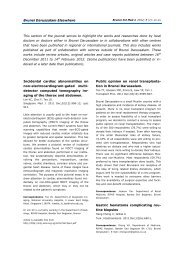
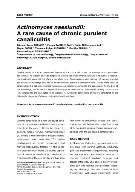
FRANCIS et al. Brunei Int Med J. 2013; 9 (2): 119Fig. 1: Chronic <strong>canaliculitis</strong> with pouting punctum.fibrillation, mitral regurgitation and sick sinussyndrome with an implanted permanent cardiacpacemaker. Examination showed a wateryleft eye with <strong>purulent</strong> discharge and mattedlashes. There was associated erythema andswelling <strong>of</strong> the inner end <strong>of</strong> the lower eyelidand medial angular congestion. The lowerpunctum was stenosed (Figure 1), and appearedred, swollen and pouting. The lowerpunctum and canaliculus was tender to gentledigital manipulation which expressed a thickwhite tenacious discharge containing solidpale-yellow sulphur granules (Figure 1). Thelower canaliculus was strikingly thickened.Probing <strong>of</strong> the left upper canaliculus revealeda ‘hard stop’ whilst that <strong>of</strong> the lower canaliculuswas gritty in nature with a ‘s<strong>of</strong>t stop’.Chronic dacryocystitis was also identified inthe same eye as evidenced by regurgitation <strong>of</strong>mucoid substance when the upper punctumwas irrigated with saline. revealed immaturethe lower canaliculus sample revealed Grampositive cocci in small groups and pairs withlarge numbers <strong>of</strong> gram positive branching filaments(Figure 2), while that from the uppercanaliculus showed chain forms <strong>of</strong> gram positivecocci. Culture <strong>of</strong> the lower canaliculussample showed pr<strong>of</strong>use growth <strong>of</strong> A. <strong>naeslundii</strong>along with Staphylococcus aureus whilstthat <strong>of</strong> the upper canaliculus showed a moderategrowth <strong>of</strong> Streptococci Lancefield groupF.Based on the antibiotic sensitivity, shewas started on oral doxycycline, 100mg twicedaily with topical cipr<strong>of</strong>loxacin drops fourtimes a day. However doxycycline had to bediscontinued two days later following the development<strong>of</strong> dyspepsia and was replaced withcataracts in both eyes and evidence <strong>of</strong> drynessin the right eye.A working diagnosis <strong>of</strong> left <strong>chronic</strong>dacryocystitis with ipsilateral lower <strong>purulent</strong><strong>canaliculitis</strong> was made. The expressed <strong>purulent</strong>materials from upper and lower canaliculiwere separately sent for bacteriological analysisand sensitivity testing. Gram-staining <strong>of</strong>Fig. 2: <strong>Actinomyces</strong> <strong>naeslundii</strong> under gram stain,branching filaments are indicated by white arrow(H&E stain Magnification x40).
FRANCIS et al. Brunei Int Med J. 2013; 9 (2): 120The post-operative period was uneventfuland she was discharged on the secondday. She was started on oral Cipr<strong>of</strong>loxacin500mg twice daily. for one month. The woundhealed well and the sutures were removedafter seven days. She was followed-up in theclinic for six months after surgery and she hasnot shown any recurrence or complications.Fig. 3: Canaliculus slit open showingsulphur granules.oral amoxicillin-clavulanic acid 625mg twicedaily for 10 days. Although some improvementwas noted, the infection did not completelysubside as evidenced by continuingdischarge <strong>of</strong> pus and granules. She was alsoadditionally treated with bi-weekly intracanalicularlavage with 5% povidone iodine for herlower canaliculus for three weeks.DISCUSSION<strong>Actinomyces</strong> are cast-forming Gram-positive,non-acid fast, non-spore bearing anaerobicrods, characterised by filamentous branchingat acute angles, and are difficult to isolate,identify and culture. Although these organismsare normal commensals <strong>of</strong> the mouth,gastrointestinal tract and the female genitaltract, they may <strong>cause</strong> infectious disease(Actinomycosis) <strong>of</strong> which oral and cervic<strong>of</strong>acialActinomycosis are the commonest. ThoughA single-snip lower canaliculotomyunder local anaesthesia with sedation waswhereby daily lavage with povidone iodinewas performed for three days pre-operatively.Intra-operatively, the stenosed punctum wasdilated gently and the canaliculus was irrigatedwith iodine solution. The lower punctumand canaliculus were then probed and slit,then opened with a single snip along its posterioraspect. Exploration revealed a sausagelikegross dilatation <strong>of</strong> the lower canaliculuswith the distal end blocked by greyish fibroustissue. The canalicular mucosa was oedematousand thickened. Immediately beyond thepunctum, there was a diverticulum inferiorly,which was studded with large sulphur granules(Figure 3). The granules were removed,the mucosa was curetted, and sent for histopathologicalstudies. Considering the highrisks involved, surgery was completed with adacryocystectomy on the same side.<strong>rare</strong>, pelvic Actinomycosis, is a known complication<strong>of</strong> intra-uterine contraceptive devices(IUCD). When it affects the abdominal cavity,Actinomycosis may present with a neoplasmlikemass. It is known to <strong>cause</strong> liver, splenic,colon and caecal abscesses.These organisms are also known to<strong>cause</strong> opportunistic infections within hollowspaces, like the canaliculus, producing indolentand slowly progressive infection with yellowsulphur granules. 2 The granules are colonies<strong>of</strong> intertwined branching <strong>Actinomyces</strong>filaments, solidified by tissue exudates andpus cells. <strong>Actinomyces</strong> <strong>canaliculitis</strong> accountsfor approximately 2% <strong>of</strong> all lacrimal drainagesystem diseases presenting with epiphora. 2, 5Majority <strong>of</strong> reported cases involvedfemales and the lower canaliculus.3, 6, 7Though complete cure has been reported with
FRANCIS et al. Brunei Int Med J. 2013; 9 (2): 121conservative management, our case neededadditional surgical intervention namely, punctoplastyand snip canaliculotomy with evacuation<strong>of</strong> the sulphur granules and curettage.The organisms, although usually sparse in thepus, are concentrated in the sulphur granulesalmost always form part <strong>of</strong> a mixed infection.<strong>Actinomyces</strong> <strong>naeslundii</strong> have similarmorphological features as other <strong>Actinomyces</strong>strains. PubMed search <strong>of</strong> the literatureshowed no previously reported cases <strong>of</strong> A.<strong>naeslundii</strong>-related <strong>chronic</strong> <strong>canaliculitis</strong>. Hence,we believe this the first report <strong>of</strong> such an infection.Various treatment options have beenreported with some advocating that completecure can be achieved with conservative line <strong>of</strong>treatment alone but others are <strong>of</strong> the opinionsthat surgical treatment namely punctoplasty,canaliculotomy and expression <strong>of</strong> granuleswith or without curettage is the line <strong>of</strong> treatment.1, 8 Hussain et al. reported that medicaltherapy alone could not eradicate the infection.9 Surgical procedures such as punctoplastyand canaliculotomy are safe procedure.1, 10, 11Our patient had <strong>chronic</strong> <strong>canaliculitis</strong>with stenosed punctum as well as <strong>chronic</strong>dacryocystitis on the same side, requiringboth medical and surgical management.In conclusion, primary <strong>chronic</strong> <strong>purulent</strong><strong>canaliculitis</strong> is a <strong>rare</strong> form <strong>of</strong> unilaterallacrimal system pathology. However, it shouldbe considered in the differential diagnosis <strong>of</strong>epiphora and <strong>chronic</strong> or recurrent conjunctivitis.Our case highlights a <strong>rare</strong> <strong>cause</strong> <strong>of</strong> <strong>chronic</strong><strong>purulent</strong> <strong>canaliculitis</strong>, which can also be difficultto isolate and identify. A complete eradi-cation <strong>of</strong> the canalicular <strong>Actinomyces</strong> infectioncannot be achieved unless and until all theconcretions are removed.REFERENCES1: McKellar MJ, Aburn NS. Cast-forming actinomycesisraeli <strong>canaliculitis</strong>. Aust N Z J Ophthalmol.1997; 25:301-3.2: Pande M, Mathew R, Ramprakash M, Kalyani M.Actinomycotic lacrimal <strong>canaliculitis</strong>. Indian J PatholMicrobiol. 2010; 53:864-5.3: Sathananthan N, Sullivan TJ, Rose GE, MoseleyIF. Intubation dacryocystography in patients with aclinical diagnosis <strong>of</strong> <strong>chronic</strong> <strong>canaliculitis</strong>(“Streptothrix”). Br J Radiol. 1993; 66:389-93.4: Bharathi MJ, Ramakrishnan R, Meenakshi R,Vasu S. Nocardia asteroides <strong>canaliculitis</strong>: a casereport <strong>of</strong> uncommon aetiology. Indian J Ophthalmol.2004; 22:123-5.5: Vagarali MA, Karadesai SG, Dandur MS. Lacrimal<strong>canaliculitis</strong> due to actinomyces – a <strong>rare</strong> entity.Indian J Pathol. 2011; 54:661-3.6: Lee MJ, Choung HK, Kim NJ, Khwarg SI. OneSnip Punctoplasty and Canalicular curettagethrough the punctum. Ophthalmology. 2009;116:2027-307: Lin SC, Kao SC, Tsai CC, et al. Clinical characteristicsand Factors associated the outcome <strong>of</strong> lacrimal<strong>canaliculitis</strong>. Acta Ophthalmologica. 2011;89:759–63.8: Al-Mujaini A, Wali U, Al-Senawi R. Canaliculitis:Are we missing the diagnosis? Oman J Ophthalmol.2009; 2:145-6.9: Hussain I, Bonshek RE, Loudon K, Armstrong M,Tullo AB. Canalicular infection by actinomyces. Eye(Lond). 1993; 7:542-4.10: Briscoe D, Edelstein E, Zacharopoulos I, KenessY, Kilman A, Assia EL. <strong>Actinomyces</strong> <strong>canaliculitis</strong>:diagnosis <strong>of</strong> a masquerading disease. Graefes ArchClin Exp ophthalmol. 2004; 242: 682-6.11: Anand S, Hollingworth K, Kumar V, SandramouliS. Canaliculitis: the incidence <strong>of</strong> long-termepiphora following canaliculotomy. Orbit. 2004; 23:19-26.