General Anatomy of Skeletal Muscle
General Anatomy of Skeletal Muscle
General Anatomy of Skeletal Muscle
- No tags were found...

Create successful ePaper yourself
Turn your PDF publications into a flip-book with our unique Google optimized e-Paper software.
<strong>General</strong> anatomy <strong>of</strong> skeletal muscle,its innervation and blood supply,<strong>General</strong> anatomy <strong>of</strong> spinal nerve,<strong>General</strong> terms <strong>of</strong> angiology and lymphologyMiloš GrimInstitute <strong>of</strong> <strong>Anatomy</strong>, First Faculty <strong>of</strong> Medicine,Charles University in Praguewinter semester 2013/2014
How to study skeletal muscles:origo, insertion, position (scheme,tables), identification, muscle groups,innervation, function,oste<strong>of</strong>ascial spaces (compartments),transverse sections <strong>of</strong> limb segments,dissection
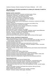
eneral <strong>Anatomy</strong> <strong>of</strong> <strong>Skeletal</strong> <strong>Muscle</strong>rigin and development <strong>of</strong> muscles, molecular mechanismseneral features <strong>of</strong> striated muscle, its auxiliary structuresotor end plate, motor unit,muscle spindle, Golgi tendon organ),otor and proprioceptive innervationttachments <strong>of</strong> skeletal muscles – origin, insertionuscle fibres, my<strong>of</strong>ibrils, sarcomeresaming <strong>of</strong> muscleshape and fibre architecture, pennationhe endomysial and perimysial sheathshe interface between muscle and connective tissuey<strong>of</strong>ibrilar proteinsliding filament mechanism <strong>of</strong> contractionendons, Aponeurosesynovial sheaths and bursaeascia, intermuscular septa, oste<strong>of</strong>ibrous spaces
Attachments <strong>of</strong> skeletal muscles – origin, insertion,endomysial and perimysial sheaths, fascia
http://fig.cox.miami.edu/~cmallery/150/neuro/muscle.htmstriated muscle fibres
muscle fibre,my<strong>of</strong>ibril,sarcomeresarcoplasmicreticulum,T-tubules,triadsmitochondria,sarcolemma,basal lamina
Posture, isometric contraction
Motion, isotonic contraction
myo-tendinous junction
synovial sheath, synovial bursa
visualization <strong>of</strong> motor end plates
pennation <strong>of</strong> musclesarrangement <strong>of</strong> parallelrunning muscle fibres
Naming <strong>of</strong> <strong>Muscle</strong>sShape:deltoid (= triangular), quadratus (= square), rhomboid(= diamond-shaped)teres (= round), gracilis (= slender), rectus (= straight),lumbrical (= worm-like)Size : major, minor, longus (= long), brevis (= short),latissimus (= broadest), longissimus (= longest)Number <strong>of</strong> Heads or Bellies:biceps (= 2 heads), triceps (= 3 heads), quadriceps (= 4 heads)digastric (= 2 bellies), biventer (= 2 bellies),Position:anterior, posterior, interosseus (= between bones)supraspinatus (= above spine <strong>of</strong> scapula),infraspinatus (= below spine),dorsi (= <strong>of</strong> the back), abdominis (= <strong>of</strong> the abdomen)pectoralis (= <strong>of</strong> the chest), brachii (= <strong>of</strong> the arm)femoris (= <strong>of</strong> the thigh), oris (= <strong>of</strong> the mouth)
Naming <strong>of</strong> <strong>Muscle</strong>sDepth:superficialis (= superficial), pr<strong>of</strong>undus (= deep),externus (or externi), internus (or interni)Attachment:sternocleidomastoid(from sternum and clavicle to mastoid process)coracobrachialis (from the coracoid process to the arm)Action:extensor, flexor, abductor, adductor,levator (= lifter), depressor,supinator, pronator, constrictor, dilator
Fascia, intermuscular septum, oste<strong>of</strong>ibrous spaces
Innervation <strong>of</strong> skeletal muscleNeurovascular hilumBlood supplyMotor innervationmotoneurons: slow and fast alfa motoneurons,gamma motoneurons, motor end-plate, AChmotor unit, zone <strong>of</strong> motor end-plates,polyneural innervation, segmental innervationSensory (proprioceptive) innervationmuscle spindle, Golgi tendon organ,proprioceptive reflexes
tendons, aponeuroses, neuro-vascular hilum (motor point)
Innervation <strong>of</strong> skeletal muscle: motoneurons, motor units,motor end- plates, acetylcholine, proprioceptive neurons,muscle spindles, Golgi tendon organs
visualization <strong>of</strong> motor end plates and axons,acetylcholinesterase in subneural apparatus
synaptic vesicles containing acetylcholine(neurotransmitter) in axon terminal <strong>of</strong> motor end-plate;curare blocks the transmission
<strong>Muscle</strong> spindleGolgi tendon organElektromyography (EMG)
A young women with sensory neuropathy <strong>of</strong> unknownorigin who completely lost proprioceptive sensation:She could not stand without watching her feet,she could not held anything in her hands, and theywandered around without her awareness…„Something awful´s happened, I can´t feel my body.I feel weird-disembodied“, she said, and „I may losemy arms. I think they´re one place and I find they´reanother“.After having proprioception explained, she said:„This proprioception is like the eyes <strong>of</strong> the body,the way the body sees itself. And if it goes, as it´s gonewith me, it´s like the body is blind…so I haveto watch it - be its eyes. Right?“
Fibre Types <strong>of</strong> <strong>Skeletal</strong> <strong>Muscle</strong>• Type 1 fibres are slow-contracting and fatigue-resistant• Type 2A fibres are fast-contracting and fatigue-resistant• Type 2X (B) fibres are fast-contracting and susceptibleto fatigueType I SO Type IIa FOG Type IIx FGmyosin ATPase, dehydrogenase, glycogen phosphorylase
FG IIx FOG IIa SO I
FG IIx FOG IIa SO ICapillaries <strong>of</strong> skeletal muscle
Development and Differentiation<strong>of</strong> <strong>Skeletal</strong> <strong>Muscle</strong>Myogenesis,Myogenic Determination FactorsMyf-5, myogenin, MyoD and Myf-6 (herculin)MyostatinGrowth <strong>of</strong> <strong>Skeletal</strong> <strong>Muscle</strong>Hypertrophy, not hyperplasiaAnabolic SteroidsRegeneration <strong>of</strong> <strong>Skeletal</strong> <strong>Muscle</strong>Satellite cells
MyogenesisMyoblastMyotube<strong>Muscle</strong> fibreSatellite cellMyogenicDeterminationFactorsMyostatin
Mutation <strong>of</strong> myostatine gene resulting in overproduction <strong>of</strong> myogenic cells
Development <strong>of</strong> skeletal muscles
ParaxialmesodermNonsegmentedin the head,somites in thetrunkMyogenic cellsGrowth andmigration<strong>of</strong> myogeniccells fromsomitesinto the bodywalland limbprimordia
MyoDHH 25
Epaxial and hypaxial musculature and its innervationfrom dorsal and ventral branches <strong>of</strong> spinal nerves
Epaxial and hypaxial musculature and its innervationfrom dorsal and ventral branches <strong>of</strong> spinal nerves
C DK HH 22
Colonization<strong>of</strong> the limb bud(myogenic cells,Schwann cells,angioblasts,ingrowingAxons)
Extra-ocular muscles(Innervation III.,IV. VI.)<strong>Muscle</strong>s <strong>of</strong> branchial(pharyngeal) arches<strong>Muscle</strong>s <strong>of</strong> auditory ossicles(BA 1,2.- V., VII.)Masticatory muscles(BA 1 -V.)Facial muscles (BA 2 -VII.)Musculi palati mollis et atfaucium (IX., X.)<strong>Muscle</strong>s from occipital somitesMusculi linguae (XII.),Musculi laryngis (X.)M. trapezius,M. sternocleidomastoideus(XI.)
Regeneration <strong>of</strong> skeletal muscle
Activation <strong>of</strong> satellite scells in muscle regeneration
Fate <strong>of</strong> the free muscle graft in the rat60. day Intact muscle30. day1.day7. day3. day5. day
<strong>General</strong> anatomy <strong>of</strong> peripheral nervous system (PNS)Cranial nerves Spinal nerves Autonomic nervous systém-sympathetic and parasympathetis partNeural crest cells and their differentiationVentral and dorsal spinal nerve root, dorsal root ganglion, drawgeneral structure <strong>of</strong> the spinal nerve and its branches, autonomicfibers <strong>of</strong> spinal nerveSegmental innervation, radicular areas, dermatomes, Head´szones (zones <strong>of</strong> reffered visceralpain), sensory receptors, peripheral nerve regenerationneuron, neuroglia (Schwann cells) nerve fibres,endoneurium, perineurium, epineurium, synapse, ganglion
Development <strong>of</strong> nervous system - neurulationB.M. Carlson (1999)Neural tube withbrain vesiclesNeural crestNeural placodes
Derivatives <strong>of</strong> neural crest cellsHNK-1Neurons <strong>of</strong> spinal ganglia, <strong>of</strong> autonomic ganglia, enteric neurons,Schwann cells, pigment cells, cells <strong>of</strong> adrenal medulla
Migration <strong>of</strong> NC cells <strong>of</strong> the headED4 chickN. LeDouarin (1999)Ectomesenchyme:osteoblasts, fibroblasts,chondroblasts, smoothmuscle cells, odontoblastsCardiac NC (R4-R8):for cardiac coutflow tract
Wnt1-cre / R26RXgalBgal
Spinal nervetrunk <strong>of</strong> spinal nerve - mixed nerve, rootlets,anterior root - motor root,posterior root - sensory root, spinal ganglion
Marking <strong>of</strong> motoneurons using retrograde transportation <strong>of</strong>horseradish peroxidase (HRP) injected into the muscleQ PL inj. HRPC/Q ED 10C PL inj. HRP
Localization <strong>of</strong> motoneurons for individual muscle groupson transverse section <strong>of</strong> cervical spinal cord
spinal nerve plexuses,autonomic plexuses,perivascular plexuses
Segmental innervation - areae aradiculares (dermatomes)Kandel et al:Principles <strong>of</strong> neuralscience,2000 McGraw-Hill
Regeneration <strong>of</strong> interrupted nerve fibre
peripheral nerve, endoneurium, perineurium,epineuriumNerve graft bridging the partial defect,suture <strong>of</strong> perineurium
Terms<strong>of</strong> general angiologyVascular development, structure<strong>of</strong> arteries, veins, lymphaticvessel, collateral circulationblood vessels,arteriesies, , veins, , capillariesies,arteriovenous anastomosis,collateral vessels,venous plexuseses,hepatic portal vein
Venous valves
Fetal circulation before birthCirculation after birth
Terms <strong>of</strong> general anatomy <strong>of</strong> lymphatic systemTerms <strong>of</strong> general anatomy <strong>of</strong> lymphatic systemLymph, lymph node, lymph tissue in the organs, mainlymphatic trunks, lymphatic vessels and ducts
Literature:Platzer: Color Atlas and Textbook <strong>of</strong> Human <strong>Anatomy</strong> –Vol.1 Locomotor System, Thieme 2008Kahle, Frotscher: Color Atlas and Textbook <strong>of</strong> Human <strong>Anatomy</strong>– Vol. 3 Nervous System and Sensory Organs, Thieme 2010Stingl, Grim, Druga: Regional <strong>Anatomy</strong>, 2012Netter: Atlas <strong>of</strong> Human <strong>Anatomy</strong>, Icon 2003Sobotta: Atlas <strong>of</strong> Human <strong>Anatomy</strong> Vol.1+2, Williams and Wilkins2000Sadler: Langman´s Medical Embryology, 11th Edit. 2009Carlson: Human embryology and developmental biologyMescher: Junqueira´s Basic Histology 12th Edit., 2010Different original publications in scientific journalsAuthor´s own figs. and illustrations
Sources <strong>of</strong> used illustrations:Gray´s <strong>Anatomy</strong>,Sobotta: Atlas der Anatomie des MenschenStingl, Grim, Druga: Regional <strong>Anatomy</strong>, 2012Benningh<strong>of</strong>f, Drenckhahn: Anatomie I., II.Sadler: Langman´s Medical Embryology, 11th Edit. 2009Carlson: Human embryology and developmental biologyČihák: Anatomie 1Different original publications in scientific journalsAuthor´s own figs. and illustrations