AtREV1, a Y-Family DNA Polymerase in ... - Plant Physiology
AtREV1, a Y-Family DNA Polymerase in ... - Plant Physiology
AtREV1, a Y-Family DNA Polymerase in ... - Plant Physiology

Create successful ePaper yourself
Turn your PDF publications into a flip-book with our unique Google optimized e-Paper software.
<strong>AtREV1</strong>, a Y-<strong>Family</strong> <strong>DNA</strong> <strong>Polymerase</strong> <strong>in</strong> Arabidopsis, Has<br />
Deoxynucleotidyl Transferase Activity <strong>in</strong> Vitro 1[W]<br />
Sh<strong>in</strong>ya Takahashi 2,3 , Ayako N. Sakamoto 2 *, Atsushi Tanaka, and Kikuo Shimizu<br />
Radiation-Applied Biology Division, Japan Atomic Energy Agency, Takasaki, Gunma 370–1292, Japan<br />
(S.T., A.N.S., A.T.); and Radioisotope Research Center, Osaka University, Suita, Osaka 565–0871, Japan (K.S.)<br />
To clarify the functions of the Arabidopsis thaliana REV1 (<strong>AtREV1</strong>) prote<strong>in</strong>, we expressed it <strong>in</strong> Escherichia coli and purified it to<br />
near homogeneity. The deoxynucleotidyl transferase activity of the recomb<strong>in</strong>ant <strong>AtREV1</strong> was exam<strong>in</strong>ed <strong>in</strong> vitro us<strong>in</strong>g a primer<br />
extension assay. The recomb<strong>in</strong>ant <strong>AtREV1</strong> transferred one or two nucleotides to the primer end. It efficiently <strong>in</strong>serted dCMP<br />
regardless of the opposite base. <strong>AtREV1</strong> also <strong>in</strong>serted a dCMP opposite an apur<strong>in</strong>ic/apyrimid<strong>in</strong>ic site, which is physiologically<br />
generated or <strong>in</strong>duced by various <strong>DNA</strong>-damag<strong>in</strong>g agents. In contrast, <strong>AtREV1</strong> had no <strong>in</strong>sertion activities aga<strong>in</strong>st UV-<strong>in</strong>ducible<br />
<strong>DNA</strong> lesions as reported <strong>in</strong> yeast or mammalian system. Although the substrate specificity of <strong>AtREV1</strong> was rather narrow <strong>in</strong> the<br />
presence of magnesium ion, it widened <strong>in</strong> the presence of manganese ion. These results suggest that <strong>AtREV1</strong> serves as a<br />
deoxycytidyl transferase <strong>in</strong> plant cells.<br />
<strong>Plant</strong>s are cont<strong>in</strong>uously exposed to harmful UV-B<br />
(290–320 nm) as well as the photosynthetic light (400–<br />
700 nm). Therefore, an <strong>in</strong>crease of UV-B is predicted to<br />
<strong>in</strong>hibit plant growth <strong>in</strong> a large area, caus<strong>in</strong>g the disruption<br />
of ecosystems. UV-B radiation <strong>in</strong>duces various<br />
lesions on plant <strong>DNA</strong>. The vast majority of UV-B<strong>in</strong>duced<br />
damage is cyclobutane pyrimid<strong>in</strong>e dimer<br />
(CPD), which corresponds to 75% to 78% of total<br />
damage (Mitchell, 1995; Cadet et al., 2005). The 6-4<br />
photoproducts (6-4PP) and Dewar isomer, a derivative<br />
of 6-4PP, correspond to most of the rema<strong>in</strong>der (20%–<br />
24%; Mitchell, 1995; Cadet et al., 2005). Other types of<br />
<strong>DNA</strong> damage, <strong>in</strong>clud<strong>in</strong>g 8-oxoG, pyrimid<strong>in</strong>e hydrate,<br />
thym<strong>in</strong>e glycol, <strong>DNA</strong> s<strong>in</strong>gle-strand break, etc., are<br />
generated by UV-B radiation at very low yield<br />
(,0.1%–2%; Mitchell, 1995; Gao and Murphy, 2001;<br />
Song et al., 2002; Cadet et al., 2005; Friedberg et al.,<br />
2006). On the other hand, apur<strong>in</strong>e/apyrimid<strong>in</strong>e (AP)<br />
sites are among the most abundant <strong>DNA</strong> lesions generated<br />
spontaneously (Boiteux and Guillet, 2004). For<br />
example, approximately 10,000 AP sites arise spontaneously<br />
<strong>in</strong> a mammalian cell per day (Nakamura et al.,<br />
1 This work was supported <strong>in</strong> part by the M<strong>in</strong>istry of Education,<br />
Science, Sports and Culture of Japan (grants-<strong>in</strong>-aid 15201010 and<br />
19570049 for scientific research).<br />
2 These authors contributed equally to the article.<br />
3 Present address: <strong>Plant</strong> Functional Genomics Research Team,<br />
<strong>Plant</strong> Functional Genomics Research Group, <strong>Plant</strong> Sciences Center,<br />
RIKEN Yokohama Institute, Suehiro-cho 1–7–22, Tsurumi, Yokohama,<br />
Kanagawa 230–0045, Japan.<br />
* Correspond<strong>in</strong>g author; e-mail sakamoto.ayako@jaea.go.jp.<br />
The author responsible for distribution of materials <strong>in</strong>tegral to<br />
the f<strong>in</strong>d<strong>in</strong>gs presented <strong>in</strong> this article <strong>in</strong> accordance with the policy<br />
described <strong>in</strong> the Instructions for Authors (www.plantphysiol.org) is:<br />
Ayako N. Sakamoto (sakamoto.ayako@jaea.go.jp).<br />
[W] The onl<strong>in</strong>e version of this article conta<strong>in</strong>s Web-only data.<br />
www.plantphysiol.org/cgi/doi/10.1104/pp.107.101980<br />
1998). The AP site also is generated when exposed to<br />
the various <strong>DNA</strong>-damag<strong>in</strong>g agents, such as ioniz<strong>in</strong>g<br />
radiation and alkylat<strong>in</strong>g chemicals, through the reaction<br />
of <strong>DNA</strong> N-glycosylases that remove the damaged<br />
base (Friedberg et al., 2006). The AP sites are rarely<br />
formed after UV irradiation directly (Song et al., 2002)<br />
or as a consequence of a cellular process remov<strong>in</strong>g the<br />
damaged base (Friedberg et al., 2006). Such lesions can<br />
lead to <strong>in</strong>corporation of the wrong base or can <strong>in</strong>hibit<br />
<strong>DNA</strong> replication and transcription, result<strong>in</strong>g <strong>in</strong> mutations<br />
and cell death (Britt, 1999). To prevent the effects<br />
of UV and <strong>in</strong>duced <strong>DNA</strong> lesions, plants have many<br />
<strong>DNA</strong> repair mechanisms, such as photorepair by<br />
the CPD and 6-4 photolyases (Landry et al., 1997;<br />
Nakajima et al., 1998). In addition, they have nucleotide<br />
excision repair (Gallego et al., 2000; Liu et al., 2001,<br />
2003), base excision repair (García-Ortiz et al., 2001),<br />
and recomb<strong>in</strong>ation repair (Osakabe et al., 2006).<br />
Translesion synthesis (TLS) is one of the damagetolerant<br />
mechanisms prevalent <strong>in</strong> both prokaryotes<br />
and eukaryotes (Prakash et al., 2005). In TLS, a <strong>DNA</strong><br />
lesion is bypassed by the action of specialized <strong>DNA</strong><br />
polymerases (Broomfield et al., 2001; Friedberg et al.,<br />
2005). <strong>DNA</strong> synthesis dur<strong>in</strong>g TLS often results <strong>in</strong><br />
mutations because of the <strong>in</strong>tr<strong>in</strong>sic nature of TLS-type<br />
polymerases (Friedberg et al., 2000). The TLS-type<br />
polymerases <strong>in</strong>clude <strong>DNA</strong> polymerases (Pols) z, i, h,<br />
REV1, and other specialized polymerases (Prakash<br />
et al., 2005). The REV1 gene was first identified <strong>in</strong> yeast<br />
through a screen<strong>in</strong>g for reversionless mutants (Lemontt,<br />
1971). Subsequent analyses showed that REV1 is a<br />
Y-family <strong>DNA</strong> polymerase. In vitro analyses showed<br />
that yeast Rev1 <strong>in</strong>serts a C opposite the AP site (Nelson<br />
et al., 1996a), and then the mismatched end is efficiently<br />
extended by Pol z (Nelson et al., 1996b; L<strong>in</strong><br />
et al., 1999). Thus, Rev1 and <strong>DNA</strong> Pol z are thought to<br />
be major components of the error-prone TLS (Nelson<br />
et al., 1996a, 1996b).<br />
1052 <strong>Plant</strong> <strong>Physiology</strong>, November 2007, Vol. 145, pp. 1052–1060, www.plantphysiol.org Ó 2007 American Society of <strong>Plant</strong> Biologists
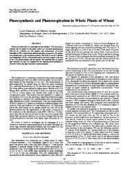
Figure 1. Structure of the recomb<strong>in</strong>ant <strong>AtREV1</strong> expression plasmid. A,<br />
<strong>AtREV1</strong> c<strong>DNA</strong> was modified to <strong>in</strong>clude restriction sites on both sides<br />
and <strong>in</strong>serted <strong>in</strong>to the pET32a(1) vector. The pET32a(1) vector <strong>in</strong>cludes<br />
a T7lac promoter (T7lac), an ampicill<strong>in</strong>-resistant gene (Ap), a lacI<br />
cod<strong>in</strong>g sequence (lacI), and pBR322 orig<strong>in</strong> (ori). B, Schematic representation<br />
of the recomb<strong>in</strong>ant <strong>AtREV1</strong> prote<strong>in</strong>. The recomb<strong>in</strong>ant prote<strong>in</strong><br />
conta<strong>in</strong>s a thioredox<strong>in</strong> tag (Trx), a His tag (His), and an S prote<strong>in</strong>b<strong>in</strong>d<strong>in</strong>g<br />
tag (S-tag) at the N term<strong>in</strong>us, and another His tag at the C<br />
term<strong>in</strong>us. The bottom <strong>DNA</strong> and am<strong>in</strong>o acid sequences represent the<br />
restriction sites (NcoI and NotI) <strong>in</strong>duced for subclon<strong>in</strong>g. The K (Lys) of<br />
the native <strong>AtREV1</strong> am<strong>in</strong>o acid sequence was changed to A (Ala; <strong>in</strong><br />
boldface) <strong>in</strong> the recomb<strong>in</strong>ant <strong>AtREV1</strong>.<br />
Little is known about the prote<strong>in</strong>s that are <strong>in</strong>volved<br />
<strong>in</strong> TLS <strong>in</strong> plants. We recently described an Arabidopsis<br />
(Arabidopsis thaliana) homolog of Pol z, whichis<br />
a heterodimer of family-B <strong>DNA</strong> polymerase REV3<br />
(Sakamoto et al., 2003) and the regulatory subunit REV7<br />
(Takahashi et al., 2005). The Arabidopsis genome has<br />
three Y-family polymerases, which are most similar to<br />
REV1, D<strong>in</strong>B1, and Rad30, respectively (Ohmori et al.,<br />
2001). The homolog of D<strong>in</strong>B1 (AtPolK) was recently<br />
characterized and its polymerase activity was analyzed<br />
<strong>in</strong> vitro (García-Ortiz et al., 2004). The homolog<br />
of Rad30 (AtPolH) was shown to suppress the UV<br />
sensitivity of the Saccharomyces cerevisiae rad30 stra<strong>in</strong><br />
(Santiago et al., 2006). However, it is still unknown<br />
whether these Y-family polymerases perform TLS and<br />
whether they are <strong>in</strong>volved <strong>in</strong> damage tolerance <strong>in</strong><br />
plants. We recently showed that a mutant with disrupted<br />
<strong>AtREV1</strong> had <strong>in</strong>creased sensitivity to UV-B and<br />
other <strong>DNA</strong>-damag<strong>in</strong>g agents (Takahashi et al., 2005),<br />
suggest<strong>in</strong>g that <strong>AtREV1</strong> has a role <strong>in</strong> the damage tolerance<br />
<strong>in</strong> Arabidopsis.<br />
Here, we show that recomb<strong>in</strong>ant <strong>AtREV1</strong> has deoxynucleotidyl<br />
transferase activity. The recomb<strong>in</strong>ant <strong>AtREV1</strong><br />
clearly <strong>in</strong>serted a nucleotide opposite AP sites <strong>in</strong> vitro.<br />
Our results suggest that <strong>AtREV1</strong> has a role <strong>in</strong> provid<strong>in</strong>g<br />
tolerance to <strong>DNA</strong> damage through the TLS pathway.<br />
RESULTS<br />
Deoxynucleotidyl Transferase Activity of <strong>AtREV1</strong><br />
Overproduction and Purification of Recomb<strong>in</strong>ant<br />
<strong>AtREV1</strong> Prote<strong>in</strong><br />
To prepare soluble and active <strong>AtREV1</strong> prote<strong>in</strong>, the<br />
<strong>AtREV1</strong> c<strong>DNA</strong> was <strong>in</strong>serted <strong>in</strong>to the pET32a vector<br />
that carries a thioredox<strong>in</strong>-tag fusion prote<strong>in</strong>, which<br />
<strong>in</strong>creases the solubility of the expressed prote<strong>in</strong> (Fig.<br />
1). Under optimized growth conditions of Escherichia<br />
coli cells (see ‘‘Materials and Methods’’), the recomb<strong>in</strong>ant<br />
<strong>AtREV1</strong> prote<strong>in</strong> was successfully expressed on a<br />
large scale. SDS-PAGE analysis showed the presence<br />
of a thick band that migrated between the 116-kD and<br />
194-kD size markers after <strong>in</strong>duction with isopropyl<br />
b-D-1-thiogalactopyranoside (IPTG; Fig. 2, lane 3).<br />
S<strong>in</strong>ce the size of the band (approximately 140 kD) was<br />
consistent with the predicted molecular mass of the recomb<strong>in</strong>ant<br />
<strong>AtREV1</strong> fusion prote<strong>in</strong> and s<strong>in</strong>ce the band<br />
cross-reacted with the anti-His tag antibody (data not<br />
shown), we concluded that the band corresponded to<br />
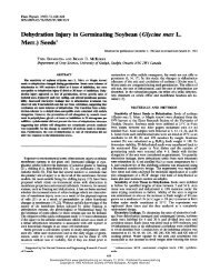
Figure 2. Purification of recomb<strong>in</strong>ant <strong>AtREV1</strong> prote<strong>in</strong>s. Overproduction<br />
and purification of recomb<strong>in</strong>ant <strong>AtREV1</strong> prote<strong>in</strong> <strong>in</strong> E. coli. Fractions <strong>in</strong><br />
each purification step were separated on a 7.5% SDS-polyacrylamide<br />
gel, then sta<strong>in</strong>ed with Coomassie Brilliant Blue. Lane 1, M r marker<br />
(Invitrogen); lanes 2 and 3, total E. coli prote<strong>in</strong>s with (lane 3) or without<br />
(lane 2) IPTG <strong>in</strong>duction; lane 4, clear lysate; lane 5, fraction eluted from<br />
Ni-aff<strong>in</strong>ity column; lane 6, fraction from a hepar<strong>in</strong> aff<strong>in</strong>ity column; lane<br />
7, purified fraction from HiTrap Q HP column. Molecular weight is<br />
shown on the left.<br />
<strong>Plant</strong> Physiol. Vol. 145, 2007 1053
Takahashi et al.<br />
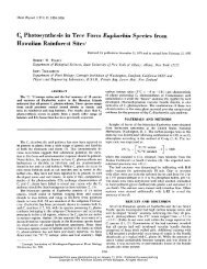
Figure 3. Deoxynucleotidyl transferase activity of the recomb<strong>in</strong>ant<br />
<strong>AtREV1</strong> prote<strong>in</strong>. The primer P13 was 32 P-labeled at the 5# end and<br />
annealed with each of the templates, prepar<strong>in</strong>g four template/primer<br />
pairs: 30G (B and F), 30A (C and G), 30T (D and H), and 30C (E and I).<br />
The nucleotide sequences around the primer term<strong>in</strong>us are shown <strong>in</strong> A.<br />
The underl<strong>in</strong>ed bases represent the variation of templates. One hundred<br />
and sixty nanograms of <strong>AtREV1</strong> recomb<strong>in</strong>ant prote<strong>in</strong> and the<br />
<strong>in</strong>dicated template/primer were <strong>in</strong>cubated with 0.1 mM of a s<strong>in</strong>gle<br />
dNTP (G, A, T, or C), 0.1 mM each of all four dNTPs (N), or no dNTP (2)<br />
under standard reaction conditions with 2 mM magnesium chloride<br />
(B–E) or 1 mM manganese chloride (F–I) at 30°C for 10 m<strong>in</strong>. The reaction<br />
products were resolved <strong>in</strong> 20% polyacrylamide gels conta<strong>in</strong><strong>in</strong>g<br />
8 M urea and visualized by autoradiography.<br />
<strong>AtREV1</strong>. After sonicat<strong>in</strong>g the E. coli cells, the recomb<strong>in</strong>ant<br />
<strong>AtREV1</strong> as well as some degradation products<br />
were detected <strong>in</strong> the soluble fraction (clear lysate; Fig.<br />
2, lane 4). The recomb<strong>in</strong>ant <strong>AtREV1</strong> prote<strong>in</strong>s were then<br />
fractionated by their aff<strong>in</strong>ity for nickel (Ni)-chelated<br />
Sepharose and successfully concentrated to a major<br />
band (Fig. 2, lane 5). To exclude the other rema<strong>in</strong><strong>in</strong>g<br />
prote<strong>in</strong>s derived from E. coli, the fraction was purified<br />
by hepar<strong>in</strong> column and anion-ion-exchange column<br />
chromatography (Fig. 2, lanes 6 and 7). The activity of<br />
<strong>AtREV1</strong> was monitored <strong>in</strong> each purification step. The<br />
fractions eluted from the hepar<strong>in</strong> column and the<br />
anion-exchange column-purified fraction were exam<strong>in</strong>ed<br />
by a primer extension assay us<strong>in</strong>g a primer<br />
template conta<strong>in</strong><strong>in</strong>g G and dCTP (template 30G; see<br />
‘‘Materials and Methods’’). As a result, the dCMP <strong>in</strong>sertion<br />
activity was detected <strong>in</strong> both fractions (data not<br />
shown). Thus, the anion-exchange column-purified<br />
recomb<strong>in</strong>ant <strong>AtREV1</strong> prote<strong>in</strong>, represented as a s<strong>in</strong>gle<br />
band (Fig. 2, lanes 6 and 7), was used to assay deoxynucleotidyl<br />
transferase activity.<br />
In Vitro Assay of Deoxynucleotidyl Transferase Activity<br />
of Recomb<strong>in</strong>ant <strong>AtREV1</strong><br />
To exam<strong>in</strong>e the substrate specificity of the recomb<strong>in</strong>ant<br />
<strong>AtREV1</strong> prote<strong>in</strong>, we performed a primer extension<br />
assay us<strong>in</strong>g four different primer templates (Fig.<br />
3A) <strong>in</strong> the presence of dGTP, dATP, dTTP, or dCTP<br />
<strong>in</strong>dividually, or all four dNTPs together. At first, the<br />
activity was exam<strong>in</strong>ed <strong>in</strong> the presence of 2 mM magnesium<br />
ion, based on previous observations (Masuda<br />
and Kamiya, 2002). The recomb<strong>in</strong>ant <strong>AtREV1</strong> prote<strong>in</strong><br />
efficiently <strong>in</strong>serted a dCMP regardless of the opposite<br />
base (Fig. 3, B–E). The prote<strong>in</strong> sometimes <strong>in</strong>serted<br />
additional dCMPs, for example, when the first template<br />
base was G, A, or T (Fig. 3, B–D). <strong>AtREV1</strong> also<br />
<strong>in</strong>serted dTMP or dGMP when the opposite base<br />
was G (Fig. 3B). Although the efficiency was low,<br />
we detected dGMP <strong>in</strong>sertions opposite G, T, and C,<br />
dAMP <strong>in</strong>sertions opposite G, A, and T, and dTMP<br />
<strong>in</strong>sertion opposite A (Fig. 3, B–E). These results <strong>in</strong>dicate<br />
that the recomb<strong>in</strong>ant <strong>AtREV1</strong> has an ability to<br />
transfer a deoxynucleotide at the end of the primer.<br />
Compar<strong>in</strong>g the nucleotide transferase activities to the<br />
four templates, we found that the recomb<strong>in</strong>ant <strong>AtREV1</strong><br />
seems to prefer template G to other templates. This<br />
is consistent with the character of yeast and human<br />
REV1s (Haracska et al., 2002; Zhang et al., 2002). Although<br />
the recomb<strong>in</strong>ant <strong>AtREV1</strong> <strong>in</strong>corporated two to<br />
four k<strong>in</strong>ds of nucleotides depend<strong>in</strong>g on the template<br />
base, a robust dCMP <strong>in</strong>corporation activity was detected<br />
at all four templates we exam<strong>in</strong>ed. Thus, we<br />
concluded that the recomb<strong>in</strong>ant <strong>AtREV1</strong> has deoxycytidyl<br />
transferase activity.<br />
Effects of Divalent Cations on Activity of<br />
Recomb<strong>in</strong>ant <strong>AtREV1</strong><br />
To exam<strong>in</strong>e the effects of divalent cations on the<br />
transferase activity of <strong>AtREV1</strong>, the assay was performed<br />
<strong>in</strong> the presence of 1 mM manganese chloride<br />
(Fig. 3, F–I). The substrate specificity of the recomb<strong>in</strong>ant<br />
<strong>AtREV1</strong> was weaker <strong>in</strong> the manganese buffer<br />
than <strong>in</strong> the magnesium buffer. Namely, <strong>in</strong> the manganese<br />
buffer, the recomb<strong>in</strong>ant <strong>AtREV1</strong> transferred almost<br />
all four dNTPs regardless of the opposite base<br />
(Fig.3,F–I).Also,uptothreenucleotideswere<strong>in</strong>serted<br />
<strong>in</strong> the manganese buffer, while a maximum of<br />
two nucleotides were <strong>in</strong>serted <strong>in</strong> the magnesium buffer.<br />
1054 <strong>Plant</strong> Physiol. Vol. 145, 2007
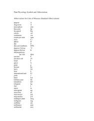
Figure 4. Deoxynucleotidyl transferase activity of the recomb<strong>in</strong>ant<br />
<strong>AtREV1</strong> prote<strong>in</strong> opposite the AP site. The primer P13 was 32 P-labeled at<br />
the 5# end and annealed with the template 30G or 30AP. The nucleotide<br />
sequences are shown <strong>in</strong> A. The underl<strong>in</strong>ed bases represent the<br />
variation of templates; O <strong>in</strong>dicates the AP site <strong>in</strong> template 30AP. One<br />
hundred and sixty nanograms of <strong>AtREV1</strong> recomb<strong>in</strong>ant prote<strong>in</strong> and the<br />
<strong>in</strong>dicated template/primer were <strong>in</strong>cubated with 0.1 mM of a s<strong>in</strong>gle<br />
dNTP (G, A, T, or C), 0.1 mM each of all four dNTPs (N), or no dNTP (2)<br />
under standard reaction conditions with 2 mM magnesium chloride (B<br />
and D) or 1 mM manganese chloride (C and D) at 30°C for 10 m<strong>in</strong>. The<br />
reaction products were resolved <strong>in</strong> 20% polyacrylamide gels conta<strong>in</strong><strong>in</strong>g<br />
8 M urea, and then visualized by autoradiography.<br />
These results suggest that the cations chelated <strong>in</strong> the<br />
active site of <strong>AtREV1</strong> affect the substrate specificity of<br />
this prote<strong>in</strong>, as reported <strong>in</strong> other polymerases (Doublie<br />
et al., 1998; Steitz, 1998).<br />
Insertion of Nucleotide Opposite the AP Site by<br />
Recomb<strong>in</strong>ant <strong>AtREV1</strong><br />
To exam<strong>in</strong>e whether <strong>AtREV1</strong> recomb<strong>in</strong>ant prote<strong>in</strong><br />
can <strong>in</strong>sert a nucleotide opposite an AP site, we performed<br />
the <strong>in</strong> vitro transferase assay with template<br />
30AP (Fig. 4A). In the presence of magnesium ion, the<br />
recomb<strong>in</strong>ant <strong>AtREV1</strong> prote<strong>in</strong> efficiently <strong>in</strong>serted only<br />
one nucleotide opposite the AP site (Fig. 4D). Among<br />
four deoxynucleotides, one dCMP was preferentially<br />
<strong>in</strong>corporated opposite the AP site when compared to<br />
the other three deoxynucleotides. Other nucleotides,<br />
dTMP and dAMP, were also <strong>in</strong>serted opposite AP, but<br />
activity of <strong>in</strong>corporation was lower than that of the<br />
dCMP <strong>in</strong>sertion. In the presence of manganese chlo-<br />
ride, the recomb<strong>in</strong>ant <strong>AtREV1</strong> prote<strong>in</strong> <strong>in</strong>serted one or<br />
two deoxynucleotides opposite the AP site (Fig. 4E).<br />
No Nucleotide Insertion Activity of <strong>AtREV1</strong> Opposite<br />
the UV Damage<br />
To exam<strong>in</strong>e whether the <strong>AtREV1</strong> recomb<strong>in</strong>ant prote<strong>in</strong><br />
can <strong>in</strong>sert a nucleotide opposite the typical UV<strong>in</strong>duced<br />
damage, we performed an <strong>in</strong> vitro transferase<br />
assay with a template conta<strong>in</strong><strong>in</strong>g CPD or 6-4PP (Fig.<br />
5A). The template CPD or 6-4PP and a primer (P16)<br />
were annealed and transferase activity was measured<br />
<strong>in</strong> the presence of all four dNTPs. As a result, no<br />
<strong>in</strong>sertion activity was detected even <strong>in</strong> a prolonged (up<br />
to 30 m<strong>in</strong>) <strong>in</strong>cubation (Fig. 5B). In contrast, the presence<br />
of ladder bands over the primer band with<br />
template 30T <strong>in</strong>dicates that the recomb<strong>in</strong>ant <strong>AtREV1</strong><br />
can replicate this template unless UV damage is present.<br />
When the templates were annealed with P17A<br />
primer that possesses a correct nucleotide (A) opposite<br />
the 3#T of CPD or 6-4PP, <strong>AtREV1</strong> did not extend this<br />
primer end (Fig. 5B). Naturally, no nucleotide <strong>in</strong>sertion<br />
activity was observed when the templates were annealed<br />
with P17G that possesses an <strong>in</strong>correct nucleotide<br />
(G) opposite the 3#T of CPD or 6-4PP, although<br />
<strong>AtREV1</strong> extended this mismatch end if there was no<br />
UV damage. The absence of nucleotide <strong>in</strong>sertion activity<br />
of <strong>AtREV1</strong> aga<strong>in</strong>st the UV damage is consistent<br />
with several reports about human or yeast REV1s<br />
(Haracska et al., 2002; Zhang et al., 2002). However, the<br />
absence of nucleotide <strong>in</strong>sertion activity conflicts with<br />
the f<strong>in</strong>d<strong>in</strong>g that an <strong>AtREV1</strong>-disrupted plant is sensitive<br />
to UV-B (Takahashi et al., 2005).<br />
DISCUSSION<br />
Deoxynucleotidyl Transferase Activity of <strong>AtREV1</strong><br />
REV1 is known as a <strong>DNA</strong> template-dependent<br />
dCMP transferase <strong>in</strong> yeast, human, and mouse (Nelson<br />
et al., 1996a; Masuda et al., 2002; Zhang et al., 2002). We<br />
previously identified <strong>AtREV1</strong>, an Arabidopsis homolog<br />
of the REV1 gene (Takahashi et al., 2005), but did<br />
not <strong>in</strong>vestigate its biochemical characteristics. In this<br />
study, we successfully purified an active recomb<strong>in</strong>ant<br />
<strong>AtREV1</strong> prote<strong>in</strong> and exam<strong>in</strong>ed its deoxynucleotidyl<br />
transferase activity us<strong>in</strong>g a primer extension assay.<br />
The recomb<strong>in</strong>ant <strong>AtREV1</strong> prote<strong>in</strong> <strong>in</strong>corporated nucleotides,<br />
especially cytos<strong>in</strong>es and thym<strong>in</strong>es, onto the<br />
primer end, regardless of the opposite template residue.<br />
In addition, the <strong>AtREV1</strong> <strong>in</strong>corporated dCTP or<br />
dTTP opposite of the AP sites. The substrate specificity<br />
of <strong>AtREV1</strong> is similar to the substrate specificities of<br />
yeast, human, and mouse REV1 prote<strong>in</strong>s (Haracska<br />
et al., 2002; Masuda and Kamiya, 2002; Masuda et al.,<br />
2002). These results <strong>in</strong>dicate that <strong>AtREV1</strong> is a deoxycytidyl<br />
transferase.<br />
Nair et al. (2005) determ<strong>in</strong>ed the crystal structure of<br />
yeast Rev1 prote<strong>in</strong> when it is bound to the template G<br />
and ready for an <strong>in</strong>com<strong>in</strong>g dCTP. In the template/<br />
REV1 complex, an <strong>in</strong>com<strong>in</strong>g dCTP b<strong>in</strong>ds to Arg-324 <strong>in</strong><br />
<strong>Plant</strong> Physiol. Vol. 145, 2007 1055
Takahashi et al.<br />
Figure 5. Deoxynucleotidyl transferase activity of the recomb<strong>in</strong>ant<br />
<strong>AtREV1</strong> prote<strong>in</strong> opposite sites of UV damage. The primer P16, P17A, or<br />
P17G was 32 P-labeled at the 5# end and annealed with template 30T,<br />
CPD, or 6-4 (A). The nucleotide sequences around the primer term<strong>in</strong>us<br />
and position of UV damage (T^T for CPD, T 5 T for 6-4PP) are shown<br />
<strong>in</strong> A. Four hundred nanograms of <strong>AtREV1</strong> recomb<strong>in</strong>ant prote<strong>in</strong> and the<br />
<strong>in</strong>dicated template/primer were <strong>in</strong>cubated with 0.1 mM of each dNTPs<br />
under standard reaction conditions with 2 mM magnesium chloride at<br />
30°C for 30 m<strong>in</strong>. The reaction products were resolved <strong>in</strong> 20% polyacrylamide<br />
gels conta<strong>in</strong><strong>in</strong>g 8 M urea and visualized by autoradiography.<br />
the N-digit, a unique subdoma<strong>in</strong> <strong>in</strong> REV1, rather than<br />
to the G <strong>in</strong> the template (Nair et al., 2005). At the same<br />
time, the G residue <strong>in</strong> the template is evicted from the<br />
<strong>DNA</strong> helix and makes a hydrogen bond with Leu-325<br />
<strong>in</strong> the N-digit. This hydrogen bond is not made if the G<br />
is replaced with an A, T, or C residue <strong>in</strong> the template.<br />
This result supports the previous observation that<br />
human REV1 has a high k cat /K m value for the template<br />
G. The Arg and the Leu residues <strong>in</strong> the N-digit of<br />
yeast, mouse, and human REV1s (Masuda et al., 2001,<br />
2002) are conserved <strong>in</strong> <strong>AtREV1</strong> (Takahashi et al., 2005).<br />
In addition, <strong>AtREV1</strong> more efficiently <strong>in</strong>corporated<br />
nucleotides <strong>in</strong>to template G than <strong>in</strong>to template A, T,<br />
or C. These results suggest that <strong>AtREV1</strong> transfers a<br />
dCMP opposite G <strong>in</strong> a REV1-specific manner, and not<br />
through Watson-Crick pair<strong>in</strong>g between dG and dCTP.<br />
The substrate specificity of <strong>AtREV1</strong> was reduced by<br />
replac<strong>in</strong>g the magnesium ion <strong>in</strong> the buffer with man-<br />
ganese ion. The specificity of human REV1 was also<br />
reduced by replac<strong>in</strong>g magnesium ions with manganese<br />
ions (Masuda and Kamiya, 2002). Vaisman et al.<br />
(2005) analyzed the effect of metal ions on the activity<br />
of a Y-family polymerase from Sulfolobus solfataricu<br />
Dpo4 based on its crystal structure and k<strong>in</strong>etics. They<br />
suggested that four conditions are required to form an<br />
active catalytic center <strong>in</strong> Dpo4: (1) 3#-OH of the primer<br />
end, (2) match<strong>in</strong>g of the template base and the <strong>in</strong>com<strong>in</strong>g<br />
nucleotide, (3) three catalytic carboxylates, and (4)<br />
two metal ions <strong>in</strong> correct coord<strong>in</strong>ation. Replac<strong>in</strong>g the<br />
magnesium ion with manganese ion, which has a relaxed<br />
coord<strong>in</strong>ation requirement, dramatically <strong>in</strong>creased<br />
the catalytic efficiency of mis<strong>in</strong>corporation by Dpo4.<br />
Therefore, magnesium ions are probably essential for<br />
limit<strong>in</strong>g mis<strong>in</strong>corporations by <strong>AtREV1</strong>.<br />
The recomb<strong>in</strong>ant <strong>AtREV1</strong> prote<strong>in</strong> <strong>in</strong>serted one or<br />
two nucleotides opposite the AP site, but did not<br />
extend from the mispaired term<strong>in</strong>i. It is generally<br />
believed that the mispaired term<strong>in</strong>i caused by the<br />
action of REV1 are extended by another polymerase<br />
(Nelson et al., 1996a; Zhang et al., 2002; Guo et al., 2004).<br />
In fact, an oligonucleotide <strong>in</strong>clud<strong>in</strong>g an AP site was<br />
efficiently replicated <strong>in</strong> vitro if the reaction mixture<br />
<strong>in</strong>cluded both REV1 and Pol z (Haracska et al., 2001;<br />
Acharya et al., 2006). Does such a two-step replication<br />
<strong>in</strong>volv<strong>in</strong>g AtREV3 occur <strong>in</strong> Arabidopsis? We do not<br />
know the properties of AtREV3 because it is too large<br />
to express as a recomb<strong>in</strong>ant prote<strong>in</strong>. However, recent<br />
evidence strongly suggests that <strong>AtREV1</strong> and AtREV3<br />
play roles <strong>in</strong> a common pathway (Takahashi et al., 2005).<br />
First, the sensitivities of double-knockout rev1rev3<br />
plants to UV-B, cisplat<strong>in</strong>, and g-rays were similar to<br />
those of AtREV3-disrupted plants (Takahashi et al.,<br />
2005). Second, when we measured the UV-<strong>in</strong>duced reversion<br />
frequencies by us<strong>in</strong>g the po<strong>in</strong>t-mutated GUS<br />
(uidA) reporter system (Kovalchuk et al., 2000), the<br />
<strong>AtREV1</strong>- and AtREV3-disrupted plants showed a similar<br />
reversion frequency on somatic cells (M. Nakagawa,<br />
S. Takahashi, and A.N. Sakamoto, unpublished data).<br />
These facts suggest that the <strong>DNA</strong> Pol z and <strong>AtREV1</strong><br />
might cooperate to bypass some classes of <strong>DNA</strong> damage<br />
<strong>in</strong> Arabidopsis.<br />
We previously showed that the <strong>AtREV1</strong>-disrupted<br />
plants are more sensitive to UV-B and g-ray irradiation<br />
than wild-type plants (Takahashi et al., 2005). It is<br />
known that AP sites are generated by various <strong>DNA</strong>damag<strong>in</strong>g<br />
agents, <strong>in</strong>clud<strong>in</strong>g ioniz<strong>in</strong>g radiation (Boiteux<br />
and Guillet, 2004). Thus, it is reasonable that the g-ray<br />
sensitivity of <strong>AtREV1</strong>-disrupted plants is due to the<br />
loss of the dCMP <strong>in</strong>sertion activity aga<strong>in</strong>st the AP site<br />
(Takahashi et al., 2005). On the other hand, the majority<br />
of UV-<strong>in</strong>duced <strong>DNA</strong> damage consists of CPDs,<br />
which are approximately 75% to 78%, while the 6-4PPs<br />
and Dewar isomers account for most of the rema<strong>in</strong>der<br />
(Mitchell, 1995; Cadet et al., 2005). The monomeric<br />
damage, such as 8-oxoG or cytos<strong>in</strong>e hydrate, which<br />
could be turned to AP sites by the action of <strong>DNA</strong><br />
N-glycosylase, is generated at very low level (Cadet<br />
et al., 2005; Friedberg et al., 2006). Based on these facts,<br />
1056 <strong>Plant</strong> Physiol. Vol. 145, 2007
it is presumable that the UV sensitivity of <strong>AtREV1</strong>disrupted<br />
plants is caused by loss of activity to bypass<br />
CPDs and/or 6-4PPs rather than the AP site. However,<br />
<strong>in</strong> this study, the recomb<strong>in</strong>ant <strong>AtREV1</strong> prote<strong>in</strong> showed<br />
no <strong>in</strong>sertion or extension activity either on CPD- or<br />
6-4PP-conta<strong>in</strong><strong>in</strong>g oligonucleotides <strong>in</strong> vitro. There are<br />
at least two possible explanations for this contradictory<br />
result. One of the possibilities is that the presence of<br />
the AP site is quite toxic for plants, even though the<br />
yield after UV irradiation is low, and that the <strong>in</strong>ability<br />
of bypass<strong>in</strong>g the AP site caused <strong>in</strong>hibition of growth.<br />
However, this hypothesis is unlikely because the AP<br />
site is abundantly generated even under no exogenous<br />
agent (Nakamura et al., 1998; Friedberg et al., 2006).<br />
Thus, if the <strong>in</strong>ability of bypass<strong>in</strong>g the AP site were<br />
essential for plants, the REV1-disrupted plant would<br />
show the severe growth defect even under natural<br />
growth condition. The <strong>in</strong>dist<strong>in</strong>guishable growth of<br />
rev1-1 plants from wild-type plants without UV treatment<br />
(Takahashi et al., 2005) suggests that this hypothesis<br />
is not appropriate to expla<strong>in</strong> the UV sensitivity of<br />
REV1-deficient plants.<br />
The alternate possibility is that <strong>AtREV1</strong> has another<br />
function(s) <strong>in</strong>dependent of dCMP transferase activity,<br />
which contributes the UV tolerance. Ross et al. (2005)<br />
showed that the human REV1 prote<strong>in</strong> with po<strong>in</strong>t<br />
mutations <strong>in</strong> its polymerase doma<strong>in</strong> completely suppresses<br />
the UV sensitivity of REV1-disrupted chicken<br />
cells. This result <strong>in</strong>dicates that transferase activity is<br />
not directly required for damage tolerance at least <strong>in</strong><br />
vertebrates. Moreover, the yeast stra<strong>in</strong> <strong>in</strong>volv<strong>in</strong>g the<br />
rev1-1 mutation, which was disrupted <strong>in</strong> the BRCT<br />
doma<strong>in</strong>, could not bypass 6-4PPs on the plasmid<br />
(Nelson et al., 2000), although the REV1-1p prote<strong>in</strong><br />
with the same mutation showed normal dCMP transferase<br />
activity <strong>in</strong> vitro. These results support the idea<br />
that the BRCT doma<strong>in</strong> of REV1s has a dist<strong>in</strong>ct function<br />
<strong>in</strong> addition to its dCMP transferase activity. Thus, by<br />
analogy, it is conceivable that the BRCT doma<strong>in</strong> of<br />
<strong>AtREV1</strong> plays some important roles <strong>in</strong> UV tolerance <strong>in</strong><br />
Arabidopsis.<br />
Deoxynucleotidyl Transferase Activity of <strong>AtREV1</strong><br />
Recently, it was reported that the proliferat<strong>in</strong>g cell<br />
nuclear antigen (PCNA) plays a major role <strong>in</strong> polymerase<br />
switch<strong>in</strong>g, which is regulated by the posttranslational<br />
modification (Stelter and Ulrich, 2003; Lehmann<br />
et al., 2007). Especially, the monoubiquit<strong>in</strong>ation of<br />
the Lys-164 residue <strong>in</strong>creases the aff<strong>in</strong>ity of PCNA to<br />
TLS-type polymerases, promot<strong>in</strong>g TLS at the replication<br />
fork (Parker et al., 2007). Consistently, all the<br />
Y-family polymerases have conserved ubiquit<strong>in</strong>-b<strong>in</strong>d<strong>in</strong>g<br />
sequences (UBM or UBZ), which are thought to provide<br />
an <strong>in</strong>terface <strong>in</strong>teract<strong>in</strong>g with monoubiquit<strong>in</strong>ated<br />
PCNA (Bienko et al., 2005). In yeast, the damagebypass<br />
activity of REV1 is enhanced <strong>in</strong> the presence of<br />
ubiquit<strong>in</strong>ated PCNA <strong>in</strong> vitro (Garg and Burgers, 2005),<br />
and this enhancement needs one of two UBM motifs<br />
<strong>in</strong> the C-term<strong>in</strong>al region of REV1 (Wood et al., 2007).<br />
A mutation <strong>in</strong> the UBM motif dim<strong>in</strong>ished the tolerance<br />
of yeast aga<strong>in</strong>st UV, methylmethane sulfate, and<br />
4-nitroqu<strong>in</strong>olene-1-oxide to the same level as the REV1knockout<br />
mutant (Guo et al., 2006; Wood et al., 2007).<br />
Similarly, the mouse REV1 gene that has a mutation <strong>in</strong><br />
the UBM failed to suppress the UV or cisplat<strong>in</strong> tolerance<br />
of REV1-deficient chicken cells (Guo et al., 2006).<br />
These results <strong>in</strong>dicate that the <strong>in</strong>teraction of REV1 to<br />
the monoubiquit<strong>in</strong>ated PCNA is commonly important<br />
for the function of REV1. The presence of putative<br />
UBMs <strong>in</strong> the C term<strong>in</strong>us of <strong>AtREV1</strong> (Supplemental Fig.<br />
S1) strongly suggests the <strong>in</strong>volvement of <strong>AtREV1</strong> with<br />
monoubiquit<strong>in</strong>ated PCNA <strong>in</strong> Arabidopsis.<br />
Murakumo et al. showed that human REV1 <strong>in</strong>teracts<br />
with the REV7 subunit of Pol z through its C-term<strong>in</strong>al<br />
doma<strong>in</strong> (Murakumo et al., 2001). Also, the C-term<strong>in</strong>al<br />
region of mouse REV1 <strong>in</strong>teracts with Pol h and Pol i,<br />
and Pol k (Guo et al., 2003). From these facts, it is<br />
further proposed that the REV1 prote<strong>in</strong> bound to the<br />
monoubiquit<strong>in</strong>ated PCNA serves as a platform of<br />
polymerase switch<strong>in</strong>g to mediate recruitment of the<br />
appropriate polymerase to the damage site (Tissier<br />
et al., 2004; Lehmann et al., 2007). Taken together,<br />
the prote<strong>in</strong>-prote<strong>in</strong> <strong>in</strong>teractions through the BRCT doma<strong>in</strong>,<br />
UBMs, and/or C-term<strong>in</strong>al doma<strong>in</strong> of REV1 seem<br />
Figure 6. Possible role of <strong>AtREV1</strong> <strong>in</strong> polymerase switch<strong>in</strong>g. A, Replication-type polymerase (white oval) bound to PCNA (gray<br />
r<strong>in</strong>g) is stalled at the damage. B, PCNA is monoubiquit<strong>in</strong>ated (Ub), which <strong>in</strong>creases its aff<strong>in</strong>ity for <strong>AtREV1</strong> (black oval with pocket<br />
that represents the UBM). C, TLS-type polymerase (hatched oval) <strong>in</strong>teracts with the PCNA-bound <strong>AtREV1</strong>. D, A nucleotide (black<br />
square) is <strong>in</strong>serted by the action of the TLS-type polymerase. See the text for more detail.<br />
<strong>Plant</strong> Physiol. Vol. 145, 2007 1057
Takahashi et al.<br />
to be <strong>in</strong>dispensable for polymerase switch<strong>in</strong>g and for<br />
damage-tolerance activity. The <strong>AtREV1</strong> prote<strong>in</strong> putatively<br />
conta<strong>in</strong>s both a BRCT doma<strong>in</strong> (Takahashi et al.,<br />
2005) and UBMs (Supplemental Fig. S1). Thus, it is conceivable<br />
that these multiple prote<strong>in</strong>-prote<strong>in</strong> <strong>in</strong>teractions<br />
through the dist<strong>in</strong>ct doma<strong>in</strong>s of <strong>AtREV1</strong> play an<br />
important role <strong>in</strong> UV tolerance <strong>in</strong> plant, although the<br />
exact position of the C-term<strong>in</strong>al polymerase-<strong>in</strong>teract<strong>in</strong>g<br />
doma<strong>in</strong> rema<strong>in</strong>s to be identified. Based on this hypothesis,<br />
we propose a model for a possible role of <strong>AtREV1</strong><br />
and polymerase switch<strong>in</strong>g at the replication fork (Fig.<br />
6). The replication fork is stalled when it encounters<br />
<strong>DNA</strong> damage (Fig. 6A). A signal from stalled replication<br />
triggers ubiquit<strong>in</strong>ation of PCNA, which leads to<br />
dissociation of the replication-type polymerase and<br />
attracts <strong>AtREV1</strong> (Fig. 6B). TLS-type polymerases are<br />
recruited to the <strong>DNA</strong> damage through <strong>in</strong>teraction with<br />
<strong>AtREV1</strong> (Fig. 6C). TLS is accomplished by the action of<br />
one or more specialized polymerases (Fig. 6D).<br />
Structure-function analysis of <strong>AtREV1</strong>, <strong>in</strong> which the<br />
BRCT, UBM, or C-term<strong>in</strong>al region is mutated, will be<br />
necessary to evaluate the function(s) of <strong>AtREV1</strong> prote<strong>in</strong><br />
<strong>in</strong> plant damage tolerance.<br />
MATERIALS AND METHODS<br />
Construction of a Recomb<strong>in</strong>ant <strong>AtREV1</strong><br />
Expression Plasmid<br />
To express the His-tagged <strong>AtREV1</strong> prote<strong>in</strong>, the <strong>AtREV1</strong> cod<strong>in</strong>g fragment,<br />
prepared previously (Takahashi et al., 2005), was reamplified by PCR us<strong>in</strong>g<br />
the REV1-pET-1B (5#-GCCCATGGCTCGTAGCTTGGGTTCAAATTC-3#) and<br />
REV1-pET-2B (5#-GCGCGGCCGCTGGTATACTCAAGCTTCCTC-3#) primers,<br />
which conta<strong>in</strong> NcoI andNotI restriction sites (underl<strong>in</strong>ed), respectively.<br />
To <strong>in</strong>sert the NcoI site at the N term<strong>in</strong>us of <strong>AtREV1</strong>, the second codon was<br />
changed from K to A (Fig. 1B). For sequenc<strong>in</strong>g, the amplified fragment was<br />
ligated <strong>in</strong>to the pGEM-T easy vector (Promega). The fragment conta<strong>in</strong>ed two<br />
base substitutions that were <strong>in</strong>troduced by PCR, but neither of them caused an<br />
am<strong>in</strong>o acid change. The plasmid was digested with NcoI and NotI, and<br />
<strong>in</strong>serted <strong>in</strong>to the correspond<strong>in</strong>g sites of the pET32a(1) vector (Novagen),<br />
which is located downstream of the T7lac promoter under the control of lacI.<br />
The result<strong>in</strong>g plasmid was named pET32a-<strong>AtREV1</strong> (Fig. 1A).<br />
Overexpression and Purification of Recomb<strong>in</strong>ant <strong>AtREV1</strong><br />
Escherichia coli BL21(DE3) cells (Novagen) carry<strong>in</strong>g pET32a-<strong>AtREV1</strong> were<br />
<strong>in</strong>oculated <strong>in</strong>to 3 mL of Luria-Bertani (LB) broth conta<strong>in</strong><strong>in</strong>g 100 mg mL 21<br />
carbenicill<strong>in</strong> and were grown at 37°C for 18 h. One milliliter of this preculture<br />
was added <strong>in</strong>to 50 mL of fresh LB broth conta<strong>in</strong><strong>in</strong>g 100 mg mL 21 carbenicill<strong>in</strong><br />
and further grown at the same temperature for 6 h. All of the culture was<br />
transferred to 0.9 L of fresh LB broth conta<strong>in</strong><strong>in</strong>g 100 mgmL 21 carbenicill<strong>in</strong> and<br />
grown at 37°C until OD 600 reached 0.6. To optimize the recovery of recomb<strong>in</strong>ant<br />
prote<strong>in</strong>, expression was <strong>in</strong>duced with IPTG for 3 h at 30°C or22°C, or<br />
overnight at 15°C. At 30°C and 22°C, recomb<strong>in</strong>ant <strong>AtREV1</strong> was predom<strong>in</strong>antly<br />
accumulated <strong>in</strong> <strong>in</strong>clusion bodies. However, the recomb<strong>in</strong>ant prote<strong>in</strong><br />
was detected <strong>in</strong> the soluble fraction when the expression was <strong>in</strong>duced at 15°C<br />
(data not shown). Therefore, the cells were grown at 37°C until OD 600 reached<br />
0.6, transferred to 15°C, <strong>in</strong>duced with 0.8 mM IPTG (f<strong>in</strong>al concentration), and<br />
cultured for additional 20 h. One liter of cell culture was centrifuged at 4°C,<br />
and the cell paste was washed once with 100 mL of buffer A (20 mM Na 2 HPO 4 ,<br />
pH 7.4, 0.5 M NaCl, 10% glycerol, 1 mM dithiothreitol) and then stored at<br />
280°C. Cell paste from 3 L of culture (approximately 11.5 g wet weight) was<br />
resuspended <strong>in</strong> 100 mL of buffer A conta<strong>in</strong><strong>in</strong>g lysozyme and the Protease<br />
<strong>in</strong>hibitor cocktail (Sigma-Aldrich), and lysed by sonication. The cell lysate was<br />
<strong>in</strong>cubated at 4°C <strong>in</strong> 0.1% Triton X-100 for 30 m<strong>in</strong>, centrifuged at 12,000g for<br />
30 m<strong>in</strong>, and then the supernatant (clear lysate) was recovered. The His-tagged<br />
recomb<strong>in</strong>ant <strong>AtREV1</strong> was separated with a Ni-aff<strong>in</strong>ity column. First, 10 mM<br />
imidazole was added to the clear lysate, then the lysate was mixed with 3 mL<br />
of Ni 21 -charged Chelat<strong>in</strong>g Sepharose Fast Flow (GE Healthcare) equilibrated<br />
<strong>in</strong> buffer B (20 mM Na 2 HPO 4 , pH 7.4, 0.5 M NaCl, 10% glycerol, 10 mM<br />
2-mercaptoethanol) conta<strong>in</strong><strong>in</strong>g 10 mM imidazole. The mixture was centrifuged<br />
at 500g, washed with 5 volumes of buffer B, and then packed <strong>in</strong> a PD-10 empty<br />
column (GE Healthcare). Bound prote<strong>in</strong>s were eluted with 3 mL of buffer B<br />
conta<strong>in</strong><strong>in</strong>g 30, 60, 100, and 300 mM imidazole and collected <strong>in</strong> 3-mL fractions.<br />
The 300 mM imidazole fraction, conta<strong>in</strong><strong>in</strong>g <strong>AtREV1</strong> prote<strong>in</strong>s, was collected (Ni<br />
fraction). Six milliliters of the Ni fraction was desalted by 5-mL HiTrap<br />
Desalt<strong>in</strong>g column (GE Healthcare), <strong>in</strong> which the buffer was replaced with<br />
buffer C (20 mM Na 2 HPO 4 , pH 7.4, 300 mM NaCl, 10% glycerol, 1 mM<br />
dithiothreitol). The desalted Ni fraction was then applied onto a 1-mL HiTrap<br />
hepar<strong>in</strong> HP column (GE Healthcare) equilibrated with buffer C. After wash<strong>in</strong>g<br />
the column with 10 mL of buffer C, prote<strong>in</strong>s were eluted with 3.5 mL of<br />
20 mM Na 2 HPO 4 buffer, pH 7.4, supplemented with 400, 450, and 500 mM NaCl.<br />
The fractions with 450 and 500 mM NaCl were collected (hepar<strong>in</strong> fraction).<br />
The hepar<strong>in</strong> fraction (7 mL) was desalted as described above; the buffer<br />
was replaced with buffer D (20 mM Tris-HCl, pH 8.0, 300 mM NaCl, 10%<br />
glycerol, 1 mM dithiothreitol). The fraction was then applied to a 1-mL HiTrap<br />
Q HP column (GE Healthcare) equilibrated with buffer D. After the column<br />
was washed with 10 mL of buffer D, prote<strong>in</strong>s were eluted with 3.5 mL of<br />
buffer D supplemented with 350 or 400 mM NaCl. The purified <strong>AtREV1</strong><br />
prote<strong>in</strong>, eluted <strong>in</strong> 350 mM NaCl, was collected. The total volume was<br />
approximately 1.5 mL and concentration was 80 mg mL 21 . Aliquots of the<br />
purified <strong>AtREV1</strong> were dialyzed aga<strong>in</strong>st buffer E (5 mM Tris-HCl, pH 8.0,<br />
0.5 mM EDTA, 50% glycerol, 1 mM dithiothreitol) overnight and stored<br />
at 220°C.<br />
Primer Extension Assay<br />
Deoxynucleotidyl transferase activity of the purified recomb<strong>in</strong>ant <strong>AtREV1</strong><br />
was assayed accord<strong>in</strong>g to Masuda and Kamiya (2002). Oligonucleotide<br />
template 5#-CTCGTCAGCATCTTCAXCATACAGTCAGTG-3# (X 5 G, 30G;<br />
A, 30A; T, 30T; C, 30C) and the primer 5#-CACTGACTGTATG-3# (P13) were<br />
purchased from Sigma Genosys Japan. The template with AP sites (same<br />
sequence as above template except for X 5 AP) was purchased from Midland<br />
Certified Reagent Company. Templates with UV damage, 5#-CTCGTCAGC-<br />
TTCATCATACAGTCAGTG-3# (TT 5 CPD or 6-4PP), were k<strong>in</strong>dly provided<br />
by Dr. Shigenori Iwai, Osaka University. The primers 5#-CACTGACTGTA-<br />
TCATG-3# (P16), 5#-CACTGACTGTATCATGA-3# (P17A), and 5#-CAC-<br />
TGACTGTATCATGG-3# (P17G) were purchased from Sigma Genosys. To<br />
prepare primer templates, the primers were labeled us<strong>in</strong>g T4 polynucleotide<br />
k<strong>in</strong>ase (Takara BIO) and [g- 32 P]ATP (GE Healthcare) and annealed to templates.<br />
The reaction mixture (20 mL) conta<strong>in</strong>ed 40 mM Tris-HCl buffer, pH 8.0,<br />
10% glycerol, 0.1 mg mL 21 bov<strong>in</strong>e serum album<strong>in</strong>, 5 mM dithiothreitol, 0.1 mM<br />
of a s<strong>in</strong>gle dNTP or 0.1 mM each of all four dNTPs, 25 nM primer template,<br />
2mM MgCl 2 or 1 mM MnCl 2 as divalent cations, and 2 mL (approximately<br />
160 ng) of <strong>AtREV1</strong>. After <strong>in</strong>cubation at 30°C for 10 m<strong>in</strong>, the reactions were<br />
term<strong>in</strong>ated with 10 mL of stop solution (30 mM EDTA, 94% formamide, 0.05%<br />
bromphenol blue, 0.05% xylene cyanol) and the products were resolved on a<br />
20% polyacrylamide gel conta<strong>in</strong><strong>in</strong>g 8 M urea. The gel was dried and used to<br />
expose an imag<strong>in</strong>g plate (Fuji Photo Film) for about 40 h. The radioactivity<br />
was visualized us<strong>in</strong>g Bio-Imag<strong>in</strong>g Analyzer BAS1800II (Fuji Photo Film).<br />
Sequence data from this article can be found <strong>in</strong> the GenBank/EMBL data<br />
libraries under accession number AB187523.<br />
Supplemental Data<br />
The follow<strong>in</strong>g materials are available <strong>in</strong> the onl<strong>in</strong>e version of this article.<br />
Supplemental Figure S1. Conserved UBM sequences <strong>in</strong> the REV1 prote<strong>in</strong>s.<br />
ACKNOWLEDGMENTS<br />
We thank Dr. Shigenori Iwai at Osaka University for his k<strong>in</strong>d gift of UV<br />
damage-conta<strong>in</strong><strong>in</strong>g oligonucleotides, and Dr. Motoshi Suzuki at Nagoya<br />
University for his technical advice on <strong>in</strong> vitro transferase assays. We also<br />
thank Ms. Chihiro Suzuki at JAEA for her technical assistance; and Drs.<br />
Katsuya Sato, Satoshi Kitamura, Yutaka Oono, and Ms. Satomi Ishii at JAEA<br />
1058 <strong>Plant</strong> Physiol. Vol. 145, 2007
and Mr. Youichirou Matuo at Osaka University for their technical advice on<br />
prote<strong>in</strong> expression and purification. We are grateful to Drs. James Raymond<br />
and Alan Clark for careful review of the manuscript.<br />
Received May 7, 2007; accepted August 31, 2007; published September 7, 2007.<br />
LITERATURE CITED<br />
Acharya N, Johnson RE, Prakash S, Prakash L (2006) Complex formation<br />
with Rev1 enhances the proficiency of Saccharomyces cerevisiae <strong>DNA</strong><br />
polymerase z for mismatch extension and for extension opposite from<br />
<strong>DNA</strong> lesions. Mol Cell Biol 26: 9555–9563<br />
Bienko M, Green CM, Crosetto N, Rudorf F, Zapart G, Coull B,<br />
Kannouche P, Wider G, Peter M, Lehmann AR, et al (2005) Ubiquit<strong>in</strong>b<strong>in</strong>d<strong>in</strong>g<br />
doma<strong>in</strong>s <strong>in</strong> Y-family polymerases regulate translesion synthesis.<br />
Science 310: 1821–1824<br />
Boiteux S, Guillet M (2004) Abasic sites <strong>in</strong> <strong>DNA</strong>: repair and biological<br />
consequences <strong>in</strong> Saccharomyces cerevisiae. <strong>DNA</strong> Repair (Amst)<br />
3: 1–12<br />
Britt AB (1999) Molecular genetics of <strong>DNA</strong> repair <strong>in</strong> higher plants. Trends<br />
<strong>Plant</strong> Sci 4: 20–25<br />
BroomfieldS,HryciwT,XiaoW(2001) <strong>DNA</strong> postreplication repair and<br />
mutagenesis <strong>in</strong> Saccharomyces cerevisiae. MutatRes486: 167–184<br />
Cadet J, Douki T, Pouget J-P, Ravanat J-L (2005) UVB and UVA <strong>in</strong>duced<br />
formation of photoproducts with<strong>in</strong> cellular <strong>DNA</strong>. In ESage,RDrou<strong>in</strong>,<br />
M Rouabhia, eds, From <strong>DNA</strong> Photolesions to Mutations, Sk<strong>in</strong> Cancer<br />
and Cell Death. RSC Publish<strong>in</strong>g, London, pp 1–14<br />
Doublie S, Tabor S, Long AM, Richardson CC, Ellenberger T (1998)<br />
Crystal structure of a bacteriophage T7 <strong>DNA</strong> replication complex at<br />
2.2 A ˚ resolution. Nature 391: 251–257<br />
Friedberg EC, Feaver WJ, Gerlach VL (2000) The many faces of <strong>DNA</strong><br />
polymerases: strategies for mutagenesis and for mutational avoidance.<br />
Proc Natl Acad Sci USA 97: 5681–5683<br />
Friedberg EC, Lehmann AR, Fuchs RPP (2005) Trad<strong>in</strong>g places: How do<br />
<strong>DNA</strong> polymerases switch dur<strong>in</strong>g translesion <strong>DNA</strong> synthesis? Mol Cell<br />
18: 499–505<br />
Friedberg EC, Walker GC, Siede W, Wood RD, Schultz RA, Ellenberger T<br />
(2006) <strong>DNA</strong> Repair and Mutagenesis, Ed 2. American Society of Microbiology<br />
Press, Wash<strong>in</strong>gton, DC<br />
Gallego F, Fleck O, Li A, Wyrzykowska J, T<strong>in</strong>land B (2000) AtRAD1, a<br />
plant homologue of human and yeast nucleotide excision repair endonucleases,<br />
is <strong>in</strong>volved <strong>in</strong> dark repair of UV damages and recomb<strong>in</strong>ation.<br />
<strong>Plant</strong> J 21: 507–518<br />
Gao MJ, Murphy TM (2001) Alternative forms of formamidopyrimid<strong>in</strong>e-<br />
<strong>DNA</strong> glycosylase from Arabidopsis thaliana. Photochem Photobiol 73:<br />
128–134<br />
García-Ortiz MV, Ariza RR, Hoffman PD, Hays JB, Roldán-Arjona TR<br />
(2004) Arabidopsis thaliana AtPOLK encodes a D<strong>in</strong>B-like <strong>DNA</strong> polymerasethatextendsmispairedprimerterm<strong>in</strong>iandishighlyexpressed<strong>in</strong>a<br />
variety of tissues. <strong>Plant</strong> J 39: 84–97<br />
García-Ortiz MV, Ariza RR, Roldán-Arjona T (2001) An OGG1 orthologue<br />
encod<strong>in</strong>g a functional 8-oxoguan<strong>in</strong>e <strong>DNA</strong> glycosylase/lyase <strong>in</strong> Arabidopsis<br />
thaliana. <strong>Plant</strong> Mol Biol 47: 795–804<br />
Garg P, Burgers PM (2005) Ubiquit<strong>in</strong>ated proliferat<strong>in</strong>g cell nuclear antigen<br />
activates translesion <strong>DNA</strong> polymerases h and REV1. Proc Natl Acad Sci<br />
USA 102: 18361–18366<br />
Guo C, Fischhaber PL, Luk-Paszyc MJ, Masuda Y, Zhou J, Kamiya K,<br />
Kisker C, Friedberg EC (2003) Mouse Rev1 prote<strong>in</strong> <strong>in</strong>teracts with<br />
multiple <strong>DNA</strong> polymerases <strong>in</strong>volved <strong>in</strong> translesion <strong>DNA</strong> synthesis.<br />
EMBO J 22: 6621–6630<br />
Guo C, Tang TS, Bienko M, Parker JL, Bielen AB, Sonoda E, Takeda S,<br />
Ulrich HD, Dikic I, Friedberg EC (2006) Ubiquit<strong>in</strong>-b<strong>in</strong>d<strong>in</strong>g motifs <strong>in</strong><br />
REV1 prote<strong>in</strong> are required for its role <strong>in</strong> the tolerance of <strong>DNA</strong> damage.<br />
MolCellBiol26: 8892–8900<br />
Guo D, Xie Z, Shen H, Zhao B, Wang Z (2004) Translesion synthesis of<br />
acetylam<strong>in</strong>ofluorene-dG adducts by <strong>DNA</strong> polymerase z is stimulated by<br />
yeast Rev1 prote<strong>in</strong>. Nucleic Acids Res 32: 1122–1130<br />
Haracska L, Unk I, Johnson RE, Johansson E, Burgers PMJ, Prakash S,<br />
Prakash L (2001) Roles of yeast <strong>DNA</strong> polymerases d and z and of Rev1 <strong>in</strong><br />
the bypass of abasic sites. Genes Dev 15: 945–954<br />
Haracska L, Prakash S, Prakash L (2002) Yeast Rev1 prote<strong>in</strong> is G templatespecific<br />
<strong>DNA</strong> polymerase. J Biol Chem 277: 15546–15551<br />
Deoxynucleotidyl Transferase Activity of <strong>AtREV1</strong><br />
Kovalchuk I, Kovalchuk O, Hohn B (2000) Genome-wide variation of the<br />
somatic mutation frequency <strong>in</strong> transgenic plants. EMBO J 19: 4431–4438<br />
Landry LG, Stapleton AE, Lim J, Hoffman P, Hays J, Walbot V, Last R<br />
(1997) An Arabidopsis photolyase mutant is hypersensitive to ultraviolet-B<br />
radiation. Proc Natl Acad Sci USA 94: 328–332<br />
Lehmann AR, Niimi A, Ogi T, Brown S, Sabbioneda S, W<strong>in</strong>g JF,<br />
Kannouche PL, Green CM (2007) Translesion synthesis: Y-family polymerasesandthepolymeraseswitch.<strong>DNA</strong>Repair(Amst)6:<br />
891–899<br />
Lemontt JF (1971) Mutants of yeast defective <strong>in</strong> mutation <strong>in</strong>duced by<br />
ultraviolet light. Genetics 68: 21–33<br />
L<strong>in</strong> W, Xu H, Zhang Y, Wu X, Yuan F, Wang Z (1999) The human REV1 gene<br />
codes for a <strong>DNA</strong> template-dependent dCMP transferase. Nucleic Acids<br />
Res 27: 4468–4475<br />
Liu J, Hall JD, Mount DW (2001) Arabidopsis UVH3 gene is a homolog of<br />
the Saccharomyces cerevisiae RAD2 and human XPG <strong>DNA</strong> repair genes.<br />
<strong>Plant</strong> J 26: 329–338<br />
LiuJ,HongSW,EscobarM,Vierl<strong>in</strong>gE,MitchellD,MountDW,HallJD<br />
(2003) Arabidopsis UVH6, ahomologofhumanXPD and yeast RAD3<br />
<strong>DNA</strong> repair genes, functions <strong>in</strong> <strong>DNA</strong> repair and is essential for plant<br />
growth. <strong>Plant</strong> Physiol 132: 1405–1414<br />
Masuda Y, Kamiya K (2002) Biochemical properties of the human REV1<br />
prote<strong>in</strong>. FEBS Lett 520: 88–92<br />
Masuda Y, Takahashi M, Fukuda S, Sumii M, Kamiya K (2002) Mechanisms<br />
of dCMP transferase reactions catalyzed by mouse Rev1 prote<strong>in</strong>.<br />
JBiolChem277: 3040–3046<br />
Masuda Y, Takahashi M, Tsunekuni N, M<strong>in</strong>ami T, Sumii M, Miyagawa K,<br />
Kamiya K (2001) Deoxycytidyl transferase activity of the human REV1<br />
prote<strong>in</strong> is closely associated with the conserved polymerase doma<strong>in</strong>.<br />
JBiolChem276: 15051–15058<br />
Mitchell DL (1995) <strong>DNA</strong> damage and repair. In WM Horspool, P-S Song,<br />
eds, CRC Handbook of Organic Photochemistry and Photobiology. CRC<br />
Press, New York, pp 1326–1331<br />
MurakumoY,OguraY,IshiiH,NumataS,IchiharaM,CroceCM,Fishel<br />
R, Takahashi M (2001) Interaction <strong>in</strong> the error-prone postreplication<br />
repair prote<strong>in</strong>s hREV1, hREV3, and hREV7. J Biol Chem 276: 35644–<br />
35651<br />
Nair DT, Johnson RE, Prakash L, Prakash S, Aggarwal AK (2005) Rev1<br />
employs a novel mechanism of <strong>DNA</strong> synthesis us<strong>in</strong>g a prote<strong>in</strong> template.<br />
Science 309: 2219–2222<br />
Nakajima S, Sugiyama M, Iwai S, Hitomi K, Otoshi E, Kim ST, Jiang CZ,<br />
Todo T, Britt A, Yamamoto K (1998) Clon<strong>in</strong>g and characterization of a<br />
gene (UVR3) required for photorepair of 6-4 photoproducts <strong>in</strong> Arabidopsis<br />
thaliana. Nucleic Acids Res 26: 638–644<br />
Nakamura J, Walker VE, Upton PB, Chiang SY, Kow YW, Swenberg JA<br />
(1998) Highly sensitive apur<strong>in</strong>ic/apyrimid<strong>in</strong>ic site assay can detect<br />
spontaneous and chemically <strong>in</strong>duced depur<strong>in</strong>ation under physiological<br />
condition. Cancer Res 58: 222–225<br />
Nelson JR, Gibbs PEM, Nowicka AM, H<strong>in</strong>kle DC, Lawrence CW (2000)<br />
Evidence for a second function for Saccharomyces cerevisiae Rev1p. Mol<br />
Microbiol 37: 549–554<br />
Nelson JR, Lawrence CW, H<strong>in</strong>kle DC (1996a) Deoxicytidil transferase<br />
activity of yeast REV1 prote<strong>in</strong>. Nature 382: 729–731<br />
Nelson JR, Lawrence CW, H<strong>in</strong>kle DC (1996b) Thym<strong>in</strong>e-thym<strong>in</strong>e dimer<br />
bypass by yeast <strong>DNA</strong> polymerase z. Science272: 1646–1649<br />
Ohmori H, Friedberg EC, Fuchs RPP, Goodman MF, Hanaoka F, H<strong>in</strong>kle D,<br />
Kunkel TA, Lawrence CW, Livneh Z, Nohmi T, et al (2001) The<br />
Y-family of <strong>DNA</strong> polymerases. Mol Cell 8: 7–8<br />
Osakabe K, Abe K, Yoshioka T, Osakabe Y, Todoriki S, Ichikawa H, Hohn<br />
B, Toki S (2006) Isolation and characterization of the RAD54 gene from<br />
Arabidopsis thaliana. <strong>Plant</strong>J48: 827–842<br />
Parker JL, Bielen AB, Dikic I, Ulrich HD (2007) Contributions of ubiquit<strong>in</strong>and<br />
PCNA-b<strong>in</strong>d<strong>in</strong>g doma<strong>in</strong>s to the activity of <strong>Polymerase</strong> h <strong>in</strong> Saccharomyces<br />
cerevisiae. Nucleic Acids Res 35: 881–889<br />
Prakash S, Johnson RE, Prakash L (2005) Eukaryotic translesion synthesis<br />
<strong>DNA</strong> polymerases: specificity of structure and function. Annu Rev<br />
Biochem 74: 317–353<br />
Ross A-L, Simpson LJ, Sale JE (2005) Vertebrate <strong>DNA</strong> damage tolerance<br />
requires the C-term<strong>in</strong>us but not BRCT or transferase doma<strong>in</strong>s of REV1.<br />
Nucleic Acids Res 33: 1280–1289<br />
Sakamoto A, Lan VTT, Hase Y, Shikazono N, Matsunaga T, Tanaka A<br />
(2003) Disruption of the AtREV3 gene causes hypersensitivity to ultraviolet<br />
B light and g-rays <strong>in</strong> Arabidopsis: implication of the presence of a<br />
translesion synthesis mechanism <strong>in</strong> plants. <strong>Plant</strong> Cell 15: 2042–2057<br />
<strong>Plant</strong> Physiol. Vol. 145, 2007 1059
Takahashi et al.<br />
Santiago MJ, Alejandre-Durán E, Ruiz-Rubio M (2006) Analysis of<br />
UV-<strong>in</strong>duced mutation spectra <strong>in</strong> Escherichia coli by <strong>DNA</strong> polymerase<br />
from Arabidopsis thaliana. MutatRes601: 51–60<br />
Song JM, Milligan JR, Sutherland BM (2002) Bistranded oxidized pur<strong>in</strong>e<br />
damage clusters: <strong>in</strong>duced <strong>in</strong> <strong>DNA</strong> by long-wavelength ultraviolet<br />
(290-400 nm) radiation? Biochemistry 41: 8683–8688<br />
Steitz TA (1998) <strong>DNA</strong> polymerases: structural diversity and common<br />
mechanisms. J Biol Chem 274: 17395–17398<br />
Stelter P, Ulrich HD (2003) Control of spontaneous and damage-<strong>in</strong>duced<br />
mutagenesis by SUMO and ubiquit<strong>in</strong> conjugation. Nature 425: 188–191<br />
Takahashi S, Sakamoto A, Sato S, Kato T, Tabata S, Tanaka A (2005) Roles<br />
of Arabidopsis <strong>AtREV1</strong> and AtREV7 <strong>in</strong> translesion synthesis. <strong>Plant</strong><br />
Physiol 138: 870–881<br />
Tissier A, Kannouche P, Reck M-P, Lehmann AR, Fuchs RPP, Cordonnier<br />
A (2004) Co-localization <strong>in</strong> replication foci and <strong>in</strong>teraction of human<br />
Y-family members, <strong>DNA</strong> polymerase pol h and REV1 prote<strong>in</strong>. <strong>DNA</strong><br />
Repair (Amst) 3: 1503–1514<br />
VaismanA,L<strong>in</strong>gH,WoodgateR,YangW(2005) Fidelity of Dpo4: effect of<br />
metal ions, nucleotide selection and pyrophospholysis. EMBO J 24:<br />
2957–2967<br />
Wood A, Garg P, Burgers MJ (2007) A ubiquit<strong>in</strong>-b<strong>in</strong>d<strong>in</strong>g motif <strong>in</strong> the<br />
translesion <strong>DNA</strong> polymerase Rev1 mediates its essential function <strong>in</strong>teraction<br />
with ubiquit<strong>in</strong>ated proliferat<strong>in</strong>g cell nuclear antigen <strong>in</strong> response<br />
to <strong>DNA</strong> damage. J Biol Chem 282: 20256–20263<br />
Zhang Y, Wu X, Rechkoblit O, Geac<strong>in</strong>tov E, Taylor JS, Wang Z<br />
(2002) Response of human REV1 to different <strong>DNA</strong> damage: preferential<br />
dCMP <strong>in</strong>sertion opposite the lesion. Nucleic Acids Res 30:<br />
1630–1638<br />
1060 <strong>Plant</strong> Physiol. Vol. 145, 2007
CORRECTIONS<br />
Vol. 145: 1052–1060<br />
Takahashi S., Sakamoto A.N., Tanaka A., and Shimizu K. <strong>AtREV1</strong>, a Y-<strong>Family</strong> <strong>DNA</strong><br />
<strong>Polymerase</strong> <strong>in</strong> Arabidopsis, Has Deoxynucleotidyl Transferase Activity <strong>in</strong> Vitro.<br />
The authors have revised the last paragraph on Page 1056 to more accurately describe a<br />
feature of <strong>AtREV1</strong>:<br />
We previously showed that the <strong>AtREV1</strong>-disrupted plants are more sensitive to UV-B and<br />
slightly more sensitive to g-ray irradiation than wild-type plants (Takahashi et al., 2005). It<br />
is known that AP sites are generated by various <strong>DNA</strong>-damag<strong>in</strong>g agents, <strong>in</strong>clud<strong>in</strong>g ioniz<strong>in</strong>g<br />
radiation (Boiteux and Guillet, 2004). Thus, the slight difference <strong>in</strong> g-ray sensitivity between<br />
the wild type and the <strong>AtREV1</strong>-disrupted plants could be due to the difference <strong>in</strong> dCMP<br />
<strong>in</strong>sertion activity aga<strong>in</strong>st the AP site (Takahashi et al., 2005).<br />
www.plantphysiol.org/cgi/doi/10.1104/pp.104.900243<br />
<strong>Plant</strong> <strong>Physiology</strong>, January 2008, Vol. 146, p. 321, www.plantphysiol.org Ó 2008 American Society of <strong>Plant</strong> Biologists 321