Avena Coleoptile Segments"2
Avena Coleoptile Segments"2
Avena Coleoptile Segments"2

Create successful ePaper yourself
Turn your PDF publications into a flip-book with our unique Google optimized e-Paper software.
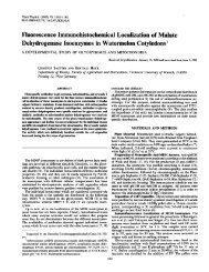
Plant Physiol. (1975) 56, 696-698<br />
Stoichiometric Correlation of Malate Accumulation with<br />
Auxin-dependent K+-H+ Exchange and Growth in<br />
<strong>Avena</strong> <strong>Coleoptile</strong> Segments"2<br />
Received for publication April 24, 1975 and in revised form July 23, 1975<br />
HANS-PETER HASCHKE AND ULRICH LUTTGE<br />
Botanisches Institut der Technischen Hochschule, D-6100 Darmstadt, Germany<br />
ABSTRACT<br />
The action of auxin in the promotion of growth has been<br />
suggested in the literature to depend on cell wall acidification.<br />
In a former investigation by the present authors the<br />
electrochemical balance in auxin-induced proton extrusion<br />
was shown to be maintained by potassium net uptake. The<br />
present paper reports data demonstrating that the elongation<br />
of <strong>Avena</strong> coleoptile segments is accompanied by an<br />
accumulation of malate, which is stoichiometrically correlated<br />
with potassium uptake. We concluded that this malate<br />
accumulation is required in a mechanism regulating intracellular<br />
pH.<br />
Auxin (IAA)-induced elongation of shoot cells is accompanied<br />
by a rapid release of protons from the cells (5, 21). External<br />
media of low pH (3-5) can mimic the action of auxin (3, 9, 25).<br />
These results have led to the suggestion that hydrogen ions play<br />
a role as second messengers in auxin action (9): acidification of<br />
the cell wall enhances its extensibility either by activating cell<br />
wall-loosening enzymes or by breaking some acid-labile links.<br />
Indoleacetic acid is also known to stimulate the uptake of<br />
K+ and Rb+ ions into different tissues (14-16). In a previous<br />
paper (10) we demonstrated that IAA-promoted proton efflux<br />
is electrochemically balanced by a stoichiometric influx of K+<br />
ions. The presence of alkali, and, in particular, of K+ ions<br />
synergistically stimulates IAA-induced growth (11).<br />
A proton efflux of the observed magnitude must be associated<br />
with a mechanism regulating cytoplasmic pH. In many tissues a<br />
proton-cation exchange (or antiport), e.g., during excess cation<br />
uptake, is associated with synthesis and subsequent accumulation<br />
of malic acid by dark C02-fixation via P-enole-pyruvate carboxylases<br />
(12). A similar mechanism is reported to be involved in<br />
K+-H+-exchange regulating stomatal guard cell movement (1,<br />
18). On the basis of these and other results, Davies (6) and<br />
Raven and Smith (19) proposed a common mechanism for<br />
stabilization of intracellular pH, which is based on synthesis<br />
and breakdown, respectively, of organic acids, especially of<br />
malate. In this respect it may appear to be pertinent that IAA-<br />
1 This work was supported by a grant from the Deutsche Forschungsgemeinschaft.<br />
2 The result of this paper has been communicated orally at the 1974<br />
conference of the Deutsche Botanische Gesellschaft, Wurzburg,<br />
September 26, 1974.<br />
promoted growth is accompanied by consumption of CO2 (25)<br />
and that IAA enhances the fixation of H'4C03- by <strong>Avena</strong> coleoptile<br />
cells (4).<br />
The present paper demonstrates that IAA-induced elongation<br />
growth is paralleled by a stoichiometric potassium and malate<br />
accumulation.<br />
MATERIAL AND METHODS<br />
Plant Material. Seeds of <strong>Avena</strong> sativa (cv. "Flemings Krone")<br />
were dehusked and soaked for 3 hr in three changes of aerated<br />
0.5 mM CaSO4. Subsequently, the seeds were placed on a steel<br />
screen floated on aerated 0.5 mm CaSO4. Germination, preparation<br />
of tissues, and experiments were carried out under dim<br />
green light at 28 + 1 C, except for a 3-hr exposure to red light<br />
at the beginning of the germination period.<br />
Experiments. Using a double blade cutter 10-mm coleoptile<br />
segments were obtained from 96-hr-old seedlings (30-40 mm in<br />
length). The top 3 mm of the coleoptile were discarded and the<br />
next 10 mm were used in the experiments. The segments were<br />
preincubated in 0.5 mm CaSO4 for 2 hr, randomized, and transferred<br />
to the test solutions, usually with 30 segments in 100 ml of<br />
solution. The solutions contained 1 mm tris + 1 mm KCl with<br />
or without 10 4M IAA; pH 7 was established by addition of<br />
some drops of 0.1 N H2SO4. At the end of the incubation time,<br />
the tissue was blotted dry and transferred to 100 ml of ice-cold<br />
bidistilled H20 for 15 min to remove adhering ions, and then<br />
frozen.<br />
Measurements. For growth measurements coleoptile segments<br />
were photographed in transmitted light before freezing, and<br />
their lengths were determined on the negatives projected onto a<br />
screen. The frozen samples were dried at room temperature<br />
under reduced pressure. Ion contents were determined from<br />
aqueous extracts; K+ by flame photometry and C1- by electrochemical<br />
titration. Malate was estimated by recording the<br />
reduction of NAD+ in the presence of malate dehydrogenase<br />
(photometrically) at 340 nm, as described by Hohorst (13).<br />
Initial amounts of ion and malate contents were obtained by<br />
analyzing coleoptile sections which were frozen after the 2-hr<br />
period of preincubation.<br />
Statistical Treatment. All experiments were repeated three<br />
times or more with three parallels each. The significance of<br />
replicates within each individual experiment was assessed by<br />
Duncan's multiple range test at the 0.05 level. If the standard<br />
errors of the means (S.E.M.) exceeded the size of the symbols<br />
in the figures they were indicated as vertical bars. The regression<br />
line in Figure 3 was calculated using the method of least squares<br />
(r = correlation coefficient).<br />
Due to the great variability of the material between different<br />
batches of tissue used for the individual experiments, a statistical<br />
696
Plant Physiol. Vol. 56, 1975 AUXIN-INDUCED GROWTH AND MALATE INCREASE<br />
697<br />
treatment comprising results of separate individual experiments<br />
proved to be of no value. Results were considered significant,<br />
when repetitive individual experiments gave similar patterns of<br />
response, although absolute data (e.g., slopes, maxima) were<br />
varying (see two individual experiments depicted in Fig. 1 or<br />
Fig. 2).<br />
RESULTS AND DISCUSSION<br />
The time courses depicted in Figure 1 show the effect of IAA<br />
on malate accumulation of the <strong>Avena</strong> coleoptile tissue. After 6<br />
to 8 hr, a clear increase of malate levels in the tissue is observed<br />
in the presence of LAA. At shorter periods significant differences<br />
in malate levels were not obtained. In the experiment depicted<br />
by closed circles in Figure 1 malate accumulation extrapolates<br />
almost linearly to zero time. In the other experiment (open<br />
circles) there is an apparent lag. Buffering capacity of the cytoplasm<br />
may maintain the intracellular pH initially for some time<br />
without detectable accumulation of malate in spite of the very<br />
rapidly measurable proton efflux (5, 21). 14CO2 dark fixation<br />
experiments are under way to collect more reliable data describing<br />
the events during the first 4 hr. At longer time periods malate<br />
accumulation continues during the entire experiment extending<br />
over 16 hr. In the absence of IAA there is a much smaller, but<br />
still significant, increase in malate levels of the tissue during the<br />
course of the experiment. This may be due to excess cation over<br />
anion uptake. In our experiments net K+ uptake by the coleoptile<br />
tissue always considerably exceeded net C1- uptake (not shown)<br />
3<br />
>. +150<br />
-o<br />
a'<br />
-3<br />
E<br />
-.' +100-<br />
0 4 8 12 16 hr<br />
FIG. 1. Time course showing IAA effect on malate accumulation<br />
(amal) in <strong>Avena</strong> coleoptile segments (two individual experiments).<br />
The initial amounts of malate in the tissue were 162 4 teq/g dry<br />
weight in the experiment indicated by the open symbols and 210 4<br />
.ueq/g dry weight in the experiment indicated by closed symbols.<br />
irrespective of IAA treatment. This is in contrast to data reported<br />
by Rubinstein and Light (22) and Rubinstein (23).<br />
Malate accumulation is clearly correlated with growth of the<br />
coleoptile cylinders (Fig. 2). In some experiments (closed symbols<br />
in Fig. 2) growth ceased after 14 hr, whereas malate syn-<br />
E 2,0<br />
14<br />
1,0-<br />
, i<br />
0<br />
A &<br />
°0/<br />
0<br />
O/<br />
A A -IAA<br />
+ IAA<br />
-50 0 +5b 16o0 +150 +200<br />
& mal (y eq/g drywt)<br />
FIG. 2. Correlation between elongation growth (Al) and malate<br />
accumulation (Amal). Symbols are as in Figure 1.<br />
amal (,peq/g dry wt)<br />
FIG. 3. Correlation of IAA-induced changes of K+ and malate<br />
levels in <strong>Avena</strong> coleoptile segments. r = correlation coefficient.
698<br />
thesis and K+ uptake proceeded. Though malate accumulation<br />
in the absence of IAA is very small, it is accompanied by a small<br />
amount of elongation. Probably this "endogenous growth,"<br />
which does not occur in the absence of KCl in the external<br />
solution (11), reflects a proton efflux and acidification of the<br />
cell wall that balances the excess K+ over Cl- uptake.<br />
Figure 3 shows the stoichiometry between K+ and malate<br />
accumulation. All data obtained of IAA-enhanced net K+<br />
uptake and malate accumulation are used in this figure to assess<br />
this correlation. The points fit the regression line quite well (r =<br />
0.94). If net K+ uptake were balanced stoichiometrically by<br />
malic acid synthesis with extrusion of the protons and accumulation<br />
of the malate2 anions, theoretically a 1:1 relationship<br />
of Amole K+ per Aeq malV or Y2 ,umole mal2 should be expected.<br />
The regression line shown in Figure 3 is very close to<br />
this theoretical expectation.<br />
Malate synthesis and accumulation requires dark fixation of<br />
CO2. It has been observed that the enhancement in growth is<br />
followed by an increase in CO2 consumption (25). It is also well<br />
known that CO2 promotes elongation in coleoptile segments (7,<br />
17). The involvement of malate accumulation in auxin-induced<br />
elongation may account for the long term growth stimulations<br />
in response to CO2. Rapid short term effects of CO2 may arise<br />
from its acidifying effect (see also 2, 20).<br />
We believe that the H+-K+-antiport, malate synthesis, and<br />
accumulation of K-malate are closely related events in IAAstimulated<br />
growth and are probably regulated by feedback<br />
mechanisms. This leads to the speculation about where IAA<br />
might exert its primary action in this system, i.e., at the level of<br />
one of the membrane transport mechanisms (H+-K+-antiport<br />
at the plasmalemma? K-malate transport at the tonoplast?), or<br />
at the level of the enzymes involved in malate synthesis. The<br />
idea of a primary action of hormones at the membranes has<br />
recently attracted much support (ref. 8, and citations therein).<br />
An effect on the mechanism of malate synthesis cannot be excluded<br />
before the appropriate tests with enzymes isolated from<br />
<strong>Avena</strong> coleoptiles have been performed.<br />
CONCLUSIONS<br />
In combination with former results (10) the present findings<br />
suggest that IAA activates a K+-H+-exchange pump, which is<br />
correlated with a stoichiometric synthesis and accumulation of<br />
malate to maintain cytoplasmic pH and cell turgor.<br />
Acknoetledgment-We thank Professor Andre Liiuchli for reading the manuscript.<br />
HASCHKE AND LTIYGE<br />
LITERATURE CITED<br />
Plant Physiol. Vol. 56, 1975<br />
1. ALLAWAY, W. G. 1973. Accumulation of malate in guard cells of Vicia faba during<br />
stomatal opening. Planta 110: 63-70.<br />
2. BARKLEY, G. M. AND A. C. LEOPOLD. 1973. Comparative effects of hydrogen ions,<br />
C02, and auxin on pea stem segment elongation. Plant Physiol. 52: 76-78.<br />
3. BONNER, J. 1934. The relation of hydrogen ions to the growth rate of the <strong>Avena</strong><br />
coleoptile. Protoplasma 21: 406423.<br />
4. BowN, A. W. AND W. W. LAMPMAN. 1971. The presence and role of phosphopyruvate<br />
carboxylase in etiolated coleoptiles of <strong>Avena</strong> sativa. Can. J. Bot. 49: 321-326.<br />
5. CLELAND, R. 1973. Auxin-induced hydrogen ion excretion from <strong>Avena</strong> coleoptiles.<br />
Proc. Nat. Acad. Sci. U.S.A. 70: 3092-3093.<br />
6. DAVIEs, D. D. 1973. Control of and by pH. Symp. Soc. Exp. Biol. 27: 513-529.<br />
7. EVANS, M. L., P. M. RAY, AND L. REINHOLD. 1971. Induction of coleoptile elongation<br />
by C02. Plant Physiol. 47: 335-341.<br />
8. EVANS, M. L. 1974. Rapid responses to plant hormones, Annu. Rev. Plant Physiol.<br />
25: 195-223.<br />
9. HAGER, A., H. MENZEL, AND A. KRAuss, 1971. Versuche und Hypothese zur<br />
Primirwirkung des Auxins beim Streckungswachstum. Planta 100: 47-75.<br />
10. HASCHKE, H.-P. AND U. LtUTrGE. 1973. 0-IndolylessigsIure-(IES)-abhangiger<br />
K+-H+-Austauschmechanismus und Streckungswachstum bei <strong>Avena</strong>-Koleoptilen.<br />
Z. Naturforsch. 28c: 555-558.<br />
11. HASCHKE, H.-P. AND U. LirrGE. 1975. Interactions between IAA, potassium, and<br />
malate accumulation, and growth in <strong>Avena</strong> coleoptile segments. Z. Pflanzenphysiol.<br />
In press.<br />
12. Hurr, A. J. 1967. Reactions in vitro of enzymes involved in CO2 fixation accompanying<br />
salt uptake by barley roots. Z. Pflanzenphysiol. 56: 233-245.<br />
13. HOHORST, H. J. 1970. L(-)Malat, Bestimmung mit Malatdehydrogenase und<br />
NAD. In: H. U. Bergmeyer, ed., Methoden der enzymatischen Analyse, Vol. 2.<br />
Verlag Chemie. Weinheim. pp. 1544-1548.<br />
14. ILAN, I. 1962. A specific stimulatory action of IAA on potassium uptake by plant<br />
cells, with concomitant inhibition of NH4+ uptake. Nature 194: 203-204.<br />
15. ILAN, I. 1973. On auxin-induced pH drop and on the improbability of its involvement<br />
in the primary mechanism of auxin-induced growth promotion. Physiol.<br />
Plant. 28: 146-148.<br />
16. Lt`TTGE, U., N. HIGINBOTHAM, AND C. K. PALLAGHY. 1972. Electrochemical evidence<br />
of specific action of IAA on membranes in Mnium leaves. Z. Naturforsch.<br />
27b: 1239-1242.<br />
17. NITSCH, J. P. AND C. NITsCH. 1956. Studies on the growth of coleoptile and first<br />
internode sections. A new, sensitive straight-growth test for auxins. Plant<br />
Physiol. 31: 94-111.<br />
18. RASCHKE, K. AND G. D. HUMBLE. 1973. No uptake of anions required by opening<br />
stomata of Vicia faba: guard cells release hydrogen ions. Planta 115: 47-57.<br />
19. RAVEN, J. A. AND F. A. SMrrH. 1974. Significance of hydrogen ion transport in<br />
plant cells. Can. J. Bot. 52: 1035-1048.<br />
20. RAYLE, D. L. AND R. CLELAND. 1972. The in vitro acid-growth response: relationl<br />
to in vivo growth responses and auixin action. Planta 104: 282-296.<br />
21. RAYLE, D. L. 1973. Auxin-induced hydrogen ion secretion in Arena coleoptiles and<br />
its implications. Planta 114: 63-73.<br />
22. RUIBINSTEIN, B. AND E. N. LIGHT. 1973. IAA-enhanced chloride uptake into<br />
coleoptile cells. Planta 110: 43-56.<br />
23. RUBINSTEIN, B. 1974. Effect of pH and auxin on chloride uptake into <strong>Avena</strong><br />
coleoptile cells. Plant Physiol. 54: 835-839.<br />
24. STRuGGER, S. 1933. Die Beeinflussung des Wachstums und des Geotropismus<br />
durch die Wasserstoffionen. Ber. Dtsch. Bot. Ges. 50: 77-92.<br />
25. YAMAKI, T. 1954. Effect of IAA upon oxygen uptake, CO2 fixation and elongation<br />
of <strong>Avena</strong> coleoptile cylinders in the darkness. Sci. Pap. Coll. Gen. Educ. Univ.<br />
Tokyo 4: 127-154.