(DFSP) vs. dermatofibroma-fibrous histiocytoma - IHC World
(DFSP) vs. dermatofibroma-fibrous histiocytoma - IHC World
(DFSP) vs. dermatofibroma-fibrous histiocytoma - IHC World
- No tags were found...

Create successful ePaper yourself
Turn your PDF publications into a flip-book with our unique Google optimized e-Paper software.
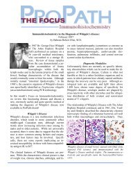
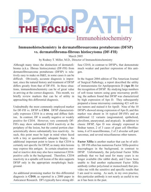
Although many times the distinction of <strong>dermatofibroma</strong>(a.k.a. <strong>fibrous</strong> <strong>histiocytoma</strong>) (DF-FH) fromdermatofibrosarcoma protuberans (<strong>DFSP</strong>) is relativelyeasy to make on H&E, in some cases it can bedifficult. Obviously, accurate diagnosis is important,since the natural history and treatment of <strong>DFSP</strong>differs greatly from that of DF-FH. In these situations,immunohistochemistry can be of great valuein arriving at the correct diagnosis. This month, webriefly review markers that can be of utility inapproaching this differential diagnosis.Undoubtedly the most commonly employed markerfor DF-FH <strong>vs</strong>. <strong>DFSP</strong> is CD34. <strong>DFSP</strong> characteristicallyexpresses CD34 in a strong and diffuse fashion.In contrast, DF is usually negative or weaklypositive for CD34. However, very commonly DF-FH's may show substantial CD34 reactivity at theperiphery of the lesion, but the central portion characteristicallyshows substantially less reactivity. Assuch, this point must be kept in mind when facedwith a tiny or questionably adequate biopsy. Anadditional important point to know is that CD34 iscertainly not specific for <strong>DFSP</strong>, as many skin lesionsmay express this antigen. In certain situations normalor reactive skin may also have numerous CD34-positive cells in the background. Therefore, CD34reactivity in a spindle cell lesion of the skin supports<strong>DFSP</strong> only in the appropriate morphologic background.ImmunohistochemistryImmunohistochemistry in dermatofibrosarcoma protuberans (<strong>DFSP</strong>)<strong>vs</strong>. <strong>dermatofibroma</strong>-<strong>fibrous</strong> <strong>histiocytoma</strong> (DF-FH)March 2005by Rodney T. Miller, M.D., Director of ImmunohistochemistryAn additional promising marker for this differentialdiagnosis is CD10, as reported in a 2000 paper inAnticancer Research. DF's typically have strong diffuseCD10, in contrast to <strong>DFSP</strong>'s, that demonstratemuch weaker and patchier expression of this antigen.In the August 2004 edition of The American Journalof Surgical Pathology, a report described the utilityof immunostains for Apolipoprotein D (Apo D) forthe recognition of <strong>DFSP</strong>. By studying large numbersof soft tissue tumors using gene microarray profiling,the authors found that <strong>DFSP</strong> was characterizedby high expression of Apo D. They subsequentlyprepared a tissue microarray containing 421 soft tissuetumors and stained it for ApoD. Nine of the 10<strong>DFSP</strong>'s showed strong expression of Apo D, and thismarker was absent in 16 typical DF-FH's and anadditional 12 variants (angiomatoid, epithelioid,plexiform, aneurysmal, and atypical). In addition toclassic <strong>DFSP</strong>, Apo D was strongly expressed inBednar tumor, 2 of 3 cases of giant cell fibroblastoma,6 of 8 neurofibromas, 2 of 2 alveolar soft partsarcomas, and several miscellaneous other tumors.Factor XIIIa is another marker that can be of utility.DF-FH often has numerous Factor XIIIa-positivemacrophages in the background, in contrast to<strong>DFSP</strong>. Unfortunately, the polyclonal Factor XIIIaantibody that we had used for many years is nolonger available (the rabbit died), and I have beenunable to find another replacement Factor XIIIaantibody (either polyclonal or monoclonal) that providesme with the sensitive and specific results thatI am used to seeing. As such, in my own practice,this particular antibody is not nearly as useful to meas it has been in the past.
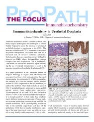
THE FOCUS - Immunohistochemistry March 2005H&EDF-FH<strong>DFSP</strong>CD34neg orDF-FHweak +<strong>DFSP</strong> strong +CD10 Apo D FXIIIa NGFRstrong +neg orweak +many+cellsneg orweak + strong + few+cellsneg orweak +modstrong+Expected immunostain results in “classic” cases of DF-FHand <strong>DFSP</strong>CD34CD10Apo DAlas, nothing is ever perfect, including the borderbetween <strong>DFSP</strong> and DF-FH. Some authors havedescribed tumors that have indeterminate or overlappingfeatures between DF-FH and <strong>DFSP</strong>, and indeedwe have seen several of these cases at ProPath overthe past few years.In most cases, the distinction of DF-FH from <strong>DFSP</strong>can be made with 1 or 2 immunostains, depending onthe particular pathologist's preferences. I usuallystart with CD34 and CD10 or ApoD and CD10, andprogress to other markers if needed.All of the markers discussed above are available atthe ProPath Immunohistochemistry Laboratory.REFERENCES:West RB et al: Apo D in soft tissue tumors. A novel marker for dermatofibrosarcomaprotuberans. Am J Surg Pathol 28(8): 1063-1069,Aug 2004.FXIIIaNGFRHigh power photomicrographs of H&E and immunostainsfrom a typical DF (left column) and <strong>DFSP</strong> (right column).Kanitakis J et al: Expression of the CD10 antigen (neutral endopeptidase)by mesenchymal tumors of the skin. Anticancer Res 20(5B):3539-3544, Sep-Oct 2000.Miettinen M: Diagnostic soft tissue pathology. Churchill-Livingstone,New York, 2003, page 56.Horenstein M et al: Indeterminate fibrohistiocytic lesions of the skin.Is there a spectrum between <strong>dermatofibroma</strong> and dermatofibrosarcomaprotuberans? Am J Surg Pathol 24(7): 996-1003, 2000.Rodney T. Miller, M.D.Director of Immunohistochemistry214-237-1631 • Fax 214-237-1770rmiller@propathlab.comPrior Editions are available for download on our Web site.Some authorities (Miettinen) have written that nervegrowth factor receptor (NGFR) can be useful to distinguish<strong>DFSP</strong> (positive for NGFR) from DF-FH(negative for NGFR), although I have not found thisto be particularly useful in my practice.The Leader in Pathology Services8267 Elmbrook Dr, Ste 100 • Dallas, Texas 75247-4009(214) 638-2000 • Fax: (214) 905-3457www.propathlab.com© 2005 ProPath. All Rights Reserved.