Forensic DNA Analysis on Microfluidic Devices: A Review
Forensic DNA Analysis on Microfluidic Devices: A Review
Forensic DNA Analysis on Microfluidic Devices: A Review
- No tags were found...

You also want an ePaper? Increase the reach of your titles
YUMPU automatically turns print PDFs into web optimized ePapers that Google loves.
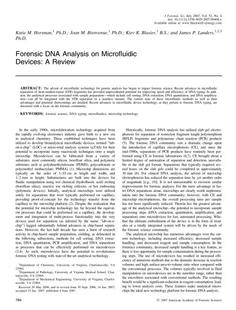
J <str<strong>on</strong>g>Forensic</str<strong>on</strong>g> Sci, July 2007, Vol. 52, No. 4doi: 10.1111/j.1556-4029.2007.00468.xAvailable <strong>on</strong>line at: www.blackwell-synergy.comKatie M. Horsman, 1 Ph.D.; Joan M. Bienvenue, 1 Ph.D.; Kiev R. Blasier, 1 B.S.; and James P. Landers, 1,2,3Ph.D.<str<strong>on</strong>g>Forensic</str<strong>on</strong>g> <str<strong>on</strong>g>DNA</str<strong>on</strong>g> <str<strong>on</strong>g>Analysis</str<strong>on</strong>g> <strong>on</strong> <strong>Microfluidic</strong><strong>Devices</strong>: A <strong>Review</strong>ABSTRACT: The advent of microfluidic technology for genetic analysis has begun to impact forensic science. Recent advances in microfluidicseparati<strong>on</strong> of short-tandem-repeat (STR) fragments has provided unprecedented potential for improving speed and efficiency of <str<strong>on</strong>g>DNA</str<strong>on</strong>g> typing. In additi<strong>on</strong>,the analytical processes associated with sample preparati<strong>on</strong>––which include cell sorting, <str<strong>on</strong>g>DNA</str<strong>on</strong>g> extracti<strong>on</strong>, <str<strong>on</strong>g>DNA</str<strong>on</strong>g> quantitati<strong>on</strong>, and <str<strong>on</strong>g>DNA</str<strong>on</strong>g> amplificati<strong>on</strong>––canall be integrated with the STR separati<strong>on</strong> in a seamless manner. The current state of these microfluidic methods as well as theiradvantages and potential shortcomings are detailed. Recent advances in microfluidic device technology, as they pertain to forensic <str<strong>on</strong>g>DNA</str<strong>on</strong>g> typing, arediscussed with a focus <strong>on</strong> the forensic community.KEYWORDS: forensic science, <str<strong>on</strong>g>DNA</str<strong>on</strong>g> typing, microfluidics, microchip technologyIn the early 1990s, microfabricati<strong>on</strong> technology acquired fromthe rapidly evolving electr<strong>on</strong>ics industry gave birth to a new erain analytical chemistry. These established techniques have beenutilized to develop bioanalytical microfluidic devices, termed ‘‘lab<strong>on</strong>-a-chip’’(LOC) or micro-total analysis systems (lTAS) for theirpotential to incorporate many macroscale techniques <strong>on</strong>to a singlemicrochip. Microdevices can be fabricated from a variety ofsubstrates, most comm<strong>on</strong>ly silic<strong>on</strong>, borofloat silica, and polymericsubstrates such as polydimethylsiloxane (PDMS), polycarb<strong>on</strong>ate orpoly(methyl methacrylate; PMMA) (1). Microchip dimensi<strong>on</strong>s aretypically <strong>on</strong> the order of 1–10 cm in length and width, and1–2 mm in height. Substructures are built into the devices forfluidic manipulati<strong>on</strong> using wet chemical (hydrofluoric acid) etching(borofloat silica), reactive i<strong>on</strong> etching (silic<strong>on</strong>), or hot embossing(polymeric devices). Initially, analytical microchips were utilizedsolely for separati<strong>on</strong>s that were typically performed <strong>on</strong> capillary,providing proof-of-c<strong>on</strong>cept for the technology transfer from thecapillary to the microchip platform (2). Despite the realizati<strong>on</strong> thatthe potential for microchip technology lay far bey<strong>on</strong>d the equivalentprocesses that could be performed <strong>on</strong> a capillary, the developmentand integrati<strong>on</strong> of multi-process functi<strong>on</strong>ality into the verydevices used for separati<strong>on</strong> (as inferred by the name ‘‘lab-<strong>on</strong>-achip’’)lagged substantially behind advances in chip-based separati<strong>on</strong>s.However, the last half decade has seen a burst of researchactivity in chip-based sample preparati<strong>on</strong>, yielding, as delineated inthe following subsecti<strong>on</strong>s, methods for cell sorting, <str<strong>on</strong>g>DNA</str<strong>on</strong>g> extracti<strong>on</strong>,<str<strong>on</strong>g>DNA</str<strong>on</strong>g> quantitati<strong>on</strong>, PCR amplificati<strong>on</strong>, and <str<strong>on</strong>g>DNA</str<strong>on</strong>g> separati<strong>on</strong>sas processes that can be effectively performed <strong>on</strong> microdevices(3,4). As such, microdevices have the potential to revoluti<strong>on</strong>izeforensic <str<strong>on</strong>g>DNA</str<strong>on</strong>g> testing with state-of-the-art analytical technology.1 Department of Chemistry, University of Virginia, Charlottesville, VA22904.2 Department of Pathology, University of Virginia Medical School, Charlottesville,VA 22904.3 Department of Mechanical Engineering, University of Virginia, Charlottesville,VA 22904.Received 20 May 2006; and in revised form 30 Sept. 2006, 14 Jan. 2007;accepted 15 Jan. 2007; published 4 June 2007.Historically, forensic <str<strong>on</strong>g>DNA</str<strong>on</strong>g> analysis has utilized slab gel electrophoresisfor separati<strong>on</strong> of restricti<strong>on</strong> fragment length polymorphism(RFLP) fragments and polymerase chain reacti<strong>on</strong> (PCR) products(5). The forensic <str<strong>on</strong>g>DNA</str<strong>on</strong>g> community saw a dramatic change up<strong>on</strong>the introducti<strong>on</strong> of capillary electrophoresis (CE), and since themid-1990s, separati<strong>on</strong>s of PCR products have routinely been performedusing CE in forensic laboratories (6,7). CE brought about alimited degree of automati<strong>on</strong> of separati<strong>on</strong> and detecti<strong>on</strong>, unavailablein the slab gel format. Separati<strong>on</strong>s that typically required severalhours <strong>on</strong> the slab gels could be completed in approximately30 min (8). For clinical <str<strong>on</strong>g>DNA</str<strong>on</strong>g> analysis, the advent of microchipelectrophoresis has reduced the separati<strong>on</strong> time by yet another orderof magnitude [e.g., (9)]. It is not unreas<strong>on</strong>able to envisi<strong>on</strong> similarimprovements for forensic analyses. For the mere advantage in faster<str<strong>on</strong>g>DNA</str<strong>on</strong>g> separati<strong>on</strong>s al<strong>on</strong>e, microchips are clearly worth implementati<strong>on</strong>into the forensic <str<strong>on</strong>g>DNA</str<strong>on</strong>g> community; however, with CE andmicrochip electrophoresis, the overall processing time per samplehas not been significantly reduced. Therein lies the greatest advantageof the analytical microchip––the potential to incorporate sampleprocessing steps (<str<strong>on</strong>g>DNA</str<strong>on</strong>g> extracti<strong>on</strong>, quantitati<strong>on</strong>, amplificati<strong>on</strong>, andseparati<strong>on</strong>) <strong>on</strong>to microdevices for fast, automated processing. Whetherthe ultimate embodiment of the device is in the form of modulesor a totally integrated system will be driven by the needs ofthe forensic science community.The analytical microchip has numerous advantages over the currenttechnology, including increased efficiency, decreased samplehandling, and decreased reagent and sample c<strong>on</strong>sumpti<strong>on</strong>. In theforensics community, decreased sample handling is a key feature, asthere is less opportunity for sample c<strong>on</strong>taminati<strong>on</strong> during the processingsteps. The use of microdevices has resulted in increased efficiencyof numerous methods due to the dramatic decrease in reacti<strong>on</strong>volumes and high surface-area-to-volume ratio when compared withthe c<strong>on</strong>venti<strong>on</strong>al processes. The volumes typically involved in fluidmanipulati<strong>on</strong> <strong>on</strong> microdevices are in the nanoliter range, rather thanthe microliters associated with c<strong>on</strong>venti<strong>on</strong>al methods. The resultingbenefit would be a significant reducti<strong>on</strong> in reagent c<strong>on</strong>sumpti<strong>on</strong>, leadingto lower analysis costs. These features make analytical microchipsthe ideal new technology platform for forensic <str<strong>on</strong>g>DNA</str<strong>on</strong>g> analysis.784 Ó 2007 American Academy of <str<strong>on</strong>g>Forensic</str<strong>on</strong>g> Sciences
HORSMAN ET AL. • <str<strong>on</strong>g>DNA</str<strong>on</strong>g> MICROFLUIDICS 785Motivati<strong>on</strong>With the backlog of <str<strong>on</strong>g>DNA</str<strong>on</strong>g> evidence to be analyzed in ournati<strong>on</strong>’s forensic laboratories, the need for a new era in forensic<str<strong>on</strong>g>DNA</str<strong>on</strong>g> analysis technology is increasingly evident. According to theNati<strong>on</strong>al Institute of Justice 2004 Annual Report, estimates placethe <str<strong>on</strong>g>DNA</str<strong>on</strong>g> backlog at approximately 800,000 c<strong>on</strong>victed offendersamples and approximately 485,000 criminal cases awaiting analysisin our nati<strong>on</strong>’s state and federal crime labs, primarily due tolimited resources, such as time and funding (10). The backlogundermines the full potential of <str<strong>on</strong>g>DNA</str<strong>on</strong>g> databases, as numeroussamples have not been typed and are, therefore, not yet included.Currently, forensic <str<strong>on</strong>g>DNA</str<strong>on</strong>g> analysis, including <str<strong>on</strong>g>DNA</str<strong>on</strong>g> extracti<strong>on</strong>,quantitati<strong>on</strong>, amplificati<strong>on</strong>, and separati<strong>on</strong>, requires approximately12–36 h of linear time to complete, much of which requires directtechnician time. An integrated forensic <str<strong>on</strong>g>DNA</str<strong>on</strong>g> analysis microdevice,however, could complete these processes <strong>on</strong> the order of 1–2 h––all in an automated system, reducing not <strong>on</strong>ly analysis time butalso user interventi<strong>on</strong>. It is the tremendous inherent potential ofmicrodevices that stands to revoluti<strong>on</strong>ize forensic <str<strong>on</strong>g>DNA</str<strong>on</strong>g> analysisand warrants attenti<strong>on</strong>.Sample Processing StepsAs with almost any form of genomic interrogati<strong>on</strong>, forensic<str<strong>on</strong>g>DNA</str<strong>on</strong>g> analysis requires numerous sample preparati<strong>on</strong> steps prior tothe analytical step that generates an STR profile, all of which couldbenefit by translati<strong>on</strong> to the microchip format. The steps discussedin detail in the following secti<strong>on</strong> include differential extracti<strong>on</strong>,<str<strong>on</strong>g>DNA</str<strong>on</strong>g> purificati<strong>on</strong>, <str<strong>on</strong>g>DNA</str<strong>on</strong>g> quantitati<strong>on</strong>, PCR, and short tandem repeat(STR) fragment separati<strong>on</strong>s. While the theory behind each processingstep remains largely unchanged, the specific methods have beenaltered to varying extents, to accommodate the microchip formatand improve performance. Although not an exhaustive discussi<strong>on</strong>of the pertinent literature, the following secti<strong>on</strong>s present the currentstate of microfluidic technology as applied to these analytical processesby alluding to select reports in the literature that we feel areparticularly relevant.Cell Sorting ⁄ Differential Extracti<strong>on</strong><str<strong>on</strong>g>Analysis</str<strong>on</strong>g> of sexual assault evidence requires the use of ‘‘differentialextracti<strong>on</strong>’’ to obtain separate fracti<strong>on</strong>s of male and female<str<strong>on</strong>g>DNA</str<strong>on</strong>g>. Differential extracti<strong>on</strong> is <strong>on</strong>e bottleneck in the automati<strong>on</strong> offorensic <str<strong>on</strong>g>DNA</str<strong>on</strong>g> analysis. While significant improvements to the other‘‘core’’ processes have been addressed to enhance throughput, differentialextracti<strong>on</strong> has advanced relatively little in the same period.The c<strong>on</strong>venti<strong>on</strong>al method involves differential lysis of sperm andepithelial cells, employing centrifugati<strong>on</strong> to separate the epithelialcell <str<strong>on</strong>g>DNA</str<strong>on</strong>g> (female fracti<strong>on</strong>) from the sperm cells (male fracti<strong>on</strong>).Although not routine, microfluidic c<strong>on</strong>trol of flow <strong>on</strong> microdeviceshas been shown possible using centrifugati<strong>on</strong> as a driving force ina compact disc-like device (11) and for clinical applicati<strong>on</strong>s thatinclude preparati<strong>on</strong> of plasma from whole blood (12). While theapproach has not been applied to forensic differential extracti<strong>on</strong>,other methods have been pursued as an alternative for obtainingmale and female fracti<strong>on</strong>s <strong>on</strong> microfluidic devices.One microscale method for differential extracti<strong>on</strong> (13) used lowpowers<strong>on</strong>icati<strong>on</strong> for selective epithelial cell lysis and a filter (poresize not indicated) to separate the epithelial cell lysate from theintact sperm cells ⁄ heads. Sperm were subsequently lysed usingmore-stringent c<strong>on</strong>diti<strong>on</strong>s, followed by <str<strong>on</strong>g>DNA</str<strong>on</strong>g> extracti<strong>on</strong> from bothcell lysates <strong>on</strong> the same device (Fig. 1). The differential extracti<strong>on</strong>process using this microfluidic-based system was fully automatedand yielded male and female fracti<strong>on</strong>s of <str<strong>on</strong>g>DNA</str<strong>on</strong>g> in
786 JOURNAL OF FORENSIC SCIENCES(a)(b)FIG. 2—Separati<strong>on</strong> of sperm and epithelial cells <strong>on</strong> a microfluidic device. (a) The sperm and epithelial cell mixture is added to the inlet reservoir of themicrodevice. (b) The epithelial cells sediment to the bottom of the inlet reservoir (t = 0). After 5 min (t = 1), flow is induced (t = 2) to mobilize the spermcells, whereas the epithelial cells are retained in the inlet reservoir, adsorbed to the glass substrate. At t = 3, the sperm cells are collected in the outlet reservoir,from which further sample processing (<str<strong>on</strong>g>DNA</str<strong>on</strong>g> extracti<strong>on</strong>, PCR amplificati<strong>on</strong>, and <str<strong>on</strong>g>DNA</str<strong>on</strong>g> separati<strong>on</strong>) can occur. Photomicrographs of the inlet reservoirand channel show the epithelial cells and sperm cells, respectively, at a late stage of the separati<strong>on</strong>. (Figure adapted from and reproduced with permissi<strong>on</strong>from Horsman et al., Analytical Chemistry 2005, 77, 742–9. Copyright 2005 American Chemical Society.)(14). The separati<strong>on</strong> was based up<strong>on</strong> differential sedimentati<strong>on</strong> ratesof the two cell types possessing distinct physicochemical properties.As shown in Fig. 2, a mixture of sperm and epithelial cells areintroduced to the inlet reservoir. After 5 min of settling time, thesperm cells were mobilized in the channel with a negative-pressure,syringe-pump driven flow. The sperm cells were then collected inthe outlet (right) reservoir, while the epithelial cells remain in theinlet (left) reservoir. A shortcoming of the approach is the potentialpresence of free <str<strong>on</strong>g>DNA</str<strong>on</strong>g> in the sample, most likely from lysed epithelialcells, although this can be addressed through modificati<strong>on</strong> ofthe microchannel surface. While the method is the <strong>on</strong>ly publishedmicroscale technique specifically addressing sexual assault evidence,other established macroscale cell-sorting techniques arepotentially applicable to this sample.A comm<strong>on</strong> cell sorting mechanism that has gained widespreaduse in the macroscale is fluorescence-activated cell sorting (FACS).The Quake lab (15,16) dem<strong>on</strong>strated FACS <strong>on</strong> the microscale, insilic<strong>on</strong> ⁄ PDMS microdevices. The technique required that <strong>on</strong>e (orboth) of the cell types be fluorescently tagged for detecti<strong>on</strong> by thesystem and, as a result, lead to a reproducible method of cell sorting.However, a major drawback was the need to fluorescently tag<strong>on</strong>e cell type and utilize an optical setup or fluorescence detecti<strong>on</strong>,which, for a stand-al<strong>on</strong>e instrument, would add substantially to thecost of the microchip method. Schoell et al. (17,18) applied flowcytometry, in the macroscale, to the separati<strong>on</strong> of sperm and epithelialcells, using fluorescently tagged antibodies to cell surfaceantigens or a <str<strong>on</strong>g>DNA</str<strong>on</strong>g> stain to distinguish the cell types. In the case ofthe <str<strong>on</strong>g>DNA</str<strong>on</strong>g> stain, the cells were distinguished via stain intensity and,thus, ploidy. The results presented were notable as they yieldedhigher sensitivity than the c<strong>on</strong>venti<strong>on</strong>al differential extracti<strong>on</strong> technique.However, a drawback to the use of antibodies is that theextent of degradati<strong>on</strong> of cell membranes in forensic samples isunknown and could substantially diminish the antibody recogniti<strong>on</strong>of cell type. In additi<strong>on</strong>, the sensitivity of the technique using the<str<strong>on</strong>g>DNA</str<strong>on</strong>g> stain is unclear when based solely <strong>on</strong> ploidy. The authorsindicated that use of the technique would require changing themethod of evidence collecti<strong>on</strong> from avaginalswabtoflushingthevagina with an isot<strong>on</strong>ic soluti<strong>on</strong>.Sperm capture was also dem<strong>on</strong>strated using dielectrophoresis, inwhich cells differentially migrate toward a c<strong>on</strong>centrated electricfield depending up<strong>on</strong> their physical properties, <strong>on</strong> a microdevice(19). The method, developed for the infertility industry, has obviousextensi<strong>on</strong> to forensic differential extracti<strong>on</strong>, as the electric field canbe utilized to trap the cells of interest, including sperm cells in acellular ⁄ biological mixture, based up<strong>on</strong> their physicochemical propertiessuch as c<strong>on</strong>ductivity and permittivity. A potential drawbackto the technique is the adherence of sperm cells to the microchipsubstrate at the trapping site. An analogous technique using anacoustic field to trap (and levitate) the sperm cells in the middle ofthe microchannel (rather than at the microchannel surface) is beingdeveloped to circumvent the limitati<strong>on</strong> (Fig. 3). The particletrappingtechnique utilizing acoustic standing waves has beendeveloped by the Laurell and Nilss<strong>on</strong> groups (20,21) and wasrecently applied to forensic differential extracti<strong>on</strong> (22). Up<strong>on</strong> infusi<strong>on</strong>of the sample, sperm cells were trapped in the near field of anultras<strong>on</strong>ic res<strong>on</strong>ator above a piezoceramic microtransducer, whereasfree <str<strong>on</strong>g>DNA</str<strong>on</strong>g> (from lysed epithelial cells) was not retained but directedto a reservoir for subsequent testing. After washing the trapped
HORSMAN ET AL. • <str<strong>on</strong>g>DNA</str<strong>on</strong>g> MICROFLUIDICS 787<strong>on</strong> a microchip would be relatively straightforward, with the majorhurdles likely being the microfabricati<strong>on</strong> and potential clogging ofthe microchannel.Although not specifically developed for the microchip platform,Eisenberg (25) reported an antibody-based approach to capturesperm cells. A cocktail of antibodies to sperm cell surface antigenswas anchored to magnetic beads over which the sample flows. Thebeauty of the approach is the ability to exploit the specificity ofAb–Ag binding to selectively capture sperm cells from evidentiarymaterial that may be comprised of a mixture of sperm cells, whiteblood cells, epithelial cells, cell lysates, etc. The major drawback,as discussed with the FACS method above, is the unknown levelof sperm cell membrane degradati<strong>on</strong> that will render the sperm cellunamenable to antibody complexati<strong>on</strong>. Additi<strong>on</strong>ally, the introducti<strong>on</strong>of unlysed epithelial cells into the column could significantlycompromise the efficiency of the separati<strong>on</strong>. Although the methodhas not been dem<strong>on</strong>strated <strong>on</strong> a microfluidic platform, <strong>on</strong>e couldenvisage the translati<strong>on</strong> of the separati<strong>on</strong> mechanism to a microdevice,with the provisi<strong>on</strong> that the drawbacks described could beeffectively addressed.As indicated, the translati<strong>on</strong> of c<strong>on</strong>venti<strong>on</strong>al differential extracti<strong>on</strong>to microdevices is still in its infancy. While <strong>on</strong>ly a fewmicrochip methods specifically addressing the separati<strong>on</strong> of spermand epithelial cells ⁄ <str<strong>on</strong>g>DNA</str<strong>on</strong>g> have been dem<strong>on</strong>strated to date, a numberof methods have shown potential for applicati<strong>on</strong> to the particularanalysis. These microchip-based methods, <strong>on</strong>ce fullydeveloped, will require rigorous validati<strong>on</strong> and testing with caseworksamples to fully dem<strong>on</strong>strate their capabilities and benefits.Integrati<strong>on</strong> of differential extracti<strong>on</strong> with downstream sample processingtechniques also remains to be dem<strong>on</strong>strated.FIG. 3—Acoustic trapping of cells in a microfluidic device. (a) Photographof the glass channel structure of the microdevice. The microtransducers(seen in cut-away view) are placed directly below the intersecti<strong>on</strong>s ofthe channels. A cut-away view of the center of the main channel in theacoustic trapping device is also shown. Cells flow through the microfluidicchannels and directly over the microtransducers (fabricated into a printedcircuit board layer). Up<strong>on</strong> activati<strong>on</strong> of the transducers, an acoustic standingwave is set up in the microdevice, resulting in a pressure minimum inthe center of the channel (as indicated), where cells are trapped. (b) Photomicrographillustrating the trapping of fluorescently tagged polystyrenebeads in the microdevice. The beads are trapped at the intersecti<strong>on</strong> of themain channel and a side channel. The channel walls are indicated in white.Outlined in red is the approximate size of the transducer, above which thecells are trapped. (A porti<strong>on</strong> of the figure (a, bottom) adapted with permissi<strong>on</strong>from Nilss<strong>on</strong> et al., Acoustic Trapping of Cells in a <strong>Microfluidic</strong> Format.Micro Total <str<strong>on</strong>g>Analysis</str<strong>on</strong>g> Systems 2005, Proceedings of the mTAS 2005Symposium, 9th, Bost<strong>on</strong>, MA. 2005;5. Photographs courtesy of T. Laurelland J. Nilss<strong>on</strong> of Lund University, Sweden.)cells with buffer, the sperm cells were released and directed into asec<strong>on</strong>d reservoir.A macroscale cell sorting method was also developed using an8 lm filter, which allows sperm cells to pass through the filter,while epithelial cells were retained (23). A drawback to the methodis that any free <str<strong>on</strong>g>DNA</str<strong>on</strong>g> or nuclei from lysed epithelial cells will penetratethe membrane and, thus, c<strong>on</strong>taminate the sperm cell fracti<strong>on</strong>.In an alternate manifestati<strong>on</strong> (24), a 2 lm filter was utilized to separateepithelial cell lysate from sperm cells, which circumvents theproblem encountered above with respect to lysed epithelial cell<str<strong>on</strong>g>DNA</str<strong>on</strong>g> c<strong>on</strong>taminating the sperm cell fracti<strong>on</strong>. As clogging can be aproblem with filter-based separati<strong>on</strong> mechanisms, the authors includedan 11 lm prefilter. While filter-based methods are not currentlyperformed <strong>on</strong> microdevices, implementati<strong>on</strong> of the separati<strong>on</strong><str<strong>on</strong>g>DNA</str<strong>on</strong>g> Purificati<strong>on</strong>Historically, c<strong>on</strong>venti<strong>on</strong>al methods utilized to purify <str<strong>on</strong>g>DNA</str<strong>on</strong>g> forforensic analysis have typically included an organic extracti<strong>on</strong> (e.g.,phenol chloroform) step. These protocols, although effective forrecovering high-molecular-weight <str<strong>on</strong>g>DNA</str<strong>on</strong>g> for downstream analysis,require multiple vortexing, centrifugati<strong>on</strong>, and transfer steps thatarenoteasilytranslatedtomicrofluidic platforms. More recentlydeveloped techniques, however, have shifted to the use of solidphase extracti<strong>on</strong> (SPE) methods (Qiagen, <str<strong>on</strong>g>DNA</str<strong>on</strong>g> IQ, etc.), reducingthe time required for the extracti<strong>on</strong> while maintaining recovery andsample purity and integrity. In additi<strong>on</strong> to enabling faster samplepreparati<strong>on</strong>, these protocols are more easily translated into microdeviceformats. Solid phases such as silica beads, sol-gels, or i<strong>on</strong>exchange resins can be easily packed into microdevices to create aSPE bed or column for <str<strong>on</strong>g>DNA</str<strong>on</strong>g> purificati<strong>on</strong>. Additi<strong>on</strong>ally, a varietyof novel solid phases can be created in microdevices during thefabricati<strong>on</strong> process that are suitable for sample purificati<strong>on</strong>.Although many of these methods were developed for clinical diagnosticapplicati<strong>on</strong>s, the devices and techniques should be readilytranslatable to forensic applicati<strong>on</strong>s, and a number research groupsare focusing efforts <strong>on</strong> c<strong>on</strong>cerns unique to the forensic community.The advancement of these methods is key to the establishment ofsample preparatory steps in microdevices, either as stand-al<strong>on</strong>e systemsor as an essential comp<strong>on</strong>ent of a fully integrated, microchipbasedanalysis to speed casework handling.The first instance of a truly microchip-based SPE was publishedby Christel et al. (26) in 1999. A silic<strong>on</strong> microdevice c<strong>on</strong>tainingpillars for high surface-area-to-volume ratios to increase <str<strong>on</strong>g>DNA</str<strong>on</strong>g>adsorpti<strong>on</strong> was designed by the group for the purificati<strong>on</strong> and c<strong>on</strong>centrati<strong>on</strong>of <str<strong>on</strong>g>DNA</str<strong>on</strong>g> for PCR amplificati<strong>on</strong>. As comm<strong>on</strong>ly utilizedwith other silica-based SPE methods, a chaotropic salt (guanidine
788 JOURNAL OF FORENSIC SCIENCESHCl) was used as the binding ⁄load soluti<strong>on</strong>, followed by an ethanolwash to remove proteins, and <str<strong>on</strong>g>DNA</str<strong>on</strong>g> eluti<strong>on</strong> with water. This representedthe first microchip-based <str<strong>on</strong>g>DNA</str<strong>on</strong>g> purificati<strong>on</strong> accomplished;however, the complex reactive-i<strong>on</strong>-etching protocol utilized to fabricatethe solid phase, combined with the high cost to produce sucha device, limits the potential utility of the format. In additi<strong>on</strong>, captureefficiencies reported with the device using stock lambda <str<strong>on</strong>g>DNA</str<strong>on</strong>g>were <strong>on</strong>ly 50%, limiting its utility for low copy purificati<strong>on</strong>s whereretenti<strong>on</strong> of <str<strong>on</strong>g>DNA</str<strong>on</strong>g> is imperative. Cady et al. (27) have employed asimilar design c<strong>on</strong>cept with the goal of providing a device withhigher binding capacity (>200 ng). However, it was not possible togauge the effectiveness of the device for forensic analysis as noextracti<strong>on</strong> efficiencies were reported. While efforts toward extracting<str<strong>on</strong>g>DNA</str<strong>on</strong>g> from larger volume samples are certainly needed, extracti<strong>on</strong>must yield a reas<strong>on</strong>ably c<strong>on</strong>centrated <str<strong>on</strong>g>DNA</str<strong>on</strong>g> soluti<strong>on</strong> amenableto downstream processes (e.g., PCR). With <str<strong>on</strong>g>DNA</str<strong>on</strong>g> eluted from thesolid phase in an unusually large volume (250 lL) in the work(27), the likelihood that such a method could be effectively integratedwith other microchip processes is minimized. While the extracti<strong>on</strong>efficiencies attainable with chromatographic solid phasescreated by microfabricated pillars are not readily apparent in the literature,there is no questi<strong>on</strong> that, in the future, high-surface-area,functi<strong>on</strong>alized solid phases may prove to be a robust and efficientmeans for <str<strong>on</strong>g>DNA</str<strong>on</strong>g> purificati<strong>on</strong>. As depicted in Fig. 4, functi<strong>on</strong>alizedsurfaces c<strong>on</strong>taining pillars and ⁄or pores that provide increased surfaceareas, and a c<strong>on</strong>comitant increase in capacity, for <str<strong>on</strong>g>DNA</str<strong>on</strong>g> bindingcan be microfabricated, without the issues of back pressure anddevice filling associated with packed solid phases. These newphases may provide reproducibly uniform surfaces with which toextract <str<strong>on</strong>g>DNA</str<strong>on</strong>g>.In a more direct translati<strong>on</strong> of current macroscale extracti<strong>on</strong> protocols,other microchip-based purificati<strong>on</strong> systems have focused <strong>on</strong>utilizing a silica bead or silica sol-gel matrix solid phase for <str<strong>on</strong>g>DNA</str<strong>on</strong>g>purificati<strong>on</strong>. The use of these solid phases for <str<strong>on</strong>g>DNA</str<strong>on</strong>g> extracti<strong>on</strong> wasfirst miniaturized in a capillary format to dem<strong>on</strong>strate the utility ofthe proposed method in the microscale (28). Tian et al. (28),employing a 500 nL capillary-based chamber packed with a silicaparticles, established that PCR-amplifiable <str<strong>on</strong>g>DNA</str<strong>on</strong>g> (with 80–90% ofproteins removed during the load and wash steps) could beobtained from white blood cells with high extracti<strong>on</strong> efficiencies(70%). The work dem<strong>on</strong>strated both the suitability of microminiaturizedSPE methods for a wide variety of biological species (whiteblood cells, cultured cells, whole blood) and the feasibility of incorporatingsuch methods into microfabricated devices.The microscale extracti<strong>on</strong> technique was extrapolated to silicamicrodevices by Wolfe et al. (29) who evaluated a variety of silicaand silica ⁄ sol-gel matrices for <str<strong>on</strong>g>DNA</str<strong>on</strong>g> extracti<strong>on</strong>. In the work, theauthors highlight the potential problem associated with using silicabeads or particles in a microdevice: the tendency of these particlesto pack more tightly under flow as multiple extracti<strong>on</strong>s proceed,thus affecting the reproducibility of repeated extracti<strong>on</strong>s. The challengewas circumvented in designs such as those proposed by Christel(26) and Cady (27), which c<strong>on</strong>tain microfabricated pillars thatare part of the channel. Recent work in our lab suggests that if thedevices are single use, packed silica bead solid phases are acceptablepurificati<strong>on</strong> phases for <str<strong>on</strong>g>DNA</str<strong>on</strong>g> extracti<strong>on</strong>. Alternatively, sol-gels,liquid colloidal suspensi<strong>on</strong>s of silica-based materials that can beacid or base catalyzed to gel in place, have been dem<strong>on</strong>strated asefficient, reusable solid phases as described by Wolfe et al. (29).These soluti<strong>on</strong>s are simply flowed into microchambers as liquidsand allowed to gel to form a porous <str<strong>on</strong>g>DNA</str<strong>on</strong>g> extracti<strong>on</strong> bed. The catalyzedreacti<strong>on</strong> can be c<strong>on</strong>trolled to create pores that allow enoughsurface area for the binding of <str<strong>on</strong>g>DNA</str<strong>on</strong>g>, as dem<strong>on</strong>strated by Wu et al.(30), who utilized the phase to extract <str<strong>on</strong>g>DNA</str<strong>on</strong>g> from bacterial(anthrax), viral (varicella zoster and herpes simplex), and human(blood) sources, with >65% extracti<strong>on</strong> efficiency from blood. In alater translati<strong>on</strong> of the work, Wen et al. (31) utilized a photopolymerizablesol-gel m<strong>on</strong>olith to extract <str<strong>on</strong>g>DNA</str<strong>on</strong>g> in a capillary-based system,which was further modified for microchip-based extracti<strong>on</strong>(32). The photopolymerizati<strong>on</strong> step allows for easy and precise formati<strong>on</strong>of the solid phase within the microdevice, without the useof retaining weirs or other microfabricated features. In additi<strong>on</strong>, thesolid phase has recently been incorporated into a novel two-stagemicrodevice that was developed for <str<strong>on</strong>g>DNA</str<strong>on</strong>g> extracti<strong>on</strong> from blood––aC18 reverse phase column for protein capture (stage 1) in serieswith a m<strong>on</strong>olithic column for <str<strong>on</strong>g>DNA</str<strong>on</strong>g> extracti<strong>on</strong> (stage 2) (32). Thedevice had a high capacity for <str<strong>on</strong>g>DNA</str<strong>on</strong>g> in blood (>240 ng) and wasfound to achieve 70% extracti<strong>on</strong> efficiency. Further, the sol-gelextracti<strong>on</strong> medium can be not <strong>on</strong>ly be used al<strong>on</strong>e, but also as aglue, to immobilize a silica bead phase, maintaining a reproducibleextracti<strong>on</strong> column from run to run. The latter solid phase was evaluatedbyBreadmoreetal.(33),whooptimized flow rates and loadingpH to affect a sample purificati<strong>on</strong> in 15 min from bacterialsources (anthrax and salm<strong>on</strong>ella) and whole blood. In additi<strong>on</strong>,intra- and interdevice reproducibility was dem<strong>on</strong>strated, with ashigh as 79% extracti<strong>on</strong> efficiency achieved.The utility of this solid phase and extracti<strong>on</strong> protocol was alsodem<strong>on</strong>strated for <str<strong>on</strong>g>DNA</str<strong>on</strong>g> purificati<strong>on</strong> from sperm cells, with a view(a)(b)FIG. 4—An example of a high surface area structure microfabricated out of plastic (polymethylmethacrylate). These structures can be functi<strong>on</strong>alized forefficient capture of nucleic acids, providing large surface areas and increased binding capacity for <str<strong>on</strong>g>DNA</str<strong>on</strong>g> extracti<strong>on</strong>s, as well as reproducible and low-backpressurecolumns for purificati<strong>on</strong>. (a) A channeled structure where the individual channels are 30 lM in diameter. (b) Complementary (inverse) structure tothat shown in (a). (Photo courtesy of HT Microanalytical, Inc. [Albuquerque, NM].)
HORSMAN ET AL. • <str<strong>on</strong>g>DNA</str<strong>on</strong>g> MICROFLUIDICS 789(a)(c)(b)FIG. 5—A microdevice setup for extracti<strong>on</strong> of <str<strong>on</strong>g>DNA</str<strong>on</strong>g> from sperm cells. This includes a syringe pump, c<strong>on</strong>nected to the microdevice with narrow-bore tubing,and held in place by a homemade plexiglass mount (a). Silica beads 5–30 lm in diameter are packed against a weir (b and c) in the channel (200 lm deep,420 lm wide) and adhered into place using sol-gel. (Figure reproduced with permissi<strong>on</strong> from Bienvenue et al., Journal of <str<strong>on</strong>g>Forensic</str<strong>on</strong>g> Sciences 2006, 51(2):266–73. Copyright 2006 Blackwell Publishing, Inc.)toward analysis of sexual assault evidence. Also, excess <str<strong>on</strong>g>DNA</str<strong>on</strong>g> templateadded to the PCR can result in off-scale signal and other PCRartifacts. Bienvenue et al. (34) describe the use of a single-channelextracti<strong>on</strong> device and modificati<strong>on</strong>s to the Breadmore et al. (33) celllysis buffer to allow for <strong>on</strong>-chip sperm cell lysis and <str<strong>on</strong>g>DNA</str<strong>on</strong>g> purificati<strong>on</strong>(Fig. 5). The resultant <str<strong>on</strong>g>DNA</str<strong>on</strong>g> was of suitable purity and c<strong>on</strong>centrati<strong>on</strong>for subsequent STR amplificati<strong>on</strong>, with comprehensive STRprofiles generated. These results dem<strong>on</strong>strate the potential of microchip-basedextracti<strong>on</strong> methods for forensic analysis.Another unique approach to microscale <str<strong>on</strong>g>DNA</str<strong>on</strong>g> extracti<strong>on</strong> currentlyunder development utilizes a serpentine channel design combinedwith an immobilized silica bead solid phase and fluidic oscillati<strong>on</strong>.The method, developed by Chung et al. (35), relies <strong>on</strong> an immobilizedsilica solid phase. Instead of a solid column of packed phase,the silica beads were immobilized <strong>on</strong> the plasma-oxidized surfaceof the PMMA channels. Following bead immobilizati<strong>on</strong>, the soluti<strong>on</strong>srequired for <str<strong>on</strong>g>DNA</str<strong>on</strong>g> binding, purificati<strong>on</strong>, and release were flowedback and forth through the device. By oscillating fluid flowover the immobilized phase, marked improvement of recovery andextracti<strong>on</strong> efficiency over the same extracti<strong>on</strong> methods with freebeads was dem<strong>on</strong>strated. The method represents yet another microchip-basedsystem for <str<strong>on</strong>g>DNA</str<strong>on</strong>g> purificati<strong>on</strong> with potential for forensicapplicati<strong>on</strong>.The development of large-scale, commercial, SPE protocolsenabled the facile translati<strong>on</strong> of <str<strong>on</strong>g>DNA</str<strong>on</strong>g> extracti<strong>on</strong> into microfluidicsystems. The methods described here represent the first examplesof microchip-based sample preparati<strong>on</strong> for downstream sample processingand a major step toward the development of microfluidicsystems capable of accepting crude samples for <str<strong>on</strong>g>DNA</str<strong>on</strong>g> analysis. Asadvancement of these technologies c<strong>on</strong>tinues, the focus will likelyturn from basic method development to the incorporati<strong>on</strong> of extracti<strong>on</strong>techniques with other microfluidic processes (cell sorting,<str<strong>on</strong>g>DNA</str<strong>on</strong>g> quantificati<strong>on</strong> PCR amplificati<strong>on</strong>, <str<strong>on</strong>g>DNA</str<strong>on</strong>g> separati<strong>on</strong> and detecti<strong>on</strong>).The implementati<strong>on</strong> of microfluidic <str<strong>on</strong>g>DNA</str<strong>on</strong>g> extracti<strong>on</strong> forforensic analysis will allow the processing of a wide variety ofsamples and is an important comp<strong>on</strong>ent of future fully automatedgenetic analysis.<str<strong>on</strong>g>DNA</str<strong>on</strong>g> Quantitati<strong>on</strong><str<strong>on</strong>g>DNA</str<strong>on</strong>g> quantitati<strong>on</strong> is a necessity in forensic <str<strong>on</strong>g>DNA</str<strong>on</strong>g> analysis, inc<strong>on</strong>trast to most clinical analyses where the sample size is morec<strong>on</strong>sistent and <str<strong>on</strong>g>DNA</str<strong>on</strong>g> c<strong>on</strong>tent more reproducible. Because of the predominanceof samples c<strong>on</strong>taining low c<strong>on</strong>centrati<strong>on</strong>s of <str<strong>on</strong>g>DNA</str<strong>on</strong>g>,quantitati<strong>on</strong> must be carried out prior to PCR in order to minimizestochastic fluctuati<strong>on</strong>s that could cause allelic dropout from unequalamplificati<strong>on</strong> of the two alleles. For forensic use, <str<strong>on</strong>g>DNA</str<strong>on</strong>g> quantitati<strong>on</strong>must be sensitive (mass detecti<strong>on</strong> limit <strong>on</strong> the order of picograms)and specific for human <str<strong>on</strong>g>DNA</str<strong>on</strong>g>.To date, minimal effort has been directed at addressing pre-PCR<str<strong>on</strong>g>DNA</str<strong>on</strong>g> quantitati<strong>on</strong> <strong>on</strong> microdevices. As dem<strong>on</strong>strated with a commercialmicrochip electrophoresis unit that uses an intercalatingagent for detecti<strong>on</strong>, quantitati<strong>on</strong> of <str<strong>on</strong>g>DNA</str<strong>on</strong>g> (typically PCR products)can be accomplished based <strong>on</strong> the separati<strong>on</strong> and detecti<strong>on</strong> ofknown marker c<strong>on</strong>centrati<strong>on</strong>s. While the limit of quantitati<strong>on</strong> is50 pg ⁄ lL (36), it is not practical for forensic samples where<str<strong>on</strong>g>DNA</str<strong>on</strong>g> c<strong>on</strong>centrati<strong>on</strong>s are often substantially below the detecti<strong>on</strong>limit. Direct quantitati<strong>on</strong> of <str<strong>on</strong>g>DNA</str<strong>on</strong>g> <strong>on</strong> microdevices for forensic analysiswill likely require the use of real time PCR which, c<strong>on</strong>venti<strong>on</strong>ally,allows for quantitati<strong>on</strong> of samples in the picogram t<strong>on</strong>anogram per microliter c<strong>on</strong>centrati<strong>on</strong> range. Although PCR hasbeen completed extensively <strong>on</strong> microdevices (as described in thesubsequent secti<strong>on</strong>), the dem<strong>on</strong>strati<strong>on</strong> of quantitative PCR (qPCR)<strong>on</strong> microdevices is still in its infancy.The first reported development of micro-quantitative PCR(l-qPCR) was in 1998, for detecti<strong>on</strong> of viral, bacterial, and human<str<strong>on</strong>g>DNA</str<strong>on</strong>g>. Using silic<strong>on</strong> reacti<strong>on</strong> chambers with thin-film heaters, Northrupet al. (37) dem<strong>on</strong>strated the use of both intercalating dye
790 JOURNAL OF FORENSIC SCIENCES(ethidium bromide) and 5¢-nuclease (Taqman Ò , Applied Biosystems,Foster City, CA) detecti<strong>on</strong> methods. A subsequent paper (38) dem<strong>on</strong>strateddetecti<strong>on</strong> limits of
HORSMAN ET AL. • <str<strong>on</strong>g>DNA</str<strong>on</strong>g> MICROFLUIDICS 791In 1998, a novel method of l-PCR was dem<strong>on</strong>strated using adevice with three heated z<strong>on</strong>es, correlating to the annealing, extensi<strong>on</strong>,and dissociati<strong>on</strong> temperatures of thermal cycling (43). Thereacti<strong>on</strong> mixture was flowed through each z<strong>on</strong>e in <strong>on</strong>e c<strong>on</strong>tinuouschannel. Hold times at each temperature were, c<strong>on</strong>sequently, basedup<strong>on</strong> the flow rate of the mixture through the z<strong>on</strong>es. In subsequentimprovements to the flow through PCR method, Hashimoto et al.(44) reported PCR amplificati<strong>on</strong> of a 99 bp fragment of lambda<str<strong>on</strong>g>DNA</str<strong>on</strong>g> in <strong>on</strong>ly 1.7 min (20 cycles). Unlike many other l-PCR methods,this method is not highly amenable to integrati<strong>on</strong> with othersample processing steps. Direct integrati<strong>on</strong> with other sample processingsteps, such as the electrophoretic separati<strong>on</strong> of PCR productsis a hallmark of microfluidic devices and has beendem<strong>on</strong>strated in the literature (45–49).Microchip PCR integrated with microchip electrophoresis(l-PCR-ME) was first reported in 1996 by Mathies and coworkers(46). Unfortunately, the design of the PCR chamber made itimpractical for further integrati<strong>on</strong> of processing steps <strong>on</strong> the fr<strong>on</strong>tend, such as <str<strong>on</strong>g>DNA</str<strong>on</strong>g> extracti<strong>on</strong>. In additi<strong>on</strong>, large volumes (50 lL)were utilized for PCR, which have been shown subsequently bynumerous laboratories to be unnecessary. Using a resistive heater,30 sec cycles were achieved, with an overall assay time of approximately15 min for 30 cycles. The entire process of <str<strong>on</strong>g>DNA</str<strong>on</strong>g> amplificati<strong>on</strong>and separati<strong>on</strong> required approximately 45 min in the device(46). Importantly, however, in 2001, Mathies et al. (48) dem<strong>on</strong>stratedl-PCR-ME from single <str<strong>on</strong>g>DNA</str<strong>on</strong>g> templates. Although the stochasticeffects at these low copy numbers are problematic for theforensic analyst, the work dem<strong>on</strong>strates that no loss in sensitivity isencountered by the transiti<strong>on</strong> to the microscale.As an alternative to the c<strong>on</strong>tact heating described in all of theaforementi<strong>on</strong>ed methods, Oda et al. (50) developed the first totallyn<strong>on</strong>c<strong>on</strong>tact heating method for PCR in capillaries using infraredradiati<strong>on</strong> (IR). The IR light source was used to selectively heat thewater in soluti<strong>on</strong> rather than the microdevice substrate, resulting infaster cycling times than with the Peltier and resistive heatingmethods. Initially, the work was completed in 28 lL volumes,although subsequent manifestati<strong>on</strong>s of the method used nanoliterreacti<strong>on</strong> volumes (51). A 240 sec PCR (15 cycles) was dem<strong>on</strong>stratedin polymeric microdevices with a 2 lL volume using IR heatingas a result of the well-matched thermal properties of thepolyimide to the heating source (52). In borosilicate glass microdevices,Easley (53) dem<strong>on</strong>strated 25-cycle amplificati<strong>on</strong> of a500 bp lambda phage <str<strong>on</strong>g>DNA</str<strong>on</strong>g> fragment in 5 min in a 130 nL reacti<strong>on</strong>.This represents <strong>on</strong>e of the fastest amplificati<strong>on</strong> reported todate and is approaching the biological limit of Taq polymerase.Unfortunately, fast amplificati<strong>on</strong>s represent <strong>on</strong>ly a porti<strong>on</strong> of thechallenge. Multiple PCR chambers (for simultaneous reacti<strong>on</strong>s) andmultiplex amplificati<strong>on</strong>s (such as the STR kits) must also be developedto replicate the c<strong>on</strong>venti<strong>on</strong>al technology.On the macroscale, numerous PCR reacti<strong>on</strong>s can be performedsimultaneously, as most c<strong>on</strong>venti<strong>on</strong>al thermocyclers hold 24 or 96polypropylene tubes. Therefore, to compete with c<strong>on</strong>venti<strong>on</strong>althermocyclers in terms of throughput, microdevices must allow forseveral reacti<strong>on</strong>s to be completed simultaneously, most importantlypositive and negative c<strong>on</strong>trols. In the forensic arena, it also may behelpful if several items of evidence from a single case could beprocessed c<strong>on</strong>currently, therefore, multiple PCR chambers <strong>on</strong> amicrodevice is an important c<strong>on</strong>siderati<strong>on</strong>, particularly if the forensic<str<strong>on</strong>g>DNA</str<strong>on</strong>g> analysis microdevices were to be of a modular design(see Integrated versus Modular secti<strong>on</strong> below). The major limitati<strong>on</strong>here is that uniform heating and cooling rates must be dem<strong>on</strong>stratedin each chamber in order to ensure the fidelity and lack of biasin each amplificati<strong>on</strong>, which is a particular c<strong>on</strong>cern in some methods(e.g., IR heating) more than others. Simultaneous amplificati<strong>on</strong>in multiple PCR chambers <strong>on</strong> a single microdevice has been shownby Waters et al. (54) (four chambers).In additi<strong>on</strong> to multiple PCR amplificati<strong>on</strong>s <strong>on</strong> <strong>on</strong>e device, for aPCR microchip to be viable in the forensics laboratory, it must becapable of amplifying multiple loci simultaneously, as accomplishedusing commercial STR kits. Multiplex l-PCR has been dem<strong>on</strong>stratedby Lagally et al. (45,47) and Waters et al. (55), through amplificati<strong>on</strong>of both amelogenin fragments, Legendre et al. (56) withAmpFlSTR Ò COfiler TM and Profiler TM amplificati<strong>on</strong>s (AppliedBiosystems, Foster City, CA) <strong>on</strong> microdevices (Fig. 7), and Schmidt(b)(a)PCRchamberinlet outletRFU400030002000AmelD3S1358TH01TPOX COfiler ®D16S539CSF1PO1000D7S8202.5 cm2.5 cm0100 150 200 250 300Fragment size (bp)(c)Amel D8S1179Profiler ®4000 D3S1358VWA3000D21S11 D18S512000D5S8181000D13S317 D7S820FGA0 100 150 200 250 300Fragment size (bp)RFUFIG. 7—STR amplificati<strong>on</strong> <strong>on</strong> a glass microdevice. (a) An example of a microdevice for multiple STR amplificati<strong>on</strong>s <strong>on</strong> a single device. The chip c<strong>on</strong>tains fivechambers: <strong>on</strong>e for temperature c<strong>on</strong>trol and four PCR chambers for parallel amplificati<strong>on</strong> (<strong>on</strong>ly <strong>on</strong>e is labeled in the figure). Amplificati<strong>on</strong> of (b) Profiler Ò and(c) COfiler Ò multiplex STR loci was performed simultaneously <strong>on</strong> the microdevice using
792 JOURNAL OF FORENSIC SCIENCESet al. (57) using PowerPlex Ò (Promega Corp., Madis<strong>on</strong>, WI) 16amplificati<strong>on</strong>s <strong>on</strong> microdevices in 1 lL reacti<strong>on</strong> volumes. Legendreet al. (56) reported the first multiple loci STR amplificati<strong>on</strong> <strong>on</strong> amicrochip, in <strong>on</strong>ly 200 nL. Thermocycling was completed <strong>on</strong> a c<strong>on</strong>venti<strong>on</strong>althermocycler in 100 min. No attempt was made to furtherdecrease the cycling time, but there is every reas<strong>on</strong> to believe that amove to an improved heating method would decrease thermocyclingtime significantly––in a manner similar to cycling timesobserved for other PCR amplificati<strong>on</strong>s <strong>on</strong> microdevices.PCR has been a comm<strong>on</strong> pursuit am<strong>on</strong>g analytical microchipresearchers, as dem<strong>on</strong>strated by the wealth of literature <strong>on</strong> the topic(42). However, for forensic <str<strong>on</strong>g>DNA</str<strong>on</strong>g> analysis, there remain a numberof avenues for development. Extensive work has not been shownusing the commercially available forensic STR kits or, further, multipleSTR amplificati<strong>on</strong>s <strong>on</strong> a single device. When fully developed,however, microchip PCR will undoubtedly be a c<strong>on</strong>siderable timeand cost savings to the forensic community.<str<strong>on</strong>g>DNA</str<strong>on</strong>g> Separati<strong>on</strong>s<str<strong>on</strong>g>Forensic</str<strong>on</strong>g> <str<strong>on</strong>g>DNA</str<strong>on</strong>g> profiling and genotyping is dependent up<strong>on</strong> theability to separate STR sequences. Historically, these separati<strong>on</strong>shave been performed using electrophoresis in slab gels c<strong>on</strong>structedof polyacrylamide (5). Although slab gels do allow for multiplesamples to be run in parallel, there are drawbacks associated withthese techniques. First, preparati<strong>on</strong> is time and labor intensive, butmore importantly, and fundamentally, slab gels are vulnerable toJoule heating, which limits the voltage that may be applied to thegel, thereby limiting the rate at which the separati<strong>on</strong>s may beaccomplished. As a result of this limitati<strong>on</strong>, capillary electrophoresis-basedseparati<strong>on</strong>s in fused-silica capillaries have becomea viable alternative to the slab-gel format. The high surface-tovolumeratio inherent to the capillary (compared with that of theslab gel), more effectively dissipates Joule heating and, thus, allowshigh separati<strong>on</strong> voltages to be applied, resulting in faster separati<strong>on</strong>times. In additi<strong>on</strong>, capillaries can be interfaced easily with <strong>on</strong>linedetecti<strong>on</strong> systems (laser-induced fluorescence, UV), permitting forhigh-resoluti<strong>on</strong> separati<strong>on</strong>s with rapid, accurate detecti<strong>on</strong> and sizingof alleles. As a result, capillary electrophoresis has become awidely accepted method for STR analysis.Just as the forensic <str<strong>on</strong>g>DNA</str<strong>on</strong>g> analysis community witnessed the shiftin technology from slab gels to capillaries in the mid-1990s, thetranslati<strong>on</strong> of STR separati<strong>on</strong>s to the microchip format is anticipated,with its accompanying analytical improvements. Sample andreagent volumes needed in microchips are reduced further from thatneeded by capillaries, thereby decreasing the cost of each analysisand reducing sample c<strong>on</strong>sumpti<strong>on</strong>. In additi<strong>on</strong>, faster separati<strong>on</strong>smaybeachievedinmicrochipsbecause higher separati<strong>on</strong> voltagesmay be applied before encountering Joule heating (58). Multiplexedformats for <str<strong>on</strong>g>DNA</str<strong>on</strong>g> separati<strong>on</strong>s have also been dem<strong>on</strong>strated to allowhigh-throughput processing and, hence, truly c<strong>on</strong>tend with the c<strong>on</strong>venti<strong>on</strong>almethods. Perhaps more significantly, however, microchips,unlike capillaries, afford the distinct opportunity to integrateother sample preparati<strong>on</strong> steps of the forensic analysis (such as<str<strong>on</strong>g>DNA</str<strong>on</strong>g> extracti<strong>on</strong> and PCR amplificati<strong>on</strong>) in an automated fashi<strong>on</strong>[e.g., (59)].For the development of any microchip system for STR separati<strong>on</strong>sto be successful, a number of requirements must be met. Naturally,these are highly dependent up<strong>on</strong> the ultimate design of thedevice (i.e., if the device is modular [high-throughput, single functi<strong>on</strong>]or totally integrated [multiplexed with other sample preparatorysteps]). For multi-use, multi-sample separati<strong>on</strong> platforms, a<str<strong>on</strong>g>DNA</str<strong>on</strong>g> sieving matrix of low viscosity is necessary so that it can bereplaced between separati<strong>on</strong>s to ensure no carryover of <str<strong>on</strong>g>DNA</str<strong>on</strong>g>. Inadditi<strong>on</strong>, the separati<strong>on</strong> length must be adequate to obtain singlebase-pairresoluti<strong>on</strong> while remaining compact enough to keep thedevice small. Also, to compete with the c<strong>on</strong>venti<strong>on</strong>al capillarycounterpart, the separati<strong>on</strong>s must be fast (
HORSMAN ET AL. • <str<strong>on</strong>g>DNA</str<strong>on</strong>g> MICROFLUIDICS 793alleles was achieved (61). Baseline resoluti<strong>on</strong> of all alleles in thePowerPlex Ò 16 amplificati<strong>on</strong> kit was reported by Mitnik et al. (63)in an 11.5 cm microchannel (35 min separati<strong>on</strong>). In 2004, utilizing4% LPA, Ehrlich reported the development of a 16-channel microdevicefor STR separati<strong>on</strong>s (65). In the device, 16 simultaneousseparati<strong>on</strong>s were completed in approximately 40 min with singlebase-pairresoluti<strong>on</strong> and four-color laser-induced fluorescencedetecti<strong>on</strong>, as dem<strong>on</strong>strated using a PowerPlex Ò 16 allelic ladder.However, the work utilized prepurificati<strong>on</strong> of the PCR product andseparati<strong>on</strong> times that are essentially equivalent to current CE methods,lending little impetus to move to the microchip methods.Mathies et al. (66,77–81) have also sought high-resoluti<strong>on</strong> <str<strong>on</strong>g>DNA</str<strong>on</strong>g>separati<strong>on</strong>s <strong>on</strong> microdevices, although primarily for sequencingreacti<strong>on</strong>s. In 2002, a 96-channel device (15 cm in diameter) capableof single-base-pair resoluti<strong>on</strong>upto430bp,whichexceedsthatnecessary for the commercially available STR multiplexes, wasdeveloped (77). More recently, STR profiling was reported <strong>on</strong> asimilar device, with the necessary single-base-pair resoluti<strong>on</strong> of the9.3 ⁄ 10 TH01 alleles, in
794 JOURNAL OF FORENSIC SCIENCES(a)(c)(b)(d)Time (min)Time (min)FIG. 9—(a) Design for the 96-channel radial capillary array electrophoresis microdevice by Mathies and coworkers. The device is comprised of 48 doubletstructures, each c<strong>on</strong>taining two electrophoresis lanes that share comm<strong>on</strong> cathode and waste reservoirs. (b) Magnified view of the doublet structure, highlightingthe ‘‘hyperturn’’ (also called ‘‘tapered turn’’), which prevents band-broadening dispersi<strong>on</strong> as the <str<strong>on</strong>g>DNA</str<strong>on</strong>g> molecules traverse the turn, thereby enabling highresoluti<strong>on</strong><str<strong>on</strong>g>DNA</str<strong>on</strong>g> separati<strong>on</strong>s in a compact device. Electropherograms of (c) Promega PowerPlex Ò 16 allelic ladder and (d) an STR profile obtained from0.17 ng template <str<strong>on</strong>g>DNA</str<strong>on</strong>g> amplified using Promega PowerPlex Ò 16 obtained <strong>on</strong> the micro capillary array electrophoresis (lCAE) microdevice. (Figure adaptedand reproduced with permissi<strong>on</strong> from Yeung et al., Journal of <str<strong>on</strong>g>Forensic</str<strong>on</strong>g> Sciences 2006, 51(4), 740–7. Copyright 2006 Blackwell Publishing, Inc.)current multiplexed capillary systems and, thus, a viable alternativeto existing separati<strong>on</strong> technologies. As a result, widespread use ofthese devices in forensic laboratories for STR separati<strong>on</strong>s is anticipatedinthenearfuture.Integrated Versus ModularIn developing microdevices for forensic applicati<strong>on</strong>s, a numberof issues must be addressed with respect to the overall design ofthe devices. Included in these is whether the system should be‘‘modular’’ (different chips for different sample preparati<strong>on</strong> methodsand for analysis) or ‘‘integrated,’’ with sample preparati<strong>on</strong> seamlesslymated with analysis <strong>on</strong> the same device in a way that wasoriginally c<strong>on</strong>ceptualized for lTAS or LOC systems.LOC systems with ‘‘sample-in-answer-out’’ capabilities havebeen promised now for more than a decade. Only recently has amicrodevice with this capability been dem<strong>on</strong>strated using clinicalapplicati<strong>on</strong>s (59). The device begins with introducti<strong>on</strong> of
HORSMAN ET AL. • <str<strong>on</strong>g>DNA</str<strong>on</strong>g> MICROFLUIDICS 795detecti<strong>on</strong> point. A distinct advantage of an integrated system isthe minimal manual sample handling, which diminishes opportunitiesfor laboratory sources of c<strong>on</strong>taminati<strong>on</strong>. However, a totallyintegrated system requires fabricati<strong>on</strong> of additi<strong>on</strong>al features andsignificant complexity into the microchip and requires precise c<strong>on</strong>trolof fluidics in various regi<strong>on</strong>s of the chip, while remainingcost-effective for disposability.An alternate manifestati<strong>on</strong> of microchips in forensic laboratoriesis the modular system, where separate devices may be used fordifferential extracti<strong>on</strong> ⁄<str<strong>on</strong>g>DNA</str<strong>on</strong>g> extracti<strong>on</strong>, <str<strong>on</strong>g>DNA</str<strong>on</strong>g> quantitati<strong>on</strong> andamplificati<strong>on</strong>, and STR separati<strong>on</strong>. Modular devices provide anumber of characteristics that are particularly advantageous forforensic analysis, including the opportunity to isolate each processingstep <strong>on</strong> a different microchip. While this requires less fluidicc<strong>on</strong>trol, the seamless (and efficient) transfer of material from<strong>on</strong>e module to the next must be addressed. Additi<strong>on</strong>ally, themodular design provides the flexibility of using different microchipsubstrates for each module, particularly those best suited tothe process (i.e., plastic for PCR, glass for separati<strong>on</strong>s, etc.). Theyalso offer the opportunity to store evidence <strong>on</strong>-chip at virtuallyany (or all) steps of the analysis for further processing at a laterdate. Finally, the modular design (e.g., Fig. 11) provides moreflexibility for multiplex analysis of samples for high throughputapplicati<strong>on</strong>s.The advancement of <str<strong>on</strong>g>DNA</str<strong>on</strong>g> technology to microdevices affordsforensic scientists with a yet unaddressed issue––to what extentshould the portability of these devices be harnessed? That is, shoulda totally integrated device (with accompanying instrumentati<strong>on</strong>) beused in the field in order to obtain profiling results rapidly and,thus, aid in investigative decisi<strong>on</strong>-making? Alternatively, should asample-preparati<strong>on</strong> module be utilized by crime scene techniciansto store a probative sample for subsequent analysis in the laboratory?These, am<strong>on</strong>g other issues, result from the new technologyand must be addressed by the forensic community in parallel withits development.In both modular and integrated designs, disposability of thedevices will be necessary in most instances. That is, any samplepreparati<strong>on</strong> modules would likely be deemed single-use, althoughthe separati<strong>on</strong> module may be designed for repeated use, in a manneranalogous to the current capillary-based methods used for STRproduct analysis. While each design has its inherent pros and c<strong>on</strong>s,the forensic community is at a unique juncture to help shape thefuture research directi<strong>on</strong>s by making their opini<strong>on</strong>s and needsknown with regard to the ultimate manifestati<strong>on</strong> of a microdevicefor forensic <str<strong>on</strong>g>DNA</str<strong>on</strong>g> analysis.Other <str<strong>on</strong>g>Forensic</str<strong>on</strong>g> Device Applicati<strong>on</strong>sThe scope of semi-automated evidence processing <strong>on</strong> microdevicesis not limited to STR analysis. Single nucleotide polymorphism(SNP) analysis of forensic <str<strong>on</strong>g>DNA</str<strong>on</strong>g> evidence has also garnered attenti<strong>on</strong>in the forensics community (92,93); however, a more thoroughdiscussi<strong>on</strong> of the technology is outside the scope of this review. Inbrief, SNP analysis can be achieved by numerous means, includinghybridizati<strong>on</strong> arrays (94), and while these are commercially available(primarily for basic biomedical research), they are not routinelyintegrated with sample preparati<strong>on</strong> steps, a major benefit of microfluidicsystems. Other more classical microfluidic approaches ofSNP analysis, likely lower in throughput capability, have been dem<strong>on</strong>stratedas well (95,96). Additi<strong>on</strong>ally, the analysis of RNA <strong>on</strong>microdevices is foreseeable as well. Although few examples ofmicrofluidic analysis of RNA exist (97–100), it is easy to envisi<strong>on</strong>the applicati<strong>on</strong> of microfluidic devices to the purificati<strong>on</strong> andreverse transcriptase PCR amplificati<strong>on</strong> of RNA. Disposable, selfc<strong>on</strong>tainedmicrochips may even enable a more efficient analysis ofnuclease-susceptible RNA, preventing c<strong>on</strong>taminati<strong>on</strong> and preservinga sterile envir<strong>on</strong>ment for these applicati<strong>on</strong>s.The reach of microchip technology to forensic analyses bey<strong>on</strong>dnucleic acid evidence is becoming increasingly evident. In the areaof illicit drugs and toxicology, microchip assays have been developedfor blood alcohol testing (101) as well as separati<strong>on</strong> and identificati<strong>on</strong>of amphetamines (102), barbiturates (103), and tricyclic antidepressantsin serum (104). With the latter two examples, the microdeviceswere designed for detecti<strong>on</strong> by electrospray i<strong>on</strong>izati<strong>on</strong> mass spectrometry.Microdevices for use in counterterrorism efforts are also currentlyunder development, including the detecti<strong>on</strong> of explosives(105–109), chemical warfare agents (110,111), and biowarfare agents(33,112,113). See reference (108) for a focused review of the literature.These applicati<strong>on</strong>s of microchip separati<strong>on</strong>s, in combinati<strong>on</strong>with miniaturized mass spectrometry (114), make c<strong>on</strong>firmatory identificati<strong>on</strong>of these analytes possible in the field. It is foreseeable thatany sample processing steps prior to separati<strong>on</strong>, such as SPE of smallmolecules, can be integrated into a microdevice for automati<strong>on</strong> ofthe entire process. A number of research groups are utilizing microchipsin this manner (115–117), similar to the integrati<strong>on</strong> of <str<strong>on</strong>g>DNA</str<strong>on</strong>g>extracti<strong>on</strong>, PCR, and <str<strong>on</strong>g>DNA</str<strong>on</strong>g> separati<strong>on</strong>s highlighted above. (59)Module 1: Module 2: Module 3:Cell sorting, <str<strong>on</strong>g>DNA</str<strong>on</strong>g> extracti<strong>on</strong>PCRSTR separati<strong>on</strong>and <str<strong>on</strong>g>DNA</str<strong>on</strong>g> quantitati<strong>on</strong>and detecti<strong>on</strong>FIG. 11—Proposed modular microdevices for forensic <str<strong>on</strong>g>DNA</str<strong>on</strong>g> analysis. In this manifestati<strong>on</strong>, the sample processing steps are divided into three modules: (1)Cell sorting ⁄ <str<strong>on</strong>g>DNA</str<strong>on</strong>g> extracti<strong>on</strong> ⁄ quantitati<strong>on</strong>, (2) PCR, and (3) STR separati<strong>on</strong>. Seamless interfacing of these modules would be required for total analysis to beaccomplished, necessitating microfluidic interc<strong>on</strong>nects between each device. The modular design would allow for easy storage of the biological material ateach step and may allow a slower c<strong>on</strong>versi<strong>on</strong> to microfluidic technology by permitting the introducti<strong>on</strong> and implementati<strong>on</strong> of the devices into crime labs in astepwise fashi<strong>on</strong>. A modular device design, however, may require more user interventi<strong>on</strong> and also provide more entry points for potential c<strong>on</strong>taminati<strong>on</strong>.
796 JOURNAL OF FORENSIC SCIENCESC<strong>on</strong>clusi<strong>on</strong>sThe rapid development of microdevices over the past decade hasforged the way for their successful applicati<strong>on</strong> to forensic geneticanalyses. As improvements to the individual microscale sampleprocessing steps, such as <str<strong>on</strong>g>DNA</str<strong>on</strong>g> purificati<strong>on</strong>, amplificati<strong>on</strong>, andseparati<strong>on</strong> for both clinical and forensic purposes c<strong>on</strong>tinues, thepotential of microfluidic systems for genetic interrogati<strong>on</strong> hasbecome increasingly more evident. Because of the potential impactof microchips <strong>on</strong> the forensic community, scientists have recentlybegun to address the unique caveats inherent to the analysis of forensicsamples. As a result, the applicati<strong>on</strong>, development, and validati<strong>on</strong>of these devices for forensic <str<strong>on</strong>g>DNA</str<strong>on</strong>g> analysis has growntremendously and leaves little doubt that a paradigm shift is occurringin forensic <str<strong>on</strong>g>DNA</str<strong>on</strong>g> testing.A number of scientific and engineering hurdles must be overcomebefore microfluidic devices can become universally implementedin forensic laboratories. Foremost, there is a need toeffectively bridge the gap between the macroscale and microscaleregimes. More specifically, the fr<strong>on</strong>t end of the device must be ableto accommodate ‘‘large’’ (hundreds of microliters) volumes forinput of the biological material, whereas the volumes typicallymanipulated <strong>on</strong> chip are
HORSMAN ET AL. • <str<strong>on</strong>g>DNA</str<strong>on</strong>g> MICROFLUIDICS 79715. Fu AY, Chou HP, Spence C, Arnold FH, Quake SR. An integratedmicrofabricated cell sorter. Anal Chem 2002;74(11):2451–7.16. Fu AY, Spence C, Scherer A, Arnold FH, Quake SR. A microfabricatedfluorescence-activated cell sorter. Nat Biotechnol1999;17(11):1109–11.17. Schoell WM, Klintschar M, Mirhashemi R, Strunk D, Giuliani A,Bogensberger G, et al. Separati<strong>on</strong> of sperm and vaginal cells based <strong>on</strong>ploidy, MHC class I-, CD45-, and cytokeratin expressi<strong>on</strong> for enhancementof <str<strong>on</strong>g>DNA</str<strong>on</strong>g> typing after sexual assault. Cytometry 1999;36(4):319–23.18. Schoell WM, Klintschar M, Mirhashemi R, Pertl B. Separati<strong>on</strong> ofsperm and vaginal cells with flow cytometry for <str<strong>on</strong>g>DNA</str<strong>on</strong>g> typing after sexualassault. Obstet Gynecol 1999;94(4):623–7.19. Fuhr G, Muller T, Baukloh V, Lucas K. High-frequency electric fieldtrapping of individual human spermatozoa. Hum Reprod1998;13(1):136–41.20. Lilliehorn T, Nilss<strong>on</strong> M, Simu U, Johanss<strong>on</strong> S, Almqvist M, Nilss<strong>on</strong> J,et al. Dynamic arraying of microbeads for bioassays in microfluidicchannels. Sens Actuators B Chem 2005;106(2):851–8.21. Lilliehorn T, Simu U, Nilss<strong>on</strong> M, Almqvist M, Stepinski T, Laurell T,et al. Trapping of microparticles in the near field of an ultras<strong>on</strong>ic transducer.Ultras<strong>on</strong>ics 2005;43(5):293–303.22. Horsman K, Evander M, Easley C, Nilss<strong>on</strong> J, Laurell T, Landers J.Using acoustic differential extracti<strong>on</strong> to enhance analysis of sexualassault evidence <strong>on</strong> a valveless glass microdevice. Proceedings of the10th Internati<strong>on</strong>al C<strong>on</strong>ference <strong>on</strong> Miniaturized Systems for Chemistryand Life Sciences; 2006 November; Tokyo, Japan. San Diego, CA:Transducer Research Foundati<strong>on</strong>, 2006.23. Chen J, Kobilinsky L, Wolosin D, Shaler R, Baum H. A physicalmethod for separating spermatozoa from epithelial cells in sexualassault evidence. J <str<strong>on</strong>g>Forensic</str<strong>on</strong>g> Sci 1998;43(1):114–8.24. Garvin AM. Filtrati<strong>on</strong> based <str<strong>on</strong>g>DNA</str<strong>on</strong>g> preparati<strong>on</strong> for sexual assault cases.J <str<strong>on</strong>g>Forensic</str<strong>on</strong>g> Sci 2003;48(5):1084–7.25. Eisenberg AJ. Development of a spermatozoa capture system for thedifferential extracti<strong>on</strong> of sexual assault evidence. <str<strong>on</strong>g>DNA</str<strong>on</strong>g> <str<strong>on</strong>g>Forensic</str<strong>on</strong>g>s; 27–28 June 2002; Washingt<strong>on</strong>, DC. Needham, MA: Cambridge HealthtechInstitute, 2002.26. Christel LA, Petersen K, McMillan W, Northrup MA. Rapid, automatednucleic acid probe assays using silic<strong>on</strong> microstructures for nucleicacid c<strong>on</strong>centrati<strong>on</strong>. J Biomech Eng 1999;121(1):22–7.27. Cady NC, Stelick S, Batt CA. Nucleic acid purificati<strong>on</strong> using microfabricatedsilic<strong>on</strong> structures. Biosens Bioelectr<strong>on</strong> 2003;19(1):59–66.28. Tian H, Huhmer AF, Landers JP. Evaluati<strong>on</strong> of silica resins for directand efficient extracti<strong>on</strong> of <str<strong>on</strong>g>DNA</str<strong>on</strong>g> from complex biological matrices in aminiaturized format. Anal Biochem 2000;283(2):175–91.29. Wolfe KA, Breadmore MC, Ferrance JP, Power ME, C<strong>on</strong>roy JF, NorrisPM, et al. Toward a microchip-based solid-phase extracti<strong>on</strong> method forisolati<strong>on</strong> of nucleic acids. Electrophoresis 2002;23(5):727–33.30. Wu Q, Bienvenue JM, Hassan BJ, Kwok YC, Giordano BC, NorrisPM, et al. Microchip-based macroporous silica sol-gel m<strong>on</strong>olith forefficient isolati<strong>on</strong> of <str<strong>on</strong>g>DNA</str<strong>on</strong>g> from clinical samples. Anal Chem2006;78(16):5704–10.31. Wen J, Guillo C, Ferrance JP, Landers JP. <str<strong>on</strong>g>DNA</str<strong>on</strong>g> extracti<strong>on</strong> using atetramethyl orthosilicate-grafted photopolymerized m<strong>on</strong>olithic solidphase. Anal Chem 2006;78(5):1673–81.32. Wen J, Guillo C, Ferrance JP, Landers JP. <strong>Microfluidic</strong>-based nucleicacid purificati<strong>on</strong> in a two-stage, dual-phase microchip c<strong>on</strong>taining a derivatizedphoto-polymerized m<strong>on</strong>olithic. Proceedings of the 10th Internati<strong>on</strong>alC<strong>on</strong>ference <strong>on</strong> Miniaturized Systems for Chemistry and LifeSciences; 2006 November; Tokyo, Japan. San Diego, CA: TransducerResearch Foundati<strong>on</strong>, 2006.33. Breadmore MC, Wolfe KA, Arcibal IG, Leung WK, Dicks<strong>on</strong> D, GiordanoBC, et al. Microchip-based purificati<strong>on</strong> of <str<strong>on</strong>g>DNA</str<strong>on</strong>g> from biologicalsamples. Anal Chem 2003;75(8):1880–6.34. Bienvenue JM, Duncalf N, Marchiarullo D, Ferrance JP, Landers JP.Microchip-based cell lysis and <str<strong>on</strong>g>DNA</str<strong>on</strong>g> extracti<strong>on</strong> from sperm cells forapplicati<strong>on</strong> to forensic analysis. J <str<strong>on</strong>g>Forensic</str<strong>on</strong>g> Sci 2006;51(2):266–73.35. Chung YC, Jan MS, Lin YC, Lin JH, Cheng WC, Fan CY. <strong>Microfluidic</strong>chip for high efficiency <str<strong>on</strong>g>DNA</str<strong>on</strong>g> extracti<strong>on</strong>. Lab Chip 2004;4(2):141–7.36. Mueller O, Hahnenberger K, Dittmann M, Yee H, Dubrow R, Nagle R,et al. A microfluidic system for high-speed reproducible <str<strong>on</strong>g>DNA</str<strong>on</strong>g> sizingand quantitati<strong>on</strong>. Electrophoresis 2000;21(1):128–34.37. Northrup MA, Benett B, Hadley D, Landre P, Lehew S, Richards J,et al. A miniature analytical instrument for nucleic acids based <strong>on</strong>micromachined silic<strong>on</strong> reacti<strong>on</strong> chambers. Anal Chem 1998;70(5):918–22.38. Ibrahim MS, Lofts RS, Jahrling PB, Henchal EA, Weedn VW,Northrup MA, et al. Real-time microchip PCR for detecting singlebasedifferences in viral and human <str<strong>on</strong>g>DNA</str<strong>on</strong>g>. Anal Chem 1998;70(9):2013–7.39. Lin YC, Li M, Chung MT, Wu CY, Young KC. Real-time microchippolymerase-chain-reacti<strong>on</strong> system. Sens Mater 2002;14(4):199–208.40. Liu J, Enzelberger M, Quake S. A nanoliter rotary device for polymerasechain reacti<strong>on</strong>. Electrophoresis 2002;23(10):1531–6.41. Lee TM, Carles MC, Hsing IM. Microfabricated PCR-electrochemicaldevice for simultaneous <str<strong>on</strong>g>DNA</str<strong>on</strong>g> amplificati<strong>on</strong> and detecti<strong>on</strong>. Lab Chip2003;3(2):100–5.42. Roper MG, Easley CJ, Landers JP. Advances in polymerase chain reacti<strong>on</strong><strong>on</strong> microfluidic chips. Anal Chem 2005;77(12):3887–93.43. Kopp MU, Mello AJ, Manz A. Chemical amplificati<strong>on</strong>: c<strong>on</strong>tinuousflowPCR <strong>on</strong> a chip. Science 1998;280(5366):1046–8.44. Hashimoto M, Chen PC, Mitchell MW, Nikitopoulos DE, Soper SA,Murphy MC. Rapid PCR in a c<strong>on</strong>tinuous flow device. Lab Chip2004;4(6):638–45.45. Lagally ET, Simps<strong>on</strong> PC, Mathies RA. M<strong>on</strong>olithic integrated microfluidic<str<strong>on</strong>g>DNA</str<strong>on</strong>g> amplificati<strong>on</strong> and capillary electrophoresis analysis system.Sens Actuators B Chem 2000;63(3):138–46.46. Woolley AT, Hadley D, Landre P, deMello AJ, Mathies RA, NorthrupMA. Functi<strong>on</strong>al integrati<strong>on</strong> of PCR amplificati<strong>on</strong> and capillary electrophoresisin a microfabricated <str<strong>on</strong>g>DNA</str<strong>on</strong>g> analysis device. Anal Chem1996;68(23):4081–6.47. Lagally ET, Emrich CA, Mathies RA. Fully integrated PCR-capillaryelectrophoresis microsystem for <str<strong>on</strong>g>DNA</str<strong>on</strong>g> analysis. Lab Chip2001;1(2):102–7.48. Lagally ET, Medintz I, Mathies RA. Single-molecule <str<strong>on</strong>g>DNA</str<strong>on</strong>g> amplificati<strong>on</strong>and analysis in an integrated microfluidic device. Anal Chem2001;73(3):565–70.49. Bienvenue J, Legendre L, Easley C, Karlinsey J, Roper M, Merkel T,et al. A fully integrated microdevice for clinical analysis. Proceedingsof the 9th Internati<strong>on</strong>al C<strong>on</strong>ference <strong>on</strong> Miniaturized Systems for Chemistryand Life Sciences; 2005 October; Bost<strong>on</strong>, MA. San Diego, CA:Transducer Research Foundati<strong>on</strong>, 2005.50. Oda RP, Strausbauch MA, Huhmer AF, Bors<strong>on</strong> N, Jurrens SR, CraigheadJ, et al. Infrared-mediated thermocycling for ultrafast polymerasechain reacti<strong>on</strong> amplificati<strong>on</strong> of <str<strong>on</strong>g>DNA</str<strong>on</strong>g>. Anal Chem 1998;70(20):4361–8.51. Huhmer AF, Landers JP. N<strong>on</strong>c<strong>on</strong>tact infrared-mediated thermocyclingfor effective polymerase chain reacti<strong>on</strong> amplificati<strong>on</strong> of <str<strong>on</strong>g>DNA</str<strong>on</strong>g> in nanolitervolumes. Anal Chem 2000;72(21):5507–12.52. Giordano BC, Ferrance J, Swedberg S, Huhmer AF, Landers JP.Polymerase chain reacti<strong>on</strong> in polymeric microchips: <str<strong>on</strong>g>DNA</str<strong>on</strong>g> amplificati<strong>on</strong>in less than 240 sec. Anal Biochem 2001;291(1):124–32.53. Easley CJ. Development and applicati<strong>on</strong> of microfluidic genetic analysissystems [dissertati<strong>on</strong>]. Charlottesville, VA: University of Virginia,2006.54. Waters LC, Jacobs<strong>on</strong> SC, Kroutchinina N, Khandurina J, Foote RS,Ramsey JM. Multiple sample PCR amplificati<strong>on</strong> and electrophoreticanalysis <strong>on</strong> a microchip. Anal Chem 1998;70(24):5172–6.55. Waters LC, Jacobs<strong>on</strong> SC, Kroutchinina N, Khandurina J, Foote RS,Ramsey JM. Microchip device for cell lysis, multiplex PCR amplificati<strong>on</strong>,and electrophoretic sizing. Anal Chem 1998;70(1):158–62.56. Legendre LL, Bienvenue JM, Ferrance JP, Landers JP. Multiplexmicrochip PCR for STR analysis. The 15th Internati<strong>on</strong>al Symposium<strong>on</strong> Human Identificati<strong>on</strong>; 2004 October; Phoenix, AZ. Madis<strong>on</strong>, WI:Promega Corp., 2004.57. Schmidt U, Lutz-B<strong>on</strong>engel S, Weisser HJ, Sanger T, Pollak S, Sch<strong>on</strong>U, et al. Low-volume amplificati<strong>on</strong> <strong>on</strong> chemically structured chipsusing the PowerPlex16 <str<strong>on</strong>g>DNA</str<strong>on</strong>g> amplificati<strong>on</strong> kit. Int J Legal Med2006;120(1):42–8.58. Effenhauser CS, Paulus A, Manz A, Widmer HM. High-speed separati<strong>on</strong>of antisense olig<strong>on</strong>ucleotides <strong>on</strong> a micromachined capillary electrophoresisdevice. Anal Chem 1994;66(18):2949–53.59. Easley CJ, Karlinsey JM, Bienvenue JM, Legendre LA, Roper MG,Feldman SH, et al. A fully integrated microfluidic genetic analysis systemwith sample-in-answer-out capability. Proc Natl Acad Sci USA2006;103:19272–7.60. Schmalzing D, Koutny L, Adourian A, Belgrader P, Matsudaira P,Ehrlich D. <str<strong>on</strong>g>DNA</str<strong>on</strong>g> typing in thirty sec<strong>on</strong>ds with a microfabricated device.Proc Natl Acad Sci U S A 1997;94(19):10273–8.61. Schmalzing D, Koutny L, Chisholm D, Adourian A, Matsudaira P,Ehrlich D. Two-color multiplexed analysis of eight short tandem repeatloci with an electrophoretic microdevice. Anal Biochem1999;270(1):148–52.
798 JOURNAL OF FORENSIC SCIENCES62. Medintz IL, Berti L, Emrich CA, Tom J, Scherer JR, Mathies RA.Genotyping energy-transfer-cassette-labeled short-tandem-repeat amplic<strong>on</strong>swith capillary array electrophoresis microchannel plates. ClinChem 2001;47(9):1614–21.63. Mitnik L, Carey L, Burger R, Desmarais S, Koutny L, Wernet O, et al.High-speed analysis of multiplexed short tandem repeats with an electrophoreticmicrodevice. Electrophoresis 2002;23(5):719–26.64. Shi Y, Anders<strong>on</strong> RC. High-resoluti<strong>on</strong> single-stranded <str<strong>on</strong>g>DNA</str<strong>on</strong>g> analysis <strong>on</strong>4.5 cm plastic electrophoretic microchannels. Electrophoresis2003;24(19–20):3371–7.65. Goedecke N, McKenna B, El-Difrawy S, Carey L, Matsudaira P, EhrlichD. A high-performance multilane microdevice system designed forthe <str<strong>on</strong>g>DNA</str<strong>on</strong>g> forensics laboratory. Electrophoresis 2004;25(10–11):1678–86.66. Crouse CA, Yeung S, Greenspo<strong>on</strong> S, McGuckian A, Sikorsky J, Ban J,et al. Improving efficiency of a small forensic <str<strong>on</strong>g>DNA</str<strong>on</strong>g> laboratory: validati<strong>on</strong>of robotic assays and evaluati<strong>on</strong> of microcapillary array device.Croat Med J 2005;46(4):563–77.67. Goedecke N, McKenna B, El-Difrawy S, Gism<strong>on</strong>di E, Swens<strong>on</strong> A,Carey L, et al. Microdevice <str<strong>on</strong>g>DNA</str<strong>on</strong>g> forensics by the simple tandemrepeat method. J Chromatogr A 2006;1111(2):206–13.68. Ugaz VM, Burke DT, Burns MA. Microdevice-based measurements ofdiffusi<strong>on</strong> and dispersi<strong>on</strong> in cross-linked and linear polyacrylamide<str<strong>on</strong>g>DNA</str<strong>on</strong>g> sequencing gels. Electrophoresis 2002;23(16):2777–87.69. Ugaz VM, Brahmasandra SN, Burke DT, Burns MA. Cross-linkedpolyacrylamide gel electrophoresis of single-stranded <str<strong>on</strong>g>DNA</str<strong>on</strong>g> for microfabricatedgenomic analysis systems. Electrophoresis2002;23(10):1450–9.70. Xu F, Baba Y. Polymer soluti<strong>on</strong>s and entropic-based systems for double-stranded<str<strong>on</strong>g>DNA</str<strong>on</strong>g> capillary electrophoresis and microchip electrophoresis.Electrophoresis 2004;25(14):2332–45.71. Gao QF, Yeung ES. A matrix for <str<strong>on</strong>g>DNA</str<strong>on</strong>g> separati<strong>on</strong>: genotyping andsequencing using poly(vinylpyrrolid<strong>on</strong>e) soluti<strong>on</strong> in uncoated capillaries.Anal Chem 1998;70(7):1382–8.72. Nock T, Dove J, McCord B, Mao D. Temperature and pH studies ofshort tandem repeat systems using capillary electrophoresis at elevatedpH. Electrophoresis 2001;22(4):755–62.73. Hartzell B, Graham K, McCord B. Resp<strong>on</strong>se of short tandem repeatsystems to temperature and sizing methods. <str<strong>on</strong>g>Forensic</str<strong>on</strong>g> Sci Int2003;133(3):228–34.74. Biega LA, Duceman BW. Substituti<strong>on</strong> of H 2 O for formamide in thesample preparati<strong>on</strong> protocol for STR analysis using the capillary electrophoresissystem: the effects <strong>on</strong> precisi<strong>on</strong>, resoluti<strong>on</strong>, and capillarylife. J <str<strong>on</strong>g>Forensic</str<strong>on</strong>g> Sci 1999;44(5):1029–31.75. Hjerten S. High-performance electrophoresis – eliminati<strong>on</strong> of electroendosmosisand solute adsorpti<strong>on</strong>. J Chromatogr A 1985;347(2):191–8.76. Ruizmartinez MC, Berka J, Belenkii A, Foret F, Miller AW, KargerBL. <str<strong>on</strong>g>DNA</str<strong>on</strong>g>-sequencing by capillary electrophoresis with replaceable linearpolyacrylamide and laser-induced fluorescence detecti<strong>on</strong>. AnalChem 1993;65(20):2851–8.77. Paegel BM, Emrich CA, Wedemayer GJ, Scherer JR, Mathies RA.High throughput <str<strong>on</strong>g>DNA</str<strong>on</strong>g> sequencing with a microfabricated 96-lanecapillary array electrophoresis bioprocessor. Proc Natl Acad Sci USA2002;99(2):574–9.78. Paegel BM, Yeung SH, Mathies RA. Microchip bioprocessor for integratednanovolume sample purificati<strong>on</strong> and <str<strong>on</strong>g>DNA</str<strong>on</strong>g> sequencing. AnalChem 2002;74(19):5092–8.79. Yeung SH, Greenspo<strong>on</strong> SA, McGuckian A, Crouse CA, Emrich CA,Ban J, et al. Rapid and high-throughput forensic short tandem repeattyping using a 96-lane microfabricated capillary array electrophoresismicrodevice. J <str<strong>on</strong>g>Forensic</str<strong>on</strong>g> Sci 2006;51(4):740–7.80. Woolley AT, Mathies RA. Ultra-high-speed <str<strong>on</strong>g>DNA</str<strong>on</strong>g> fragment separati<strong>on</strong>susing microfabricated capillary array electrophoresis chips. Proc NatlAcad Sci U S A 1994;91(24):11348–52.81. Blazej RG, Kumaresan P, Mathies RA. Microfabricated bioprocessorfor integrated nanoliter-scale Sanger <str<strong>on</strong>g>DNA</str<strong>on</strong>g> sequencing. Proc Natl AcadSci U S A 2006;103(19):7240–5.82. Schmalzing D, Adourian A, Koutny L, Ziaugra L, Matsudaira P, EhrlichD. <str<strong>on</strong>g>DNA</str<strong>on</strong>g> sequencing <strong>on</strong> microfabricated electrophoretic devices.Anal Chem 1998;70(11):2303–10.83. Shi Y. <str<strong>on</strong>g>DNA</str<strong>on</strong>g> sequencing and multiplex STR analysis <strong>on</strong> plastic microfluidicdevices. Electrophoresis 2006;27(19):3703–11.84. Bienvenue JM, Wils<strong>on</strong> KL, Landers JP, Ferrance JP. Evaluati<strong>on</strong> ofsieving polymers for fast, reproducible electrophoretic analysis ofshort tandem repeats (STR) in capillaries. J <str<strong>on</strong>g>Forensic</str<strong>on</strong>g> Sci 2005;50(4):842–8.85. Zhang NY, Yeung ES. Simultaneous separati<strong>on</strong> and genetic typingof four short tandem repeat loci by capillary electrophoresis.J Chromatogr A 1997;768(1):135–41.86. Koutny L, Schmalzing D, Salas-Solano O, El-Difrawy S, Adourian A,Bu<strong>on</strong>ocore S, et al. Eight hundred base sequencing in a microfabricatedelectrophoretic device. Anal Chem 2000;72(14):3388–91.87. Paegel BM, Hutt LD, Simps<strong>on</strong> PC, Mathies RA. Turn geometry forminimizing band broadening in microfabricated capillary electrophoresischannels. Anal Chem 2000;72(14):3030–7.88. Griffiths SK, Nils<strong>on</strong> RH. Low dispersi<strong>on</strong> turns and juncti<strong>on</strong>s for microchannelsystems. Anal Chem 2001;73(2):272–8.89. Dutta D, Leight<strong>on</strong> DT. A low dispersi<strong>on</strong> geometry for microchip separati<strong>on</strong>devices. Anal Chem 2002;74(5):1007–16.90. Jacobs<strong>on</strong> SC, Hergenroder R, Koutny LB, Warmack RJ, Ramsey JM.Effects of injecti<strong>on</strong> schemes and column geometry <strong>on</strong> the performanceof microchip electrophoresis devices. Anal Chem 1994;66(7):1107–13.91. Culberts<strong>on</strong> CT, Jacobs<strong>on</strong> SC, Ramsey JM. Dispersi<strong>on</strong> sources for compactgeometries <strong>on</strong> microchips. Anal Chem 1998;70(18):3781–9.92. Divne AM, Allen M. A <str<strong>on</strong>g>DNA</str<strong>on</strong>g> microarray system for forensic SNP analysis.<str<strong>on</strong>g>Forensic</str<strong>on</strong>g> Sci Int 2005;154(2–3):111–21.93. Borsting C, Sanchez JJ, Morling N. Multiplex PCR, amplic<strong>on</strong> size andhybridizati<strong>on</strong> efficiency <strong>on</strong> the NanoChip electr<strong>on</strong>ic microarray. Int JLegal Med 2004;118(2):75–82.94. Wang DG, Fan JB, Siao CJ, Berno A, Young P, Sapolsky R, et al.Large-scale identificati<strong>on</strong>, mapping, and genotyping of single-nucleotidepolymorphisms in the human genome. Science1998;280(5366):1077–82.95. Schmalzing D, Belenky A, Novotny MA, Koutny L, Salas-Solano O,El-Difrawy S, et al. Microchip electrophoresis: a method for highspeedSNP detecti<strong>on</strong>. Nucleic Acids Res 2000;28(9):E43.96. Grover WH, Mathies RA. An integrated microfluidic processor for singlenucleotide polymorphism-based <str<strong>on</strong>g>DNA</str<strong>on</strong>g> computing. Lab Chip2005;5(10):1033–40.97. Hawtin P, Hardern I, Wittig R, Mollenhauer J, Poustka A, Salowsky R,et al. Utility of lab-<strong>on</strong>-a-chip technology for high-throughput nucleicacid and protein analysis. Electrophoresis 2005;26(19):3674–81.98. Jiang G, Harris<strong>on</strong> DJ. mRNA isolati<strong>on</strong> in a microfluidic device foreventual integrati<strong>on</strong> of c<str<strong>on</strong>g>DNA</str<strong>on</strong>g> library c<strong>on</strong>structi<strong>on</strong>. Analyst2000;125(12):2176–9.99. Lagally ET, Lee SH, Soh HT. Integrated microsystem for dielectrophoreticcell c<strong>on</strong>centrati<strong>on</strong> and genetic detecti<strong>on</strong>. Lab Chip2005;5(10):1053–8.100. Nishikawa F, Arakawa H, Nishikawa S. Applicati<strong>on</strong> of microchipelectrophoresis in the analysis of RNA aptamer-protein interacti<strong>on</strong>s.Nucleosides Nucleotides Nucleic Acids 2006;25(4–6):369–82.101. Steigert J, Rieger L, Grumann M, Brenner T, Harter J, Zengerle R,et al. Rapid alcohol testing in whole blood by disk-based real-timeabsorpti<strong>on</strong> measurement. Proceedings of the 9th Internati<strong>on</strong>al C<strong>on</strong>ference<strong>on</strong> Miniaturized Systems for Chemistry and Life Sciences; 2005October; Bost<strong>on</strong>, MA. San Diego, CA: Transducer Research Foundati<strong>on</strong>,2005.102. Wallenborg SR, Lurie IS, Arnold DW, Bailey CG. On-chip chiral andachiral separati<strong>on</strong> of amphetamine and related compounds labeled with4-fluoro-7-nitrobenzofurazane. Electrophoresis 2000;21(15):3257–63.103. Jiang Y, Wang PC, Locascio LE, Lee CS. Integrated plastic microfluidicdevices with ESI-MS for drug screening and residue analysis. AnalChem 2001;73(9):2048–53.104. Deng Y, Zhang H, Heni<strong>on</strong> J. Chip-based quantitative capillary electrophoresis⁄ mass spectrometry determinati<strong>on</strong> of drugs in human plasma.Anal Chem 2001;73(7):1432–9.105. Wang J, Pumera M, Chatrathi MP, Escarpa A, Musameh M, Collins G,et al. Single-channel microchip for fast screening and detailed identificati<strong>on</strong>of nitroaromatic explosives or organophosphate nerve agents.Anal Chem 2002;74(5):1187–91.106. Hilmi A, Lu<strong>on</strong>g JH. Electrochemical detectors prepared by electrolessdepositi<strong>on</strong> for microfabricated electrophoresis chips. Anal Chem2000;72(19):4677–82.107. Wallenborg SR, Bailey CG. Separati<strong>on</strong> and detecti<strong>on</strong> of explosives <strong>on</strong>a microchip using micellar electrokinetic chromatography and indirectlaser-induced fluorescence. Anal Chem 2000;72(8):1872–8.108. Wang J. Microchip devices for detecting terrorist weap<strong>on</strong>s. AnalyticaChimica Acta 2004;507(1):3–10.109. Wang J, Tian BM, Sahlin E. Micromachined electrophoresis chips withthick-film electrochemical detectors. Anal Chem 1999;71(23):5436–40.110. Hadd AG, Jacobs<strong>on</strong> SC, Ramsey JM. <strong>Microfluidic</strong> assays of acetylcholinesteraseinhibitors. Anal Chem 1999;71(22):5206–12.
HORSMAN ET AL. • <str<strong>on</strong>g>DNA</str<strong>on</strong>g> MICROFLUIDICS 799111. Wang J, Pumera M, Collins GE, Mulchandani A. Measurements ofchemical warfare agent degradati<strong>on</strong> products using an electrophoresismicrochip with c<strong>on</strong>tactless c<strong>on</strong>ductivity. Anal Chem 2002;74(23):6121–5.112. McBride MT, Gamm<strong>on</strong> S, Pitesky M, O’Brien TW, Smith T, AldrichJ, et al. Multiplexed liquid arrays for simultaneous detecti<strong>on</strong> ofsimulants of biological warfare agents. Anal Chem 2003;75(8):1924–30.113. Fruetel JA, Renzi RF, VanderNoot VA, Stamps J, Horn BA, WestJAA, et al. Microchip separati<strong>on</strong>s of protein biotoxins using an integratedhand-held device. Electrophoresis 2005;26(6):1144–54.114. Badman E, Cooks R. Miniature mass analyzers. J Mass Spectrom2000;35(6):659–71.115. Jemere AB, Oleschuk RD, Ouchen F, Fajuyigbe F, Harris<strong>on</strong> DJ. Anintegrated solid-phase extracti<strong>on</strong> system for sub-picomolar detecti<strong>on</strong>.Electrophoresis 2002;23(20):3537–44.116. Yang Y, Li C, Lee KH, Craighead HG. Coupling <strong>on</strong>-chip solid-phaseextracti<strong>on</strong> to electrospray mass spectrometry through an integratedelectrospray tip. Electrophoresis 2005;26(19):3622–30.117. Masaki H, Hoshi J, Amano S, Sasaki Y, Korenaga T. Integrati<strong>on</strong> of thecleanup process: pretreatment of polycyclic aromatic hydrocarb<strong>on</strong>sfrom diesel exhaust particles <strong>on</strong> silica gel beads in a microchannel.Anal Sci 2006;22(3):345–8.118. Liu CN, Toriello NM, Mathies RA. Multichannel PCR-CE microdevicefor genetic analysis. Anal Chem 2006;78(15):5474–9.119. Koh CG, Tan W, Zhao MQ, Ricco AJ, Fan ZH. Integrating polymerasechain reacti<strong>on</strong>, valving, and electrophoresis in a plastic device for bacterialdetecti<strong>on</strong>. Anal Chem 2003;75(17):4591–8.120. Matsubara Y, Kerman K, Kobayashi M, Yamamura S, Morita Y, TakamuraY, et al. On-chip nanoliter-volume multiplex TaqMan polymerasechain reacti<strong>on</strong> from a single copy based <strong>on</strong> counting fluorescencereleased microchambers. Anal Chem 2004;76(21):6434–9.121. Guttenberg Z, Muller H, Habermuller H, Geisbauer A, Pipper J, FelbelJ, et al. Planar chip device for PCR and hybridizati<strong>on</strong> with surfaceacoustic wave pump. Lab Chip 2005;5(3):308–17.Additi<strong>on</strong>al informati<strong>on</strong> and reprint requests:James P. Landers, Ph.D.Department of ChemistryUniversity of VirginiaBox 400319Charlottesville, VA 22901E-mail: landers@virginia.edu