Symbiotic Root Nodules of the Actinorhizal Plant Datisca glomerata ...
Symbiotic Root Nodules of the Actinorhizal Plant Datisca glomerata ...
Symbiotic Root Nodules of the Actinorhizal Plant Datisca glomerata ...

You also want an ePaper? Increase the reach of your titles
YUMPU automatically turns print PDFs into web optimized ePapers that Google loves.
<strong>Plant</strong> Physiology, June 1999, Vol. 120, pp. 411–420, www.plantphysiol.org © 1999 American Society <strong>of</strong> <strong>Plant</strong> Physiologists<br />
<strong>Symbiotic</strong> <strong>Root</strong> <strong>Nodules</strong> <strong>of</strong> <strong>the</strong> <strong>Actinorhizal</strong> <strong>Plant</strong><br />
<strong>Datisca</strong> <strong>glomerata</strong> Express Rubisco Activase mRNA 1<br />
Patricia A. Okubara 2 , Katharina Pawlowski 3 , Terence M. Murphy, and Alison M. Berry*<br />
Department <strong>of</strong> Environmental Horticulture (P.A.O., A.M.B.), and <strong>Plant</strong> Biology Section (T.M.M.), University <strong>of</strong><br />
California, Davis, California 95616; and Department <strong>of</strong> Molecular Biology, Wageningen Agricultural University,<br />
6703HA Wageningen, The Ne<strong>the</strong>rlands (K.P.)<br />
N 2-fixing symbiotic root nodules <strong>of</strong> <strong>the</strong> actinorhizal host <strong>Datisca</strong><br />
<strong>glomerata</strong> express Dgrca (D. <strong>glomerata</strong> Rubisco activase) mRNA, a<br />
transcript usually associated with photosyn<strong>the</strong>tic organs or tissues.<br />
In nor<strong>the</strong>rn blots a mature, 1700-nucleotide Dgrca mRNA was<br />
detected in green plant organs (leaves, flowers, and developing<br />
fruits) and in nodules but was not detected in roots. A second<br />
message <strong>of</strong> 3000 nucleotides was observed only in nodules. Both<br />
size classes <strong>of</strong> transcripts were polyadenylated. The larger transcript<br />
was 2- to 5-fold more abundant than <strong>the</strong> mature mRNA; it was<br />
hybridized to an intronic probe, indicating that a stable, incompletely<br />
spliced transcript was accumulating. Treatment with light on<br />
excised nodules did not alter <strong>the</strong> relative abundance <strong>of</strong> <strong>the</strong> two<br />
species. In in situ hybridizations <strong>the</strong> Dgrca message was expressed<br />
intensely in <strong>the</strong> nuclei <strong>of</strong> infected cells. The Dgrca transcripts also<br />
accumulated at lower levels in uninfected cortical cells adjacent to<br />
<strong>the</strong> periderm and <strong>the</strong> vascular cylinder. mRNA encoding <strong>the</strong> large<br />
subunit <strong>of</strong> Rubisco (DgrbcL) was abundant in mature infected cells<br />
and in <strong>the</strong> amyloplast-rich sheath <strong>of</strong> uninfected cortical cells lying<br />
between <strong>the</strong> infected cells and nodule periderm. The proteins<br />
Rubisco activase, Rubisco, and <strong>the</strong> 33-kD O 2-evolving complex<br />
subunit did not accumulate to detectable levels, indicating that a<br />
functional photosyn<strong>the</strong>tic apparatus was not prevalent in nodule<br />
tissue. Signals or factors required for <strong>the</strong> transcription <strong>of</strong> Dgrca<br />
appeared to be present in nodules, but efficient splicing and translation<br />
<strong>of</strong> <strong>the</strong> message were not observed in Frankia-infected tissue<br />
where transcript accumulation was highest.<br />
Organogenesis <strong>of</strong> N 2-fixing symbiotic root nodules represents<br />
an intricate interplay between two separate organisms:<br />
a plant and a microbial endosymbiont. Development<br />
<strong>of</strong> a functionally specialized organ, such as <strong>the</strong> symbiotic<br />
root nodule, depends on environmentally and developmentally<br />
induced cellular, physiological, and molecular<br />
events. <strong>Actinorhizal</strong> root nodules are induced by N 2-fixing<br />
actinomycetes <strong>of</strong> <strong>the</strong> genus Frankia on woody dicotyledon-<br />
1 This work was supported by <strong>the</strong> California Agricultural Experiment<br />
Station (project no. 6258 to A.M.B.) and by a Ka<strong>the</strong>rine<br />
Esau Fellowship from <strong>the</strong> University <strong>of</strong> California, Davis, to K.P.<br />
2 Present address: U.S. Department <strong>of</strong> Agriculture, Agricultural<br />
Research Station, Western Regional Research Center, 800<br />
Buchanan Street, Albany, CA 94710.<br />
3 Present address: Albrecht-von-Haller-Institut für Pflanzenwissenschaften,<br />
Abteilung Biochemie der Pflanze, Untere Karlspüle 2,<br />
37073 Göttingen, Germany.<br />
* Corresponding author; e-mail amberry@ucdavis.edu; fax<br />
ous plants belonging to eight angiosperm families (Benson<br />
and Silvester, 1993). <strong>Actinorhizal</strong> symbioses contribute<br />
substantially to <strong>the</strong> global nitrogen economy, with particular<br />
impact on land reclamation, forestry, and management<br />
<strong>of</strong> sustainable ecosystems worldwide.<br />
Functional aspects <strong>of</strong> <strong>the</strong> establishment <strong>of</strong> <strong>the</strong> endosymbiont,<br />
e.g. <strong>the</strong> infection process, N 2 assimilation, O 2 regulation,<br />
and C reduction, are shared between actinorhizal<br />
and legume nodules (for review, see Pawlowski and Bisseling,<br />
1996; Pawlowski et al., 1996). However, <strong>the</strong>re is<br />
considerable diversity in <strong>the</strong> morphological and organizational<br />
characteristics <strong>of</strong> <strong>the</strong>se two nodule types. The actinorhizal<br />
nodule is a modified lateral root, having an organized<br />
meristem that arises from <strong>the</strong> pericycle and a central<br />
vascular cylinder surrounded by cortical cells, some <strong>of</strong><br />
which contain Frankia. Legume nodules arise from interior<br />
cortical cell layers <strong>of</strong> <strong>the</strong> parent root and have peripheral<br />
vasculature (Hirsch and LaRue, 1997).<br />
Molecular and physiological studies on a number <strong>of</strong><br />
actinorhizal systems are gradually providing information<br />
on patterns <strong>of</strong> gene expression and biochemical processes<br />
relevant to nodule organogenesis. Homologs <strong>of</strong> genes coding<br />
for enzymes in primary N 2 and C fixation, including<br />
Gln syn<strong>the</strong>tase, Orn carbamoyl transferase, Suc synthase,<br />
and enolase, have been cloned from Alnus glutinosa by<br />
differential screening <strong>of</strong> cDNA libraries (Pawlowski et al.,<br />
1993). The characterization <strong>of</strong> expression <strong>of</strong> genes for<br />
leghemoglobin from Casuarina glauca (Jacobsen-Lyon et al.,<br />
1995; Gherbi et al., 1997), for a subtilisin-like protease<br />
(Ribeiro et al., 1995), for an enzyme involved in thiazole<br />
biosyn<strong>the</strong>sis (Ribeiro et al., 1996), both from A. glutinosa,<br />
and for an A. glutinosa Cys protease <strong>of</strong> <strong>the</strong> papain superfamily<br />
(Goetting-Minesky and Mullin, 1994) is helping to<br />
identify metabolic processes in young and mature actinorhizal<br />
nodules. Recent reports <strong>of</strong> glutamate-and-Pro-rich,<br />
putative cell wall protein cDNA cell wall protein cDNA<br />
(Guan et al., 1997) and two Gly- and His-rich mRNAs<br />
expressed in <strong>the</strong> early infection zone (Pawlowski et al.,<br />
1997) contribute to an emerging picture <strong>of</strong> cell- and tissuespecific<br />
gene expression and <strong>the</strong>ir corresponding biochemical<br />
processes in nodule organogenesis.<br />
To better understand actinorhizal nodule development,<br />
<strong>the</strong> relationship between nodule organization and function,<br />
and <strong>the</strong> cytological and biochemical diversity underlying<br />
1–530–752–1819. Abbreviation: OEC33, oxygen-evolving complex 33-kD subunit.<br />
411
412 Okubara et al. <strong>Plant</strong> Physiol. Vol. 120, 1999<br />
different host-microbe N 2-fixing symbioses, we initiated<br />
studies with <strong>Datisca</strong> <strong>glomerata</strong>. D. <strong>glomerata</strong> is an herbaceous<br />
perennial that grows in sandy soils <strong>of</strong> riparian ecosystems<br />
in California and nor<strong>the</strong>rn Mexico (Davidson, 1973; Liston<br />
et al., 1989). It is closely related to <strong>the</strong> Indo-European<br />
species <strong>Datisca</strong> cannabina (Davidson, 1973); it is also related<br />
to begonias and cucurbits based on molecular systematics<br />
<strong>of</strong> rbcL (Rubisco large subunit genes (Swensen et al., 1994).<br />
The herbaceous nature <strong>of</strong> <strong>the</strong> foliage and <strong>the</strong> low tissue<br />
levels <strong>of</strong> phenolics render D. <strong>glomerata</strong> amenable to molecular<br />
biological studies. D. <strong>glomerata</strong>, with Coriaria, is distinguished<br />
from o<strong>the</strong>r actinorhizal hosts in that <strong>the</strong> symbiotic<br />
N 2-fixing cells form a distinct, dense sector within <strong>the</strong><br />
nodule cortex, to one side <strong>of</strong> <strong>the</strong> central vascular cylinder<br />
(Hafeez et al., 1984).<br />
Because <strong>of</strong> our interest in gene expression in developing<br />
nodules <strong>of</strong> D. <strong>glomerata</strong>, we isolated a nodule cDNA clone,<br />
designated Dgrca (D. <strong>glomerata</strong> Rubisco activase), encoding<br />
a putative Rubisco activase. Rubisco activase normally accumulates<br />
in greening or photosyn<strong>the</strong>tic tissues expressing<br />
Rubisco. The molecular mode <strong>of</strong> action <strong>of</strong> Rubisco activase<br />
has not been completely elucidated; however, it is postulated<br />
to cause conformational changes in Rubisco that<br />
promote <strong>the</strong> ordered binding <strong>of</strong> substrates required for<br />
optimal enzymatic activity and stability (Portis, 1990; Lan<br />
and Mott, 1991). Hence, it has a regulatory role in photosyn<strong>the</strong>tic<br />
C reduction via <strong>the</strong> action <strong>of</strong> Rubisco. The significance<br />
<strong>of</strong> an mRNA encoding Rubisco activase in symbiotic<br />
root nodules <strong>of</strong> D. <strong>glomerata</strong> remains unknown, but might<br />
be attributed to: (a) a role in photosyn<strong>the</strong>sis as a minor<br />
process that occurs in some nodules; (b) a role in O 2 partitioning,<br />
whereby it activates <strong>the</strong> oxygenase function <strong>of</strong><br />
Rubisco; and (c) part <strong>of</strong> a general induction <strong>of</strong> cellular<br />
processes during nodule organogenesis. In this paper we<br />
characterize <strong>the</strong> pattern <strong>of</strong> expression <strong>of</strong> Dgrca and o<strong>the</strong>r<br />
components <strong>of</strong> <strong>the</strong> photosyn<strong>the</strong>tic apparatus to examine<br />
<strong>the</strong> possible role <strong>of</strong> Dgrca in nodules.<br />
<strong>Plant</strong> Material<br />
MATERIALS AND METHODS<br />
<strong>Datisca</strong> <strong>glomerata</strong> (Presl) Baill seeds were obtained from<br />
plants in Vaca Hills, California. <strong>Plant</strong>s were grown ei<strong>the</strong>r in<br />
liquid culture medium consisting <strong>of</strong> one-quarter-strength<br />
Hoagland solution or in a 2:1 (v/v) mixture <strong>of</strong> Perlite and<br />
sand:peat:fir bark (1:1:1, v/v). <strong>Root</strong> nodules <strong>of</strong> D. <strong>glomerata</strong><br />
were induced on greenhouse-grown seedlings by inoculation<br />
with crushed nodules <strong>of</strong> Ceanothus griseus var. horizontalis<br />
(Liu and Berry, 1991). Inoculated plants were fertilized<br />
with one-quarter-strength Hoagland medium. <strong>Nodules</strong><br />
and root tips were excised from intact root systems <strong>of</strong><br />
plants grown in soil or in liquid culture, and immediately<br />
transferred to liquid N 2. <strong>Nodules</strong> were harvested 4, 5, 7,<br />
and 11 weeks after inoculation. Enhanced greening and<br />
vigorous growth <strong>of</strong> plants at 4 to 5 weeks after inoculation<br />
correlated with nodule lobe expansion and suggested that<br />
<strong>the</strong> onset <strong>of</strong> N 2 fixation occurred at this time. Young leaves,<br />
flowers (sepals, an<strong>the</strong>rs, stamens, and styles), and fruits<br />
containing immature seed (developing fruits) were col-<br />
lected from 5-month-old plants. To examine <strong>the</strong> effect <strong>of</strong><br />
light on nodule greening and Dgrca mRNA levels, washed<br />
root systems on intact plants inoculated 17 weeks earlier<br />
were ei<strong>the</strong>r wrapped in clear plastic and placed under<br />
continuous white light at 20°C for 16 to 20 h or placed in<br />
darkness for 20 h before excision <strong>of</strong> nodules. <strong>Nodules</strong> were<br />
also harvested from light-treated roots excised from plants<br />
10 weeks after inoculation. <strong>Plant</strong> materials for RNA and<br />
DNA preparations were stored at �80°C.<br />
RNA and DNA Isolation and Blot Analysis<br />
Total RNA was isolated from D. <strong>glomerata</strong> nodules as<br />
previously described (Pawlowski et al., 1994). For blot<br />
analysis, poly(A � ) RNA was recovered after two passages<br />
over oligo(dT 25) magnetic beads (Dynal, Lake Success, NY)<br />
to remove poly(A � ) RNA. RNA was partitioned on 1%<br />
agarose containing 6% formaldehyde (Sambrook et al.,<br />
1989), transferred to a nylon membrane (Zeta Probe, Bio-<br />
Rad), and hybridized as recommended by <strong>the</strong> manufacturer.<br />
Loading <strong>of</strong> equal amounts <strong>of</strong> total RNA for nor<strong>the</strong>rn<br />
blots was determined from A 260 titers and by visualization<br />
<strong>of</strong> ethidium bromide-stained rRNA bands. For cDNA<br />
library construction and blot analysis, poly(A � ) RNA was<br />
obtained by two passes through oligo(dT)-cellulose<br />
columns (Boehringer Mannheim). Total DNA was obtained<br />
from young leaves <strong>of</strong> D. <strong>glomerata</strong>, essentially as described<br />
by Ribeiro et al. (1995), and transferred to <strong>the</strong> Zeta Probe<br />
nylon membrane. Hybridization <strong>of</strong> DNA blots was<br />
performed according to instructions (Bio-Rad). Autoradiography<br />
was carried out using preflashed Kodak XAR 5<br />
film at �80°C with an intensifying screen (Lightning II<br />
Plus, DuPont).<br />
Hybridization probes for RNA and DNA blots consisted<br />
<strong>of</strong> <strong>the</strong> 400-bp partial Dgrca cDNA insert, <strong>the</strong> full-length<br />
1.7-kb cDNA insert, or intron A (128 bp) and intron C (160<br />
bp) amplified by PCR. DNA probes were purified from<br />
agarose gels (JETSORB Gel Extraction kit, Genomed, Research<br />
Triangle Park, NC) and radiolabeled using<br />
[�- 32 P]dCTP (Amersham) and <strong>the</strong> Multiprime DNA Labeling<br />
System (Amersham) to specific activities <strong>of</strong> 3.5 to 7.0 �<br />
10 4 Bq �g �1 (2.0–4.0 � 10 6 cpm ng �1 ). The intron A probe<br />
used in Figure 3B was labeled with both [�- 32 P]dCTP and<br />
[�- 32 P]dATP (Amersham).<br />
32 P radiolabel was quantified from freshly hybridized<br />
nylon membranes using a phosphor imager (Storm, Molecular<br />
Dynamics, Sunnyvale, CA) and imaging plate (BAS<br />
IIIs, Fuji) and analyzed with ImageQuant s<strong>of</strong>tware (version<br />
4.0, Molecular Dynamics).<br />
cDNA Library Construction and Screening<br />
Two Lambda Zap cDNA libraries were made representing<br />
mRNAs from D. <strong>glomerata</strong> nodules harvested 4, 5, and<br />
7 weeks after inoculation with Frankia. cDNA syn<strong>the</strong>sis and<br />
ligation was carried out according to <strong>the</strong> protocol for<br />
Lambda Zap Express cDNA syn<strong>the</strong>sis (Stratagene).<br />
After mass excision, DNA was prepared from approximately<br />
400 randomly selected phagemids and differentially<br />
screened with ei<strong>the</strong>r radiolabeled-nodule or root cDNA.
The average insert size <strong>of</strong> <strong>the</strong> first library was 500 to 600 bp.<br />
The second cDNA library was constructed to obtain a<br />
full-length clone. Double-stranded cDNA was fractionated<br />
on Sepharose CL-4B (Pharmacia); <strong>the</strong> 1- to 2-kb fraction<br />
was ligated to <strong>the</strong> Lambda Zap Express phage vector. The<br />
average size <strong>of</strong> cDNA inserts in this library was approximately<br />
1.6 kb.<br />
Primers<br />
The oligonucleotide primers (Operon Technologies, Alameda,<br />
CA) for nucleotide sequencing <strong>of</strong> Dgrca cDNA and<br />
<strong>the</strong> Dgrca gene segment and for PCR <strong>of</strong> <strong>the</strong> Dgrca gene<br />
segment, intron A and intron C, are listed in Table I.<br />
In Situ Localization<br />
Whole nodules were harvested 6 to 8 weeks after inoculation<br />
with Frankia and fixed in buffered 4% paraformaldehyde<br />
and 0.25% glutaraldehyde under a vacuum. <strong>Nodules</strong><br />
were <strong>the</strong>n dehydrated, embedded in paraffin, and<br />
sectioned. Nodule lobe sections were hybridized to 35 Sradiolabeled<br />
RNA probes (see “RNA and DNA Isolation<br />
and Blot Analysis”), as described previously (van de Wiel<br />
et al., 1990; Ribeiro et al., 1995). Adjacent sections were<br />
used for hybridization to antisense and sense RNA probes.<br />
Sections were stained with ru<strong>the</strong>nium red and counterstained<br />
with toluidine blue.<br />
A 500-bp HindIII fragment was obtained from <strong>the</strong> fulllength<br />
clone <strong>of</strong> Dgrca and ligated to <strong>the</strong> Bluescript pBKS�<br />
vector (Stratagene). The resulting subclone was ei<strong>the</strong>r<br />
treated with SalI and transcribed with T7 RNA polymerase<br />
(antisense probe) or digested with EcoRI and transcribed<br />
with T3 RNA polymerase (sense probe). To obtain an antisense<br />
probe <strong>of</strong> DgrbcL, plasmid pDgrbcL, containing <strong>the</strong><br />
rbcL coding sequence cloned in pBKS�, was linearized<br />
with SalI and transcribed with T7 RNA polymerase. The<br />
DgrbcL sense probe was generated with BamHI and T3<br />
RNA polymerase. Probes for <strong>the</strong> nitrogenase subunit H<br />
Table I. Oligonucleotide primers used for sequencing or<br />
amplification <strong>of</strong> DNA fragments<br />
Purpose and Fragment Name Sequence (5�–3�)<br />
Rubisco Activase mRNA in <strong>Datisca</strong> <strong>glomerata</strong> <strong>Symbiotic</strong> <strong>Root</strong> <strong>Nodules</strong> 413<br />
Sequencing/primer<br />
walking<br />
Dgrca cDNA Dg195A GTCCCCCAACGTTTGAT<br />
rca3A GGATTCTACATAGCTCCAG<br />
rca3B CTGGAGCTATGTAGAATCC<br />
rca3C CGTCGTCGTAAACCCGT<br />
Dgrca gene segment rca3A GGATTCTACATAGCTCCAG<br />
rca31B GCCTTTGACACATCTG<br />
rca31C GCTTCTCCAGTGTCAT<br />
PCR amplification<br />
Intron A rcaInt3F CATGACTCTCCCCAAC<br />
rcaInt3R CCTTGACCTTTACCTC<br />
Intron C rcaInt1F AGCAGTCAACAGATCC<br />
rcaInt1R TCCAGTTCCATTGTTG<br />
Dgrca gene segment rca31C GCTTCTCCAGTGTCAT<br />
rca31D CCAACCAATGTTCAGC<br />
rca31E TCACGATACCGTTGCC<br />
gene (nifH) <strong>of</strong> Frankia were described by Ribeiro et al.<br />
(1995). Antisense and sense RNA probes were radiolabeled<br />
with [ 35 S]UTP, as described by van de Wiel et al. (1990),<br />
without a cold chase. Maximum lengths <strong>of</strong> <strong>the</strong> probes were<br />
<strong>the</strong>refore 400 to 500 nucleotides.<br />
Western Blots<br />
Protein extracts were obtained from D. <strong>glomerata</strong> roots,<br />
root-free nodules and leaves, and leaves <strong>of</strong> soybean and<br />
tobacco by <strong>the</strong> following procedure: Tissue samples were<br />
frozen at �70°C, <strong>the</strong>n homogenized with a mortar and<br />
pestle in 50 mm Tris-Cl, pH 7.4, 200 mm NaCl, and 10 mm<br />
MgCl 2 (4 mL g �1 fresh weight tissue), and centrifuged for<br />
15 min at 27,000g at 4°C. Equal amounts (by fresh weight)<br />
<strong>of</strong> clarified supernatants were used directly for SDS-PAGE<br />
(Laemmli, 1970). Protein determinations were performed<br />
according to <strong>the</strong> method <strong>of</strong> Bradford (1976). Immunodetection<br />
was performed according to <strong>the</strong> method <strong>of</strong> Sambrook<br />
et al. (1989) using rabbit polyclonal antibodies made to<br />
cotton Rubisco activase (provided by M. Salvucci, U.S.<br />
Department <strong>of</strong> Agriculture-Agricultural Research Service,<br />
Western Cotton Research Laboratory, Phoenix, AZ), to <strong>the</strong><br />
soybean large subunit protein (Murphy, 1978), and to <strong>the</strong><br />
OEC33 <strong>of</strong> pea (provided by S. Theg, University <strong>of</strong> California,<br />
Davis). Secondary anti-rabbit IgG conjugated to alkaline<br />
phosphatase was obtained from Sigma. Protein bands<br />
were visualized by staining with 5-bromo-4-chloro-3indolyl<br />
phosphate (Sigma) and nitroblue tetrazolium (Eastman<br />
Kodak). At least two western-blot analyses using independent<br />
protein extractions were conducted with each<br />
primary antiserum.<br />
RESULTS<br />
We isolated an unusually expressed full-length (1634-bp)<br />
cDNA from symbiotic root nodules <strong>of</strong> D. <strong>glomerata</strong>. In<br />
BLASTX alignments (Altschul et al., 1990), <strong>the</strong> deduced<br />
amino acid sequence <strong>of</strong> <strong>the</strong> cDNA shared up to 89% similarity<br />
with Rubisco activases from many higher plants and<br />
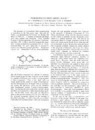
cyanobacteria. We <strong>the</strong>refore designated our clone Dgrca. For<br />
clarity, only <strong>the</strong> three highest-scoring matches to sequences<br />
<strong>of</strong> apple, Arabidopsis, and tobacco are shown (Fig. 1).<br />
The relative numbers <strong>of</strong> Dgrca clones in both libraries<br />
indicated that <strong>the</strong> corresponding mRNA was not a rareclass<br />
message in D. <strong>glomerata</strong> nodules. Initially, a 400-bp<br />
partial cDNA clone <strong>of</strong> Dgrca was isolated from approximately<br />
400 clones picked at random. A screen <strong>of</strong> approximately<br />
1.2 � 10 5 recombinant phage from a second library<br />
yielded over 20 separate clones that hybridized to a probe<br />
consisting <strong>of</strong> <strong>the</strong> partial Dgrca cDNA insert. The cDNAs<br />
ranged in size from 0.7 to 1.8 kb. The 3�-untranslated region<br />
varied in length; among five <strong>of</strong> <strong>the</strong> longest clones examined,<br />
two were found to contain 41 additional nucleotides<br />
immediately upstream <strong>of</strong> <strong>the</strong> poly(A � ) tail, suggesting that<br />
multiple polyadenylation signals in Dgrca are utilized.<br />
Two Size Classes <strong>of</strong> Dgrca RNA Are Present in <strong>Nodules</strong><br />
Dgrca mRNA was expressed in nodules and in photosyn<strong>the</strong>tic<br />
organs <strong>of</strong> D. <strong>glomerata</strong>, including leaves, flowers,
414 Okubara et al. <strong>Plant</strong> Physiol. Vol. 120, 1999<br />
Figure 1. The deduced amino acid sequence <strong>of</strong> Dgrca (Dg) is conserved<br />
with respect to Rubisco activase coding sequences from o<strong>the</strong>r<br />
photosyn<strong>the</strong>tic species, including apple (Md, P[N] � 1.8e �259 ; Watillon<br />
et al., 1993), Arabidopsis small peptide (At, P[N] � 5.8e �247 ;<br />
Orozco et al., 1993), and tobacco small peptide (Nt, P[N] �<br />
4.8e �240 ; accession no. U35111).<br />
and immature fruits (Fig. 2A). Dgrca mRNA was not detected<br />
in <strong>the</strong> root samples. In <strong>the</strong> photosyn<strong>the</strong>tic organs<br />
Dgrca mRNA was estimated to be 1700 nucleotides long.<br />
This corresponded to <strong>the</strong> size <strong>of</strong> <strong>the</strong> full-length cDNA clone<br />
(1634 bp), which was similar to Rubisco activase mRNAs<br />
from o<strong>the</strong>r plants (1650–1900 nucleotides). The data indicate<br />
that <strong>the</strong> 1700-nucleotide mRNA band represented <strong>the</strong><br />
mature, fully spliced message.<br />
The two distinct size classes <strong>of</strong> Dgrca transcripts, 3000<br />
and 1700 nucleotides, were observed in nodules harvested<br />
4 to 11 weeks after Frankia inoculation (Fig. 2A). Only <strong>the</strong><br />
1700-nucleotide mRNA was detected in <strong>the</strong> photosyn<strong>the</strong>tic<br />
organs, e.g. <strong>the</strong> flower, immature fruit, and leaves. <strong>Root</strong>s<br />
showed no detectable levels <strong>of</strong> ei<strong>the</strong>r species. The 3000nucleotide<br />
species was 2- to 5-fold more abundant than <strong>the</strong><br />
1700-nucleotide mRNA in total RNA from nodules harvested<br />
11 weeks after inoculation (data not shown). The<br />
relative abundance <strong>of</strong> <strong>the</strong> 3000-nucleotide mRNA was reduced<br />
in <strong>the</strong> older (17-week) nodule samples, but was not<br />
altered significantly by exposure to white light (Fig. 2B,<br />
lanes 1–3). However, this light treatment was sufficient to<br />
cause faint greening in <strong>the</strong> mature nodules closest to <strong>the</strong><br />
light source (data not shown).<br />
To determine whe<strong>the</strong>r both size classes <strong>of</strong> mRNA were<br />
polyadenylated, poly(A � ) and poly(A � ) fractions were hybridized<br />
to <strong>the</strong> Dgrca cDNA insert in gel blots (Fig. 2B,<br />
lanes 7 and 9). Whereas no hybridization was detected in<br />
<strong>the</strong> poly(A � ) fraction, both <strong>the</strong> 3000- and 1700-nucleotide<br />
species were observed in <strong>the</strong> poly(A � ) fraction. The abun-<br />
dance ratio, quantified by phosphor imaging (data not<br />
shown), was nearly 1:1 in <strong>the</strong> nodule poly(A � ) RNA fraction,<br />
as compared with 5:1 in total RNA. Attempts to obtain<br />
a 3-kb cDNA clone from several different poly(A � ) RNA<br />
preparations, using random amplification <strong>of</strong> cDNA 3� ends<br />
with two different primer pairs were unsuccessful.<br />
Dgrca Is a Single-Copy Gene in D. <strong>glomerata</strong><br />
To determine <strong>the</strong> size <strong>of</strong> <strong>the</strong> rca gene family in D.<br />
<strong>glomerata</strong>, we hybridized total DNA to <strong>the</strong> 400-bp partial<br />
cDNA insert. A single 8.2-kb EcoRI fragment and a single<br />
4.7-kb HindIII fragment (Fig. 3A) were radiolabeled; <strong>the</strong>re<br />
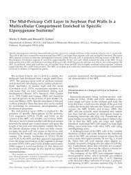
Figure 2. Expression <strong>of</strong> Dgrca mRNA in various organs <strong>of</strong> D.<br />
<strong>glomerata</strong> (A), in nodules with or without light treatment (B, lanes N e,<br />
N w, and N w) and in oligo(dT)-cellulose fractions <strong>of</strong> nodule RNA (B,<br />
lanes A� and A�). Sizes in nucleotide <strong>of</strong> rRNA species are shown on<br />
<strong>the</strong> right <strong>of</strong> each panel. A, Total RNA from leaves (lane L), flowers<br />
(lane Fl), immature fruits (lane Fr), roots (lane R), and nodules harvested<br />
4 to 11 weeks after Frankia inoculation (N 4–N 11) was hybridized<br />
to radiolabeled Dgrca cDNA insert. B, Total RNA from nodules<br />
harvested from excised roots (lane N e) or from roots <strong>of</strong> whole plants<br />
(lanes N w) after treatment with white light (Lt) or darkness (Dk) were<br />
hybridized to <strong>the</strong> full-length Dgrca cDNA probe. Total N 11 RNA<br />
(lane N 11), root RNA (lane R), and leaf RNA (lane L) are included for<br />
a comparison. The unbound fraction <strong>of</strong> total N 11 RNA (lane A�) and<br />
<strong>the</strong> bound N 11 RNA fraction (lane A�) after oligo(dT)-cellulose<br />
treatment were also hybridized to <strong>the</strong> full-length Dgrca cDNA probe.<br />
C, Total RNA from roots (lane R), N 4 nodules, or N 7 nodules was<br />
hybridized to a radiolabeled intron-A probe. The asterisk indicates<br />
<strong>the</strong> predicted position <strong>of</strong> <strong>the</strong> 1700-nucleotide mRNA species. For A<br />
and C and <strong>the</strong> first seven lanes <strong>of</strong> B, approximately 5 �g <strong>of</strong> total RNA<br />
was loaded into each lane; for lane A� in B, 2.5 �g <strong>of</strong> poly(A � ) RNA<br />
was used. Autoradiography was carried out with preflashed Kodak<br />
XAR 5 film at �80°C for 16.5 h (A), 6 d (B), and 5 d (C).
Rubisco Activase mRNA in <strong>Datisca</strong> <strong>glomerata</strong> <strong>Symbiotic</strong> <strong>Root</strong> <strong>Nodules</strong> 415<br />
was no evidence <strong>of</strong> two polymorphic Dgrca genes. We<br />
<strong>the</strong>refore conclude that Rubisco activase is encoded by a<br />
single gene or by a small, conserved gene family in D.<br />
<strong>glomerata</strong>, as in o<strong>the</strong>r plant species (Wernecke and Ogren,<br />
1989; Wernecke et al., 1989; Rundle and Zielinski, 1991;<br />
Qian and Rodermel, 1993).<br />
Occurrence <strong>of</strong> Intronic Sequences in <strong>the</strong> Dgrca Gene and<br />
3000-Nucleotide mRNA<br />
We were interested in whe<strong>the</strong>r <strong>the</strong> Dgrca gene was interrupted<br />
by introns that could account for <strong>the</strong> additional<br />
mass <strong>of</strong> <strong>the</strong> 3000-nucleotide mRNA. We examined Dgrca<br />
gene sequences from Arabidopsis (Orozco et al., 1993),<br />
spinach (Wernecke et al., 1989), and barley (Rundle and<br />
Zielinski, 1991). All three genes have four to six introns at<br />
conserved positions within <strong>the</strong> coding regions. A 1.2-kb<br />
fragment was amplified from <strong>the</strong> D. <strong>glomerata</strong> genome<br />
using PCR primers specific to <strong>the</strong> Dgrca coding region<br />
(Table I). This Dgrca gene segment contained three<br />
stretches <strong>of</strong> nucleotides, 96, 201, and 128 bp in length, that<br />
were not present in <strong>the</strong> Dgrca cDNA (Fig. 4A). The positions<br />
<strong>of</strong> <strong>the</strong>se AT-rich stretches with respect to <strong>the</strong> coding<br />
region were identical to those <strong>of</strong> <strong>the</strong> introns in <strong>the</strong> rca genes<br />
from <strong>the</strong> o<strong>the</strong>r plants (Fig. 4B). Therefore, we refer to <strong>the</strong>se<br />
genomic sequences as introns A, B, and C.<br />
To generate hybridization probes, genomic DNA template<br />
was amplified with PCR primers annealing to coding<br />
sequences that immediately flanked intron A and intron C<br />
(Table I). In both RNA and DNA blots, <strong>the</strong> intron C PCR<br />
product hybridized to a wide number <strong>of</strong> species, possibly<br />
because it carried a repetitive sequence (data not shown).<br />
However, intron A hybridized to a single 8.2-kb EcoRI<br />
DNA fragment (Fig. 3B) and hybridized weakly to <strong>the</strong><br />
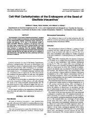
Figure 3. Total DNA <strong>of</strong> D. <strong>glomerata</strong> hybridized to <strong>the</strong> 400-bp<br />
partial cDNA insert (A) and intron-A probe (B). DNA was treated with<br />
HindIII (lane H) or EcoRI (lanes E) before transfer to nylon membrane.<br />
Sizes (in kb) <strong>of</strong> phage Lambda HindIII fragments are shown in <strong>the</strong><br />
middle. Autoradiography was carried out at �80°C for 3 (A) and<br />
5 (B) d.<br />
Figure 4. A, The coding sequence (uppercase letters) <strong>of</strong> <strong>the</strong> Dgrca<br />
genomic DNA segment is interrupted by at least three intronic sequences<br />
(lowercase letters), designated Intron A, Intron B, and Intron<br />
C. B, The positions <strong>of</strong> <strong>the</strong>se introns were highly conserved with<br />
respect to <strong>the</strong> Arabidopsis, spinach, and barley genes.<br />
3000-nucleotide mRNA in blots <strong>of</strong> total nodule RNA<br />
(Fig. 2C).<br />
Dgrca and DgrbcL mRNA Localization Patterns<br />
The mature Frankia-infected zone <strong>of</strong> <strong>the</strong> D. <strong>glomerata</strong><br />
nodule consisted <strong>of</strong> a compact region <strong>of</strong> expanded host<br />
cells filled with Frankia vesicles arrayed peripherally<br />
around a central vacuole (Fig. 5, A and E). The mature<br />
tissue was distinguished by expression <strong>of</strong> nifH, seen as<br />
silver grains localized over <strong>the</strong> Frankia vesicles in <strong>the</strong> infected<br />
cells (Fig. 5, B and F). The youngest <strong>of</strong> <strong>the</strong>se cells<br />
relative to <strong>the</strong> developmental gradient <strong>of</strong> <strong>the</strong> infection zone<br />
are indicated by white arrows in Figure 5, E through H.<br />
Beginning in <strong>the</strong> cells <strong>of</strong> <strong>the</strong> early infection zone, <strong>the</strong> host<br />
cytoplasm contained multiple nuclei (Fig. 5, A–C).<br />
In contrast, <strong>the</strong> in situ expression pattern <strong>of</strong> Dgrca mRNA<br />
was complex. High levels <strong>of</strong> expression <strong>of</strong> Dgrca were<br />
observed in Frankia-infected cells, especially at early stages<br />
<strong>of</strong> vesicle differentiation before <strong>the</strong> onset <strong>of</strong> N 2 fixation<br />
(Fig. 5, C, D, and G). The punctate appearance <strong>of</strong> <strong>the</strong> Dgrca<br />
signal (Fig. 5G) was attributed to <strong>the</strong> concentration <strong>of</strong><br />
message in <strong>the</strong> nuclei <strong>of</strong> infected cells (Fig. 5C). Dgrca<br />
message was detected at lower levels in adjacent (younger)<br />
cells in <strong>the</strong> early stages <strong>of</strong> Frankia infection. Some Dgrca<br />
RNA was also detected in <strong>the</strong> nuclei <strong>of</strong> mature, infected
416 Okubara et al. <strong>Plant</strong> Physiol. Vol. 120, 1999<br />
Figure 5. In situ localization <strong>of</strong> Dgrca, DgrcbL, and nifH mRNA in D. <strong>glomerata</strong> nodule sections. A, Bright-field micrograph<br />
<strong>of</strong> <strong>the</strong> interface between Frankia-infected cells and mature, uninfected cells. Bar � 15 �m (for A–C). B, Bright-field<br />
micrograph <strong>of</strong> nifH expression in Frankia-infected cells. Silver grains indicating hybridization to antisense probes appear as<br />
black specks, particularly dense over <strong>the</strong> Frankia vesicles in <strong>the</strong> infected cells (white asterisk). C, Bright-field micrograph<br />
showing dense accumulations <strong>of</strong> silver grains indicating hybridization <strong>of</strong> <strong>the</strong> Dgrca antisense probe to nuclei <strong>of</strong> infected cells<br />
(arrowheads). Some Dgrca transcripts are detected in adjacent younger cells <strong>of</strong> <strong>the</strong> early infection stage. Mature, uninfected<br />
cells show scattered silver grains, with no significant accumulation <strong>of</strong> Dgrca mRNA. D, Bright-field micrograph <strong>of</strong> a<br />
longitudinal nodule section hybridized to <strong>the</strong> Dgrca antisense probe. The relatively large nuclei in <strong>the</strong> meristem are darkly<br />
stained with toluidine blue, but do not have silver grains. Infected cells accumulated Dgrca transcripts, whereas uninfected<br />
cells showed much less hybridization. Bar � 30 �m. E, Bright-field micrograph <strong>of</strong> a longitudinal section. The Frankia<br />
vesicles stained red are arrayed around central vacuoles in <strong>the</strong> mature, infected cells. Bar � 500 �m (for E–H). F, Dark-field<br />
micrograph showing nifH expression. Hybridization <strong>of</strong> transcripts to <strong>the</strong> nifH antisense probe, apparent as white specks,<br />
delineates <strong>the</strong> onset <strong>of</strong> N 2 fixation in mature, infected cells (white arrows in E–H) within <strong>the</strong> developmental gradient <strong>of</strong> <strong>the</strong><br />
infection zone. �, A zone <strong>of</strong> senescence where nifH expression is reduced; <strong>the</strong> corresponding area in E has a vesiculated<br />
appearance because <strong>of</strong> degradation <strong>of</strong> <strong>the</strong> endosymbiont. G, Dark-field micrograph <strong>of</strong> DgrbcL expression in <strong>the</strong> zone before<br />
N 2 fixation and in mature, infected cells. DgrbcL mRNA was less abundant in <strong>the</strong> zone <strong>of</strong> senescence. H, Dark-field<br />
micrograph <strong>of</strong> Dgrca expression in <strong>the</strong> infected cells and in surrounding uninfected cortical cells. nu, Nuclei; UN, uninfected<br />
cells; F, Frankia-infected cells; am, amyloplasts in uninfected cells; p, periderm; m, meristem; INF, infected cells; nr, base<br />
<strong>of</strong> a nodule root.<br />
cells expressing nifH and in <strong>the</strong> cortical cells immediately<br />
adjacent to infected cells (Fig. 5H). No Dgrca expression<br />
was observed in <strong>the</strong> nodule-lobe meristem (Fig. 5D) or in<br />
<strong>the</strong> nodule-root meristem (data not shown). The nuclei<br />
in <strong>the</strong> meristem and in <strong>the</strong> cells underlying <strong>the</strong> periderm in<br />
Figure 5D stained brightly with toluidine blue, but silver<br />
grains did not accumulate. Hybridization with <strong>the</strong> Dgrca<br />
sense probe was negative (data not shown).
Rubisco Activase mRNA in <strong>Datisca</strong> <strong>glomerata</strong> <strong>Symbiotic</strong> <strong>Root</strong> <strong>Nodules</strong> 417<br />
DgrbcL mRNA was most abundant in <strong>the</strong> infected cells <strong>of</strong><br />
<strong>the</strong> zone preceding N 2 fixation (Fig. 5G). It was also detected<br />
in mature, infected cells and <strong>the</strong> uninfected cortical<br />
cells lying between <strong>the</strong> infected cells and nodule periderm.<br />
In both infected and uninfected cells, silver grains indicating<br />
DgrbcL expression were not densely clustered over a<br />
particular region <strong>of</strong> <strong>the</strong> cell, but appeared to be distributed<br />
throughout <strong>the</strong> cytoplasm. In carpels <strong>the</strong> DgrbcL antisense<br />
probe hybridized to photosyn<strong>the</strong>tic parenchyma cells as<br />
expected, but not to immature, nonphotosyn<strong>the</strong>tic an<strong>the</strong>rs<br />
(data not shown). Hybridization <strong>of</strong> Dgrca, DgrbcL, and nifH<br />
was somewhat reduced in a region where Frankia vesicles<br />
were undergoing senescence (Fig. 5, E–H; see legend). Nei<strong>the</strong>r<br />
Dgrca nor DgrbcL antisense probes hybridized to periderm<br />
or vascular tissue.<br />
Rubisco Activase and O<strong>the</strong>r Proteins Associated with <strong>the</strong><br />
Photosyn<strong>the</strong>tic Apparatus Do Not Accumulate in<br />
D. <strong>glomerata</strong> <strong>Nodules</strong><br />
We performed western analyses to determine whe<strong>the</strong>r<br />
proteins <strong>of</strong> Rubisco activase, Rubisco large subunit, and <strong>the</strong><br />
OEC33 were present in nodule extracts. All three proteins<br />
were detected in leaf extracts, but not in nodule or root<br />
extracts (Fig. 6). With polyclonal antibodies made to soybean<br />
Rubisco large subunit, a strong protein band was seen<br />
in leaf extracts at 50 to 60 kD (Fig. 6, left panel), representing<br />
<strong>the</strong> native form <strong>of</strong> <strong>the</strong> protein. We detected a protein <strong>of</strong><br />
40 kD in D. <strong>glomerata</strong> leaf extracts with antibodies made to<br />
tobacco Rubisco activase (Fig. 6, middle panel). This band<br />
was similar in size to Rubisco activase small polypeptides<br />
from o<strong>the</strong>r plant species (41–44 kD). Antibodies made to<br />
Figure 6. Western analyses for <strong>the</strong> Rubisco large subunit (left),<br />
Rubisco activase (middle), and OEC33 (right). Ten microliters <strong>of</strong><br />
extracts <strong>of</strong> D. <strong>glomerata</strong> nodule, root, and leaf protein was partitioned<br />
on SDS-PAGE before reaction with antibodies (diluted as<br />
indicated). For Rubisco, 17, 2.0, and 59 �g <strong>of</strong> protein from nodule,<br />
root, and leaf were used, respectively; for Rubisco activase and<br />
OEC33, 32, 33, and 91 �g <strong>of</strong> protein from nodule, root, and leaf were<br />
used, respectively. Protein standards (in kilodaltons) are shown at<br />
<strong>the</strong> right.<br />
OEC33 from pea detected a 33-kD protein in <strong>the</strong> leaves <strong>of</strong><br />
D. <strong>glomerata</strong> (Fig. 6, right panel).<br />
DISCUSSION<br />
We identified an mRNA encoding Rubisco activase in<br />
symbiotic root nodules <strong>of</strong> D. <strong>glomerata</strong>. Similarities between<br />
Dgrca and Rubisco activases from o<strong>the</strong>r organisms were<br />
evident in comparisons <strong>of</strong> <strong>the</strong>ir nucleotide and amino acid<br />
sequences, mRNA expression patterns in various organs,<br />
sizes <strong>of</strong> <strong>the</strong> gene families, and positions and sizes <strong>of</strong> three<br />
introns. Rubisco activase is conserved in photosyn<strong>the</strong>tic<br />
organisms across a wide range <strong>of</strong> genera, including lower<br />
eukaryotes (Roesler and Ogren, 1990; Li et al., 1993), and<br />
has no reported alternative functions. A Rubisco activaselike<br />
GA-binding protein has been identified (Komatsu et<br />
al., 1996), but <strong>the</strong> deduced amino acid sequence <strong>of</strong> this gene<br />
shares limited identity with that <strong>of</strong> Dgrca, as well as with<br />
Rubisco activases from o<strong>the</strong>r species.<br />
Rubisco activase functions as a heterodimer in most<br />
higher plants, with <strong>the</strong> exception <strong>of</strong> a monomeric form in<br />
maize (Salvucci et al., 1987). Large and small is<strong>of</strong>orms <strong>of</strong><br />
Rubisco activase reported in Arabidopsis, spinach (Wernecke<br />
et al., 1989), and barley (Rundle and Zielinski, 1991)<br />
arise from alternative splicing <strong>of</strong> a single mRNA. In o<strong>the</strong>r<br />
species such as tobacco, <strong>the</strong> polypeptides are encoded by<br />
separate genes (Qian and Rodermel, 1993). In our RNA<br />
blots we detected two Dgrca transcripts expressed in D.<br />
<strong>glomerata</strong> nodules, a full-length, mature Dgrca mRNA <strong>of</strong><br />
1700 nucleotides, and an unusually large (3000 nucleotides),<br />
abundant transcript. The 1300-nucleotide size differential<br />
between <strong>the</strong>se mRNAs was much greater than <strong>the</strong><br />
difference <strong>of</strong> approximately 300 nucleotides that results<br />
from alternative splicing in o<strong>the</strong>r species. In addition, five<br />
separate Dgrca cDNA clones that were examined had identical<br />
3� sequences. Therefore, we found no evidence from<br />
ei<strong>the</strong>r nor<strong>the</strong>rn blots or from limited nucleotide sequence<br />
data to indicate that <strong>the</strong> alternative splicing <strong>of</strong> Rubisco<br />
activase mRNA observed in o<strong>the</strong>r species is occurring for<br />
Dgrca in D. <strong>glomerata</strong> nodules. Because Dgrca appears to be<br />
a single-copy gene, <strong>the</strong> 3000-nucleotide species is not likely<br />
to arise from transcription <strong>of</strong> a second gene.<br />
Hybridization <strong>of</strong> <strong>the</strong> large transcript to intronic DNA<br />
suggested ei<strong>the</strong>r that it was a form <strong>of</strong> heteronuclear RNA<br />
or that it represented an anomalous, incompletely spliced<br />
message. This possibility was supported by in situ localization<br />
<strong>of</strong> <strong>the</strong> Dgrca RNA primarily to nuclei <strong>of</strong> infected<br />
cells. Because <strong>the</strong> D. <strong>glomerata</strong> nodule is indeterminate, a<br />
developmental gradient from meristem to N 2-fixing infected<br />
cells is present in <strong>the</strong> mature nodule, at least until 6<br />
weeks after inoculation. Both mRNA species were observed<br />
in nodules up to 11 weeks postinoculation. The<br />
relatively low level <strong>of</strong> hybridization to <strong>the</strong> intron-A probe<br />
in RNA blots and <strong>the</strong> absence <strong>of</strong> intron-A hybridization in<br />
in situ experiments was attributed to its relatively short<br />
length (96 bp <strong>of</strong> intron A-specific sequence) and its AT-rich<br />
nature. Nucleotide sequence data <strong>of</strong> a 3-kb cDNA clone<br />
would reveal <strong>the</strong> additional sequences accounting for <strong>the</strong><br />
larger transcript, but our attempts to obtain such a clone<br />
using random amplification <strong>of</strong> cDNA 3� ends were unsuc-
418 Okubara et al. <strong>Plant</strong> Physiol. Vol. 120, 1999<br />
cessful. We do not know whe<strong>the</strong>r <strong>the</strong> 3000-nucleotide<br />
Dgrca transcript represents a novel form <strong>of</strong> alternatively<br />
spliced mRNA or whe<strong>the</strong>r it is translated. If it is translated,<br />
it must give rise to a protein that is not detected by <strong>the</strong><br />
Rubisco activase antiserum used in our experiments.<br />
The 3000-nucleotide RNA species was recovered from an<br />
oligo(dT)-cellulose column, indicating that at least a portion<br />
<strong>of</strong> this size population was polyadenylated. The ratio<br />
<strong>of</strong> <strong>the</strong> 3000-nucleotide species to <strong>the</strong> 1700-nucleotide species<br />
decreased from 5:1 in total nodule RNA preparations<br />
to about 1:1 in <strong>the</strong> nodule poly(A � ) fraction, suggesting<br />
ei<strong>the</strong>r that a significant portion <strong>of</strong> <strong>the</strong> 3000-nucleotide species<br />
did not have a poly(A � ) tail and was labile, or that <strong>the</strong><br />
3000-nucleotide poly(A � ) RNA was unstable during fractionation.<br />
Accumulation <strong>of</strong> unspliced or incompletely<br />
spliced heteronuclear RNA has been postulated to result<br />
from changes in <strong>the</strong> organization <strong>of</strong> <strong>the</strong> nucleus or from<br />
changes in splicing efficiency associated with promoter<br />
structure (Cramer et al., 1997), with heteronuclear RNA<br />
structure (for review, see Simpson and Filipowicz, 1996), or<br />
with <strong>the</strong> availability or action <strong>of</strong> components <strong>of</strong> <strong>the</strong> spliceosome<br />
complex (e.g. <strong>the</strong> SR proteins; Cáceres et al., 1997).<br />
Because <strong>of</strong> <strong>the</strong> complexity <strong>of</strong> <strong>the</strong> splicing apparatus and <strong>the</strong><br />
splicing process, we have not pursued splicing as <strong>the</strong> biological<br />
basis for <strong>the</strong> presence <strong>of</strong> <strong>the</strong> 3000-nucleotide RNA.<br />
Retention <strong>of</strong> transcripts within <strong>the</strong> nucleus has been<br />
reported for specific genes in a variety <strong>of</strong> organisms. Early<br />
expression <strong>of</strong> a male germ line-specific gene, Mst40, in<br />
spermatocytes <strong>of</strong> third larval instars <strong>of</strong> Drosophila melanogaster<br />
is associated with <strong>the</strong> nucleus; Mst40 mRNA was<br />
detected later in <strong>the</strong> cytoplasm (Russell and Kaiser, 1994).<br />
The D. melanogaster Hsr-omega gene, located in a heat-shock<br />
puff, undergoes alternative transcriptional termination in<br />
response to heat shock and o<strong>the</strong>r stimuli, generating a<br />
longer, nuclear-localized form <strong>of</strong> <strong>the</strong> transcript, which is<br />
polyadenylated and in which <strong>the</strong> introns are retained<br />
(Hogan et al., 1994). The authors suggest that <strong>the</strong> nuclear<br />
transcript may interact with proteins or have a regulatory<br />
role; for Dgrca, however, this remains to be determined.<br />
The O2 (Opaque-2) transcript, encoding a bZIP transcriptional<br />
regulator <strong>of</strong> zein storage protein syn<strong>the</strong>sis in maize<br />
endosperm, is normally found in both <strong>the</strong> nucleus and<br />
cytoplasm (Dolfini et al., 1992). In one mutant an introncontaining<br />
O2 transcript accumulates in <strong>the</strong> nucleus. The<br />
efficiency <strong>of</strong> splicing <strong>of</strong> <strong>the</strong> waxy transcript in rice endosperm<br />
appears to vary naturally among cultivars, some<br />
<strong>of</strong> which accumulate a large pre-mRNA containing an intron<br />
(Wang et al., 1995). As expected, <strong>the</strong> titers <strong>of</strong> granulebound<br />
starch synthase (Waxy protein) correlate directly<br />
with <strong>the</strong> level <strong>of</strong> mature waxy mRNA and inversely with<br />
that <strong>of</strong> <strong>the</strong> pre-mRNA. Genetic data indicate that splicing <strong>of</strong><br />
<strong>the</strong> waxy intron is governed by cis-acting elements. It is<br />
curious that all <strong>of</strong> <strong>the</strong> intron-containing transcripts described<br />
above occur in organs or cells that are undergoing<br />
endoreduplication and, hence, share a common feature<br />
with <strong>the</strong> mature infected cells <strong>of</strong> D. <strong>glomerata</strong> nodules. Our<br />
findings suggest that <strong>the</strong> D. <strong>glomerata</strong> symbiotic nodule<br />
provides <strong>the</strong> signals or trans-acting factors necessary for<br />
transcription <strong>of</strong> Dgrca. However, o<strong>the</strong>r molecular components<br />
that are needed for efficient processing <strong>of</strong> <strong>the</strong> Dgrca<br />
transcript or for its transport from <strong>the</strong> nucleus do not<br />
appear to be functional or present in appropriate amounts.<br />
The mature, 1700-nucleotide Dgrca mRNA is expected to<br />
be translatable. Our western-blot data indicate that Rubisco<br />
activase protein is present in leaves <strong>of</strong> D. <strong>glomerata</strong> but not<br />
in nodules or roots. The absence <strong>of</strong> detectable protein in<br />
nodules could result from a block in <strong>the</strong> export <strong>of</strong> mRNA to<br />
<strong>the</strong> cytoplasm, from a block in translation <strong>of</strong> <strong>the</strong> mRNA,<br />
and/or from rapid protein turnover. Low amounts <strong>of</strong> protein<br />
in tissues showing high levels <strong>of</strong> <strong>the</strong> corresponding<br />
transcript have been reported for a number <strong>of</strong> genes, including<br />
fbp1, a MADS-box transcription factor in stamens<br />
<strong>of</strong> wild-type petunia (Angenent et al., 1992); Sh1, which<br />
encodes an is<strong>of</strong>orm <strong>of</strong> Suc synthase in maize embryos<br />
(Chourey and Taliercio, 1994); and an mRNA for<br />
S-adenosyl Met syn<strong>the</strong>tase in poplar (Mijnsbrugge et al.,<br />
1996). In <strong>the</strong> case <strong>of</strong> an mRNA encoding <strong>the</strong> small subunit<br />
<strong>of</strong> ADP-Glc pyrophosphorylase, relatively low levels <strong>of</strong> <strong>the</strong><br />
small subunit <strong>of</strong> ADP-Glc pyrophosphorylase in potato<br />
leaves are attributed to its instability in <strong>the</strong> absence <strong>of</strong> <strong>the</strong><br />
large subunit (Nakata and Okita, 1996). Arrest <strong>of</strong> translation<br />
can also occur in response to environmental conditions,<br />
as observed for a mitochondrial mRNA-encoding<br />
adenine nucleotide translocator in young maize roots after<br />
O 2 deprivation (Fennoy and Bailey-Serres, 1995).<br />
In some species rbcL mRNA accumulates in etioplasts<br />
before greening (Tobin and Silvethorne, 1985; Berry et al.,<br />
1990); thus its presence in plastids <strong>of</strong> nonphotosyn<strong>the</strong>tic<br />
organs is not without precedent. The localization <strong>of</strong> Dgrca<br />
transcripts coincided with that <strong>of</strong> <strong>the</strong> rbcL mRNA, but <strong>the</strong><br />
former were most abundant in <strong>the</strong> nuclei <strong>of</strong> cells <strong>of</strong> <strong>the</strong><br />
early infection zone, whereas <strong>the</strong> latter was abundant in <strong>the</strong><br />
cytoplasm <strong>of</strong> mature, vesicle-containing cells, typically<br />
those expressing nitrogenase, leghemoglobin, Gln syn<strong>the</strong>tase,<br />
and o<strong>the</strong>r proteins involved in N 2 and C assimilation<br />
and O 2 partitioning. Dgrca expression was also localized<br />
at low levels to <strong>the</strong> cytoplasm <strong>of</strong> uninfected cortical<br />
cells adjacent to <strong>the</strong> periderm and to cells surrounding <strong>the</strong><br />
vascular cylinder. Differential expression patterns for<br />
Dgrca and DgrbcL can be expected, because <strong>the</strong>se genes are<br />
transcribed in different subcellular compartments and are<br />
independently regulated in o<strong>the</strong>r developmental systems<br />
(Reski, 1994). The observed expression patterns may <strong>the</strong>refore<br />
reflect differences in <strong>the</strong> induction <strong>of</strong> nuclear- and<br />
plastid-coded genes or in plastid number and distribution<br />
within <strong>the</strong> nodule.<br />
Nodule expression <strong>of</strong> Dgrca deviated from <strong>the</strong> organspecific,<br />
light-regulated expression for rca genes observed<br />
in leaves <strong>of</strong> spinach (Orozco and Ogren, 1993) and in<br />
Arabidopsis (Liu et al., 1996). Dgrca nodule expression also<br />
deviated from <strong>the</strong> coordinate expression <strong>of</strong> o<strong>the</strong>r photosyn<strong>the</strong>tic<br />
genes during photomorphogenesis and diurnal<br />
fluxes in photosyn<strong>the</strong>tic capacity. White light treatment<br />
had no significant effect on <strong>the</strong> overall abundance <strong>of</strong> Dgrca<br />
mRNA or on <strong>the</strong> relative abundance <strong>of</strong> <strong>the</strong> two size classes<br />
<strong>of</strong> Dgrca transcripts in symbiotic root nodules. Circadian<br />
regulation <strong>of</strong> rca genes such as that observed in tomato<br />
(Martino-Catt and Ort, 1992) and Arabidopsis (Liu et al.,<br />
1996) has not yet been examined in Dgrca nodules. Because<br />
nei<strong>the</strong>r Rubisco activase nor Rubisco proteins accumulated
Rubisco Activase mRNA in <strong>Datisca</strong> <strong>glomerata</strong> <strong>Symbiotic</strong> <strong>Root</strong> <strong>Nodules</strong> 419<br />
to detectable levels in D. <strong>glomerata</strong> nodules, a role for <strong>the</strong>se<br />
proteins in photosyn<strong>the</strong>tic C reduction, oxygenase activity,<br />
or N 2 accumulation in <strong>the</strong> nodule is unlikely. In stem<br />
nodules <strong>of</strong> Sesbania rostrata, photosyn<strong>the</strong>tically competent<br />
chloroplasts were observed in cortical cells adjacent to<br />
N 2-fixing infected cells, suggesting that <strong>the</strong>y may have a<br />
role in C assimilation (James et al., 1996). In D. <strong>glomerata</strong><br />
nodules, however, <strong>the</strong> absence <strong>of</strong> OEC33, a protein associated<br />
with <strong>the</strong> O 2-evolving complex <strong>of</strong> <strong>the</strong> photosyn<strong>the</strong>tic<br />
apparatus, suggests that Dgrca expression is not accompanied<br />
by <strong>the</strong> assembly <strong>of</strong> a functional photosyn<strong>the</strong>tic apparatus<br />
or by additional chloroplast development.<br />
The nodule greening and aut<strong>of</strong>luorescence attributed to<br />
chlorophyll that we have observed in some mature D.<br />
<strong>glomerata</strong> nodules (P.A. Okubara and A.M. Berry, unpublished<br />
data) occurs much later after Frankia inoculation<br />
than does Dgrca mRNA expression. Although <strong>the</strong> relative<br />
abundance <strong>of</strong> 3000-nucleotide mRNA was not altered significantly<br />
by white light treatment, this treatment was sufficient<br />
to cause faint greening in <strong>the</strong> mature nodules closest<br />
to <strong>the</strong> light source (data not shown). We <strong>the</strong>refore postulate<br />
that <strong>the</strong> greening represents a distinct phenomenon not<br />
necessarily related to <strong>the</strong> induction <strong>of</strong> Dgrca transcription.<br />
The greening <strong>of</strong> organs derived from roots seems anomalous,<br />
yet development <strong>of</strong> functional chloroplasts in <strong>the</strong> pith<br />
cells <strong>of</strong> young poplar twigs (van Cleve et al., 1993) has been<br />
reported. Such cases <strong>of</strong> chloroplast differentiation may reflect<br />
<strong>the</strong> recruitment <strong>of</strong> stem-like traits during <strong>the</strong> development<br />
or evolution <strong>of</strong> certain organs, including <strong>the</strong> D.<br />
<strong>glomerata</strong> nodule. Greening has also been observed in some<br />
multinucleated giant cells derived from tomato roots after<br />
<strong>the</strong>y have been infected with root knot nematodes from <strong>the</strong><br />
genus Meloidogyne (V. Williamson, personal communication).<br />
The differentiation <strong>of</strong> certain root knots appears to<br />
be accompanied by chlorophyll accumulation, presumably<br />
through <strong>the</strong> ectopic expression <strong>of</strong> photosyn<strong>the</strong>sis-associated<br />
mRNAs. The molecular or cellular basis for <strong>the</strong> unusual<br />
accumulation <strong>of</strong> Dgrca transcripts in nodules may well be<br />
revealed as progress is made in understanding <strong>the</strong> extensive<br />
cellular activity, strong induction <strong>of</strong> N 2 metabolic<br />
pathways, and o<strong>the</strong>r phenomena that characterize nodule<br />
development.<br />
ACKNOWLEDGMENTS<br />
The authors thank David Neale (U.S. Department <strong>of</strong> Agriculture<br />
Forest Service, Pacific Southwest, PWS, Davis, CA) for advice and<br />
use <strong>of</strong> facilities in construction <strong>of</strong> <strong>the</strong> first D. <strong>glomerata</strong> cDNA<br />
library; David Gilchrist, Chris Mau, and o<strong>the</strong>r members <strong>of</strong> <strong>the</strong><br />
Center for Engineering <strong>Plant</strong>s for Resistance Against Pathogens,<br />
Davis, CA, for use <strong>of</strong> <strong>the</strong> facilities for construction <strong>of</strong> <strong>the</strong> second<br />
cDNA library, for autoradiography, and for sequence alignments;<br />
Rich Jorgensen (University <strong>of</strong> California, Davis) for <strong>the</strong> use <strong>of</strong> his<br />
PCR facilities; Dean Lavelle (University <strong>of</strong> California, Davis) for<br />
diligent and expert sequencing <strong>of</strong> <strong>the</strong> Dgrca full-length cDNA and<br />
<strong>the</strong> Dgrca gene segment; Tony van Kampen (Wageningen Agricultural<br />
University) for sequencing <strong>of</strong> Dgrca clones; Michael E. Salvucci<br />
for <strong>the</strong> gift <strong>of</strong> Rubisco activase antibodies and for insightful<br />
discussions; Steve Theg for <strong>the</strong> gift <strong>of</strong> OEC33 antibodies; and<br />
Susan Swensen and Beth Mullin (University <strong>of</strong> Tennessee, Knoxville)<br />
for pDgRcbL.<br />
Received November 3, 1998; accepted March 19, 1999.<br />
The accession numbers for <strong>the</strong> sequences <strong>of</strong> Dgrca mRNA and <strong>the</strong><br />
1.2-kb Dgrca gene segment reported in this article are AF047352<br />
and AF052424, respectively.<br />
LITERATURE CITED<br />
Altschul SF, Gish W, Miller W, Myers EW, Lipman JD (1990)<br />
Basic local alignment search tool. J Mol Biol 215: 403–410<br />
Angenent GC, Busscher M, Franken J, Mol JNM, van Tunen AJ<br />
(1992) Differential expression <strong>of</strong> two MADS box genes in wildtype<br />
and mutant petunia flowers. <strong>Plant</strong> Cell 4: 983–993<br />
Benson DR, Silvester WB (1993) Biology <strong>of</strong> Frankia strains, actinomycete<br />
symbionts <strong>of</strong> actinorhizal plants. Microbiol Rev 57:<br />
293–319<br />
Berry JO, Breiding DE, Klessig DF (1990) Light-mediated control<br />
<strong>of</strong> translational initiation <strong>of</strong> ribulose-1,5-bisphosphate carboxylase<br />
in amaranth cotyledons. <strong>Plant</strong> Cell 2: 795–803<br />
Bradford MM (1976) A rapid and sensitive method for <strong>the</strong> quantitation<br />
<strong>of</strong> microgram quantities <strong>of</strong> protein utilizing <strong>the</strong> principle<br />
<strong>of</strong> protein-dye binding. Anal Biochem 72: 248–254<br />
Cáceres JF, Misteli T, Screaton GR, Spector DL, Kariner AR<br />
(1997). Role <strong>of</strong> <strong>the</strong> modular domains <strong>of</strong> SR proteins in subnuclear<br />
localization and alternative splicing specificity. J Cell<br />
Biol 138: 225–237<br />
Chourey PS, Taliercio EW (1994) Epistatic interaction and functional<br />
compensation between two tissue- and cell-specific sucrose<br />
synthase genes in maize. Proc Natl Acad Sci USA 91:<br />
7917–7921<br />
Cramer P, Pesce CG, Baralle FE, Kornblihtt AR (1997) Functional<br />
association between promoter structure and transcript alternative<br />
splicing. Proc Natl Acad Sci USA 94: 11456–11460<br />
Davidson C (1973) An anatomical and morphological study <strong>of</strong><br />
<strong>Datisca</strong>ceae. ALISO 8: 49–110<br />
Dickey LF, Nguyen T-T, Allen GC, Thompson WF (1994) Light<br />
modulation <strong>of</strong> ferredoxin mRNA abundance requires an open<br />
reading frame. <strong>Plant</strong> Cell 6: 1171–1176<br />
Dolfini SF, Landoni M, Tonelli C, Bernard L, Viotti A (1992)<br />
Spatial regulation in <strong>the</strong> expression <strong>of</strong> structural and regulatory<br />
storage-protein genes in Zea mays endosperm. Dev Genet 13:<br />
264–276<br />
Fennoy SL, Bailey-Serres J (1995) Posttranscriptional regulation <strong>of</strong><br />
gene expression in oxygen-deprived roots <strong>of</strong> maize. <strong>Plant</strong> J 7:<br />
287–295<br />
Gherbi H, Duhoux E, Franche C, Pawlowski K, Nassar A, Berry<br />
AM, Bogusz D (1997) Cloning <strong>of</strong> a full-length symbiotic hemoglobin<br />
cDNA and in situ localization <strong>of</strong> <strong>the</strong> corresponding<br />
mRNA in Casuarina glauca root nodule. Physiol <strong>Plant</strong> 99: 608–616<br />
Goetting-Minesky MP, Mullin BC (1994) Differential gene expression<br />
in an actinorhizal symbiosis: evidence for a nodulespecific<br />
cysteine protease. Proc Natl Acad Sci USA 91: 9891–9895<br />
Guan C, Akkermans ADL, van Kammen A, Bissleing T,<br />
Pawlowski K (1997) ag13 is expressed in Alnus glutinosa nodules<br />
in infected cells during endosymbiont degradation and in <strong>the</strong><br />
nodule pericycle. Physiol <strong>Plant</strong> 99: 601–607<br />
Hafeez F, Akkermans ADL, Chaudhary AH (1984) Observations<br />
on <strong>the</strong> ultrastructure <strong>of</strong> Frankia sp. in root nodules <strong>of</strong> <strong>Datisca</strong><br />
cannabina L. <strong>Plant</strong> Soil 79: 383–402<br />
Hirsch AM, LaRue TA (1997) Is <strong>the</strong> legume nodule a modified<br />
root or stem or an organ sui generis? Crit Rev <strong>Plant</strong> Sci 16:<br />
361–392<br />
Hogan NC, Traverse KL, Sullivan DE, Pardue M-L (1994) The<br />
nucleus-limited Hsr-omega-n transcript is a polyadenylated<br />
RNA with a regulated intranuclear turnover. J Cell Biol 125:<br />
21–30<br />
Jacobsen-Lyon K, Jensen EO, Joergensen J-E, Marcker KA, Peacock<br />
WJ, Dennis ES (1995) <strong>Symbiotic</strong> and nonsymbiotic hemoglobin<br />
genes <strong>of</strong> Casuarina glauca. <strong>Plant</strong> Cell 7: 213–223<br />
James EK, Iannetta PPM, Nixon PJ, Whiston AJ, Peat L, Crawford<br />
RMM, Sprent JJ, Brewin NJ (1996) Photosystem II and oxygen<br />
regulation in Sesbania rostrata stem nodules. <strong>Plant</strong> Cell Environ<br />
19: 895–910
420 Okubara et al. <strong>Plant</strong> Physiol. Vol. 120, 1999<br />
Komatsu S, Masuda T, Hirano H (1996) Rice gibberellin-binding<br />
phosphoprotein structurally related to ribulose-1,5bisphosphate<br />
carboxylase/oxygenase activase. FEBS Lett 384:<br />
167–171<br />
Laemmli UK (1970) Cleavage <strong>of</strong> structural proteins during <strong>the</strong><br />
assembly <strong>of</strong> <strong>the</strong> head <strong>of</strong> bacteriophage T4. Nature 277: 680–685<br />
Lan Y, Mott KA (1991) Determination <strong>of</strong> apparent K m values for<br />
ribulose 1,5-bisphosphate carboxylase oxygenase (Rubisco) activase<br />
using <strong>the</strong> spectrophotometric assay <strong>of</strong> Rubisco activity.<br />
<strong>Plant</strong> Physiol 95: 604–609<br />
Li LA, Gibson JL, Tabita FR (1993) The rubisco activase (rca) gene<br />
is located downstream from rbcS inAnabaena sp. strain CA and<br />
is detected in o<strong>the</strong>r Anabaena Nostoc strains. <strong>Plant</strong> Mol Biol 21:<br />
753–764<br />
Liston A, Reiseberg AH, Elias TS (1989) Morphological stasis and<br />
molecular divergence in <strong>the</strong> intercontinental disjunct genus<br />
<strong>Datisca</strong> (<strong>Datisca</strong>ceae). ALISO 12: 525–542<br />
Liu Q, Berry AM (1991) The infection process and nodule initiation<br />
in <strong>the</strong> Frankia-Ceanothus root nodule symbiosis. Protoplasma<br />
163: 82–92<br />
Liu Z, Taub CC, McClung CR (1996) Identification <strong>of</strong> an Arabidopsis<br />
thaliana ribulose-1,5-bisphosphate carboxylase/oxygenase<br />
activase (RCA) minimal promoter regulated by light and <strong>the</strong><br />
circadian clock. <strong>Plant</strong> Physiol 112: 43–51<br />
Martino-Catt S, Ort DR (1992) Low temperature interrupts circadian<br />
regulation <strong>of</strong> transcriptional activity in chilling-sensitive<br />
plants. Proc Natl Acad Sci USA 89: 3731–3735<br />
Mijnsbrugge KV, Van Montagu M, Inzé D, Boerjan W (1996)<br />
Tissue-specific expression conferred by <strong>the</strong> S-adenosyl-lmethionine<br />
syn<strong>the</strong>tase promoter <strong>of</strong> Arabidopsis thaliana in transgenic<br />
poplar. <strong>Plant</strong> Cell 37: 1108–1115<br />
Murphy TM (1978) Immunochemical comparisons <strong>of</strong> ribulosebisphosphate<br />
carboxylases using antisera to tobacco and spinach<br />
enzymes. Phytochemistry 17: 439–443<br />
Nakata PA, Okita TW (1996) Cis-elements important for <strong>the</strong> expression<br />
<strong>of</strong> <strong>the</strong> ADP-glucose pyrophosphorylase small-subunit<br />
are located both upstream and downstream from its structural<br />
gene. Mol Gen Genet 250: 581–592<br />
Orozco BM, McClung CR, Wernecke JM, Ogren WL (1993) Molecular<br />
basis <strong>of</strong> <strong>the</strong> ribulose-1,5-bisphosphate carboxylase/<br />
oxygenase activase mutation in Arabidopsis thaliana is a guanineto-adenine<br />
transition at <strong>the</strong> 5�-splice junction <strong>of</strong> intron 3. <strong>Plant</strong><br />
Physiol 102: 227–232<br />
Orozco BM, Ogren WL (1993) Localization <strong>of</strong> light-inducible and<br />
tissue-specific regions <strong>of</strong> <strong>the</strong> spinach ribulose bisphosphate carboxylase<br />
oxygenase (Rubisco) activase promoter in transgenic<br />
tobacco plants. <strong>Plant</strong> Mol Biol 23: 1129–1138<br />
Pawlowski K, Bisseling T (1996) Rhizobial and actinorhizal symbioses:<br />
what are <strong>the</strong> shared features? <strong>Plant</strong> Cell 8: 1899–1913<br />
Pawlowski K, Kunze R, de Vries S, Bisseling T (1994) Isolation <strong>of</strong><br />
total, poly(A) and polysomal RNA from plant tissues. In SB<br />
Gelvin, RA Schilperoort, eds, <strong>Plant</strong> Molecular Biology Manual<br />
D5, Ed 2. Kluwer Academic Publishers, Dordrecht, The Ne<strong>the</strong>rlands,<br />
pp 1–13<br />
Pawlowski K, Ribeiro A, Bisseling T (1996) Nitrogen fixing root<br />
nodule symbioses: legume nodules and actinorhizal nodules. In<br />
MR El-Gewely, ed, Biotechnology Annual Review. Elsevier Science<br />
Publishers, Amsterdam, The Ne<strong>the</strong>rlands, pp 151–184<br />
Pawlowski K, Ribeiro A, Guan C-H, van Kammen AB, Akkermans<br />
A, Bisseling T (1993) Differential gene expression in root<br />
nodules <strong>of</strong> Alnus glutinosa. In NA Hegazi, M Fayez, M Monib,<br />
eds, Nitrogen Fixation with Non-Legumes. American University<br />
in Cairo Press, Cairo, Egypt, pp 185–190<br />
Pawlowski K, Twigg P, Dobritsa S, Guan CH, Mullin BC (1997)<br />
A nodule-specific gene family from Alnus glutinosa encodes<br />
glycine- and histidine-rich proteins expressed in <strong>the</strong> early stages<br />
<strong>of</strong> actinorhizal nodule development. Mol <strong>Plant</strong>-Microbe Interact<br />
10: 656–664<br />
Portis AR, Jr (1990) Rubisco activase. Biochim Biophys Acta 1015:<br />
15–28<br />
Qian J, Rodermel S (1993) Ribulose-1,5-bisphosphate carboxylase/oxygenase<br />
activase cDNAs from Nicotiana tabacum. <strong>Plant</strong><br />
Physiol 102: 683–684<br />
Reski R (1994) Plastid genes and chloroplast biogenesis. In DWS<br />
Mok, MC Mok, eds, Cytokinins Chemistry, Activity, and Function.<br />
CRC Press, Boca Raton, FL, pp 179–195<br />
Ribeiro A, Akkermanns ADL, van Kammen A, Bisseling T,<br />
Pawlowski K (1995) A nodule-specific gene encoding a subtilisin-like<br />
protease is expressed in early stages <strong>of</strong> actinorhizal<br />
nodule development. <strong>Plant</strong> Cell 7: 785–794<br />
Ribeiro A, Praekelt U, Akkermans ADL, Meacock PA, van<br />
Kammen A, Bisseling T, Pawlowski K (1996) Identification <strong>of</strong><br />
agthi1, whose product is involved in biosyn<strong>the</strong>sis <strong>of</strong> <strong>the</strong> thiamine<br />
precursor thiazole, in actinorhizal nodules <strong>of</strong> Alnus glutinosa.<br />
<strong>Plant</strong> J 10: 361–368<br />
Roesler KR, Ogren WL (1990) Primary structure <strong>of</strong> Chlamydomonas<br />
reinhardtii ribulose 1,5-bisphosphate carboxylase/oxygenase activase<br />
and evidence for a single polypeptide. <strong>Plant</strong> Physiol 94:<br />
1837–1841<br />
Rundle SJ, Zielinski RE (1991) Organization and expression <strong>of</strong><br />
two tandemly oriented genes encoding ribulosebisphosphate<br />
carboxylase/oxygenase activase in barley. J Biol Chem 266:<br />
4677–4685<br />
Russell SRH, Kaiser K (1994) A Drosophila melanogaster chromosome<br />
2L repeat is expressed in <strong>the</strong> male germ line. Chromosoma<br />
103: 63–72<br />
Salvucci ME, Wernecke JM, Ogren WL, Portis AR, Jr (1987)<br />
Purification and species distribution <strong>of</strong> Rubisco activase. <strong>Plant</strong><br />
Physiol 84: 930–936<br />
Sambrook J, Fritsch EF, Maniatis T (1989) Molecular Cloning: A<br />
Laboratory Manual, Ed 2. Cold Spring Harbor Laboratory Press,<br />
Cold Spring Harbor, NY<br />
Simpson GG, Filipowicz W (1996) Splicing <strong>of</strong> precursors to<br />
mRNA in higher plants: mechanism, regulation and sub-nuclear<br />
organization <strong>of</strong> <strong>the</strong> spliceosomal machinery. <strong>Plant</strong> Mol Biol 32:<br />
1–41<br />
Swensen SM, Mullin BC, Chase MW (1994) Phylogenetic affinities<br />
<strong>of</strong> <strong>Datisca</strong>ceae based on an analysis <strong>of</strong> nucleotide sequences<br />
from <strong>the</strong> plastid rbcL gene. Syst Bot 19: 157–168<br />
Tobin EM, Silverthorne J (1985) Light regulation <strong>of</strong> gene expression<br />
in higher plants. Annu Rev <strong>Plant</strong> Physiol 36: 569–593<br />
van Cleve B, Forreiter C, Sauter JJ, Apel K (1993) Pith cells <strong>of</strong><br />
poplar contain photosyn<strong>the</strong>tically active chloroplasts. <strong>Plant</strong>a<br />
189: 70–73<br />
van de Wiel C, Scheres B, Franssen H, van Lierop M-J, Van<br />
Lammeren A, Van Kammen A, Bisseling T (1990) The early<br />
nodulin transcript ENOD2 is located in <strong>the</strong> nodule parenchyma<br />
(inner cortex) <strong>of</strong> pea and soybean root nodules. EMBO J 9: 1–7<br />
Wang Z-Y, Zheng F-Q, Shen G-Z, Gao J-P, Snustad DP, Li M-G,<br />
Zhang J-L, Hong M-M (1995) The amylose content in rice endosperm<br />
is related to <strong>the</strong> post-transcriptional regulation <strong>of</strong> <strong>the</strong><br />
waxy gene. <strong>Plant</strong> J 7: 613–622<br />
Watillon B, Kettmann R, Boxus P, Burny A (1993) Developmental<br />
and circadian pattern <strong>of</strong> Rubisco activase messenger RNA accumulation<br />
in apple plants. <strong>Plant</strong> Mol Biol 23: 501–509<br />
Wernecke JM, Chatfield JM, Ogren WL (1989) Alternative mRNA<br />
splicing generates <strong>the</strong> two ribulosebisphosphate carboxylase/<br />
oxygenase activase polypeptides in spinach and Arabidopsis.<br />
<strong>Plant</strong> Cell 1: 815–825<br />
Wernecke JM, Ogren WL (1989) Structure <strong>of</strong> an Arabidopsis thaliana<br />
cDNA encoding Rubisco activase. Nucleic Acids Res 17: 2871