improving implant aesthetics: prosthetically generated ... - BHIDE Info
improving implant aesthetics: prosthetically generated ... - BHIDE Info
improving implant aesthetics: prosthetically generated ... - BHIDE Info

You also want an ePaper? Increase the reach of your titles
YUMPU automatically turns print PDFs into web optimized ePapers that Google loves.
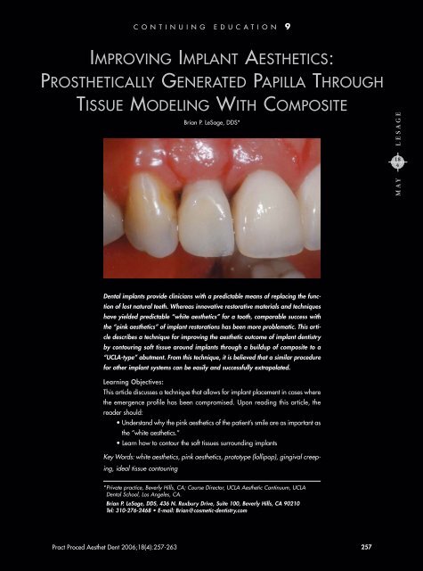
CONTINUING EDUCATION 9IMPROVING IMPLANT AESTHETICS:PROSTHETICALLY GENERATED PAPILLA THROUGHTISSUE MODELING WITH COMPOSITEBrian P. LeSage, DDS*LESAGE184MAYDental <strong>implant</strong>s provide clinicians with a predictable means of replacing the functionof lost natural teeth. Whereas innovative restorative materials and techniqueshave yielded predictable “white <strong>aesthetics</strong>” for a tooth, comparable success withthe “pink <strong>aesthetics</strong>” of <strong>implant</strong> restorations has been more problematic. This articledescribes a technique for <strong>improving</strong> the aesthetic outcome of <strong>implant</strong> dentistryby contouring soft tissue around <strong>implant</strong>s through a buildup of composite to a“UCLA-type” abutment. From this technique, it is believed that a similar procedurefor other <strong>implant</strong> systems can be easily and successfully extrapolated.Learning Objectives:This article discusses a technique that allows for <strong>implant</strong> placement in cases wherethe emergence profile has been compromised. Upon reading this article, thereader should:• Understand why the pink <strong>aesthetics</strong> of the patient’s smile are as important asthe “white <strong>aesthetics</strong>.”• Learn how to contour the soft tissues surrounding <strong>implant</strong>sKey Words: white <strong>aesthetics</strong>, pink <strong>aesthetics</strong>, prototype (lollipop), gingival creeping,ideal tissue contouring*Private practice, Beverly Hills, CA; Course Director, UCLA Aesthetic Continuum, UCLADental School, Los Angeles, CA.Brian P. LeSage, DDS, 436 N. Roxbury Drive, Suite 100, Beverly Hills, CA 90210Tel: 310-276-2468 • E-mail: Brian@cosmetic-dentistry.comPract Proced Aesthet Dent 2006;18(4):257-263 257
Practical Procedures & AESTHETIC DENTISTRYDental <strong>implant</strong>s are well-established as a predictabletreatment modality for replacing the function of lostor missing natural teeth. Improved restorative materialsand techniques have also brought a high level of qualityand predictability to the “white <strong>aesthetics</strong>” of the tooth.Comparable success with the “pink <strong>aesthetics</strong>” of an<strong>implant</strong> restoration, however, has proven to be moreproblematic. When the quantity, quality, contour, and/orform of keratinized tissue surrounding dental <strong>implant</strong>s isinadequate, the result too often is new teeth that arebeautiful in color, but emerge from a less than favorabletissue contour and have length-to-width ratios and contact/connectorissues that are unaesthetic.This article will describe a restorative technique for<strong>improving</strong> the aesthetic outcome of <strong>implant</strong> dentistry bycontouring soft tissue around <strong>implant</strong>s through a patient,progressive buildup of a composite to a standard “UCLAtype”abutment. From this well-documented technique, itis believed that a similar procedure for other <strong>implant</strong>systems can be easily and successfully extrapolated.Figure 1. Preoperative view of gingival biotype and architecture.Note apical extent of laterals verses centrals and asymmetry present.Background and ProtocolIn the 1990s, a number of developments improved the<strong>aesthetics</strong> of <strong>implant</strong> therapy. Better restorative materialsmade the replication of natural tooth morphology andcolor more attainable. At the same time, hard and softtissue graft materials and techniques, 1-9 membrane techniques,10-13 and other restorative procedures 14-18 enabledclinicians to place <strong>implant</strong>s in optimal positions close tothe missing tooth root. With enhanced <strong>implant</strong> placement,more ideal emergence profiles were made possible,and <strong>implant</strong> restorations became more lifelike. 19-24Nonetheless, <strong>implant</strong> clinicians continued to struggle toprovide soft tissue topography, contour, and form thatmet restorative ideals. The spotlight has finally turned tothe pink <strong>aesthetics</strong> and to the biological considerationsof tissue at the <strong>implant</strong> site.The tissue contour technique illustrated herein wasdeveloped by the author to help the <strong>implant</strong> team(ie, the surgeon) maximize the quantity of attached gingivaand to provide gingival scallop, zenith, and emergenceprofile mimicking tissue around the extracted toothand the adjacent teeth. Termed the “lollipop” techniquefor the shape of the provisional restoration, the clinicalprotocol progressively nurtures development of keratinizedFigure 2. Initial placement of the lollipop prototype to <strong>implant</strong> fixture.Figure 3. Deficient papillae formation is evident in view of thelollipop approximately 2 weeks after insertion.258 Vol. 18, No. 4
LeSageAFigure 4A. The composite lollipop prototype prior to modification.4B. The prototype following modification.Figure 5. View of the gingival tissue shows black triangles on mesialand distal aspects after initial modification of the prototype.Figure 6. Final lollipop exhibits proper adaptation, correct emergenceprofile, and elimination of the black triangle.Btissue at the <strong>implant</strong> site with the use of a composite “prototype”characterized by a very narrow neck.The ideal clinical strategy for optimizing pink <strong>aesthetics</strong>begins with the clinician’s preservation of soft tissueat the time of extraction. After <strong>implant</strong> placement, thesurgeon picks up the fixture with the aid of the surgicaltemplate and acrylic resin (ie, Duralay, Reliance DentalManufacturing Co, Alsip, IL) and transfers the <strong>implant</strong>impression coping to a model. The prototype is fabricatedin the laboratory with the application of composite materialsto a plastic or metal opaque UCLA-type abutment.Connection to the <strong>implant</strong> occurs as early as the day ofsecond-stage surgery and up to 2 weeks after uncovering.Experience of the surgical team, timing, and convenienceof office schedules help determine this appointment.When considering this technique in the immediate loadingcase, Glauser has shown that the stabilization of the<strong>implant</strong> is at its weakest point during the period 2 to 4weeks postoperative. 25 Following placement of the prototype,the patient is recalled at routine intervals for assessmentof gingival movement and progressive enlargementand contouring of the prototype neck with composite astissue moves into the gingival embrasure space.During the author’s early cases utilizing the protocol,patients were scheduled for office visits every3 weeks, the prototype was observed and/or removedat each appointment, and additional composite wasapplied to the neck of the restoration at most visits.Subsequent to developing the technique, Abrahamssonreported that there is an increased risk of tissue movingapically with frequent removal of a prototype from thefixture. 26,27 Although the author had not encountered thisproblem in his early trials of the technique, the protocolwas modified to eliminate any risk of inhibiting tissuedevelopment with frequent removal of the prototyperestoration. Patients now are appointed every 4 to 8weeks and the prototype is removed only when incisalcreeping of gingival papillae is observed. Tissue isallowed to fill in and is then pushed from the lingualtoward the facial to create the papillae.During this process, extreme care is taken whenevermaterial is added to the buccal aspect. If tissue is alreadyon the <strong>implant</strong> tooth at the desired height, it is inadvisableto push more tissue on the direct facial becauseasymmetry can result with the contralateral tooth. EvenPPAD 259
Practical Procedures & AESTHETIC DENTISTRYFigure 7. View of the definitive restoration two years postoperativelydemonstrates natural gingival architectureand zenith.Figure 8. View of definitive restorations at 51 monthsreveals the stability that can be attained in the gingivalcomplex over the long-term.without pushing, the gingiva will continue to move downnaturally to the point of biological consideration, 28-30 makingit possible to build out the emergence profile somewhatmore in the future if necessary. Should thesurrounding tissue blanch for more than one minute afterthe prototype is replaced, the author has found the blanchingindicates that the tissue has been overstressed. Whenthis occurs, the prototype should be removed and thenewly added composite trimmed back to relieve the tissue.While the tissue is being sculpted, the author recommendsgentle but very thorough brushing only. Mostinterproximal aids (eg, floss, interproximal brushes, toothpick)will put apical pressure on the tissue and inhibit itsability to creep more coronally to fill the intentionallyretained black triangles. Once the prototype is contouredfully and no black triangles exist, the tissue contour shouldbe picked up in the final impression. The definitive restorationmimics the prototype in <strong>aesthetics</strong>, function, biology,and phonetics, and it can then be maintained with normalbrushing and flossing.Once the papillae are well-established in optimal contour,scallop, and level relative to adjacent teeth, a precisetechnique is used to transfer the properly developedemergence profile impression without collapsing the tissue.Definitive restorations are then fabricated, tried in, andconfirmed radiographically to be seated properly; restorationsare then evaluated for parameters of white, pink, andblack <strong>aesthetics</strong>, and delivered with the patient's approval.Three to 4 months of tissue contouring and developmentfollowed by 3 to 4 months of "creeping" and stabilizationare normally required for ideal tissue contouringFigure 9. Postoperative radiograph of definitive restorationat 48 months.Figure 10. After 48 months postoperation, the papillaeheights on the mesial and distal aspects of the lateralincisors are at matching heights.utilizing the lollipop protocol. It is important that patientsbe advised during treatment planning that a minimumof 1 year is required for treatment from <strong>implant</strong> placementto definitive restoration. At the same time, patientsneed to be thoroughly reassured that they will never be260 Vol. 18, No. 4
LeSageFigure 11. Facial view of patient’s smile prior to extractionof tooth #8(11).Figure 14. Prototype with modifications only supragingivallyto aid with biological creeping to the desired gingivaland papilla formation.Figure 12. The lollipop prototype in place, replacing theextracted tooth. Patient decided to whiten her teeth afterprototype was fabricated and accepted color discrepancy.Figure 13. Facial view at 2 months reveals improved gingivalarchitecture.without a tooth replacement during the treatment period.A fee that recognizes the duration of treatment and thefrequency of clinical visits during the restorative stage isdeveloped and also communicated to the patient duringdiscussion of the treatment plan.Case PresentationsCase 1A 57-year-old female patient presented with a fracturedtooth #7(12) that had previously been endodonticallytreated (Figure 1). The tooth was removed by the periodontist,and a 3.3-mm x 15-mm standard <strong>implant</strong> (ie,NP Brånemark, Nobel Biocare, Yorba Linda, CA) wasplaced. At the same time, a connective tissue graft wasperformed to maximize the available soft tissue duringhealing (Figures 2 and 3). Tissue grafting was not arequirement for success using the lollipop protocol, butmay be used selectively when existing tissue quantity isdeemed less than adequate for the desired outcome.After 6 months of healing, the <strong>implant</strong> was uncovered,additional tissue was grafted at the <strong>implant</strong> site, andthe lollipop prototype was attached (Figures 4 and 5).Although the coronal aspect of the restoration completelyfilled the space supragingivally, the narrow neck of theprototype left ample black space into which the tissuecould be manipulated, shaped, and contoured to optimize<strong>aesthetics</strong>. There was no buccolingual cantileverwith this prototype. The prototype filled the entire coronalaspect of the missing tooth and, in doing so, wasused to evaluate <strong>aesthetics</strong>, function, and phonetics. Asthe prototype was not wider buccolingually, there wasno more cantilever than exists with the natural tooth.Additionally, ideal positioning of the <strong>implant</strong> allowed forlingual screw access through the cingulum.Since the patient was employed in the author’s dentaloffice, she was recalled more frequently than is typicalfor contouring the tissue to the desired aestheticPPAD 261
Practical Procedures & AESTHETIC DENTISTRYand biologic outcome. After placement of the prototype,the patient was seen every 3 weeks for the first 3 months.Composite was added at most visits. Between 3 and 6months, the site was observed without further enlargementof the prototype to allow tissue to continue movingcoronally. The papillae were allowed to mature biologicallyto fill the space and to develop scaffolding aroundthe prototype (Figures 6 and 7). A metal UCLA standardabutment was used because the gingival biotypewas of adequate thickness and form and because itimproved the match to the contralateral existingporcelain-fused-to-metal restoration on tooth #10(22),which had a gold-cast post and core. The result was arestoration in which the quality of the pink <strong>aesthetics</strong>enhanced the quality of the white <strong>aesthetics</strong> (Figures 8through 10).Figure 15. View of lollipop reveals minimal transtissuemodification 1 month following the prior compositecontour change.Case 2A 25-year-old woman presented with tooth #8(11)having been severely compromised by root resorption(Figure 11). Although preservation of the natural toothis a priority in a patient of this age, a clinical team thatincluded a periodontist and an endodontist concludedthat salvaging the tooth would create defects that likelywould compromise adjacent teeth #7(12) and #9(21).The tooth was extracted and a 4.3-mm x 15-mm standard<strong>implant</strong> (ie, NP Brånemark, Nobel Biocare, YorbaLinda, CA) was placed.At uncovering, a lollipop prototype was connectedto the <strong>implant</strong> (Figure 12). During the ensuing 4 months,the prototype was removed four times and compositewas added on each occasion (Figures 13 through 15).For the subsequent 3 months, the papillae were allowedto mature and stabilize. Because available soft tissuewas adequate for a correct soft tissue profile, no graftingwas performed in this case. Since the gingival biotypewas thin and scalloped, however, an abutment (ie,Procera, Nobel Biocare, Yorba Linda, CA) with a pressedceramic crown (ie, IPS Empress, Ivoclar Vivadent,Amherst, NY) was used. Again, the resulting restorationwas one in which the quality of the pink <strong>aesthetics</strong>enhanced the quality of the white <strong>aesthetics</strong> (Figures 16through 18). While this technique can be performed withsuccess and predictability, similar results can now beachieved through the use of milled abutments, where oneFigure 16. Radiograph reveals typical bone loss to firsttread and nice adaptation of abutment/crown complexto fixture.can scan the prototype and precisely reproduce the abutmentthree dimensionally.ConclusionNatural soft tissue <strong>aesthetics</strong> are equally as importantas the contour and color of the restoration in providingaesthetic, long-term, comprehensive care of the dental<strong>implant</strong> patient. The use of a narrow-neck prototype thatis progressively and patiently widened during anextended period following second-stage <strong>implant</strong> surgerypromotes the development of adequate attached gingivafor ideal <strong>implant</strong> <strong>aesthetics</strong>. Named for the prototypeitself, this lollipop technique can be utilized with all commercial<strong>implant</strong> systems and with angled or one-pieceabutments. The lollipop technique provides a prostheticmeans of predictably achieving desirable white and pinkaesthetic outcomes in <strong>implant</strong> therapy. No contraindicationsfor its use have been identified.262 Vol. 18, No. 4
LeSageFigure 17. Nearly ideal gingival contour, zenith, andpapilla formation architecture can be seen.Figure 18. Sagittal view of the definitive restoration.AcknowledgmentThe author mentions his gratitude for the <strong>implant</strong> placementand connective tissue grafts provided by SaschaJovanovic, DDS, UCLA Dental School, Los Angeles, CA.References1. Henderson RD, Greenwell H, Drisko C, et al. Predictable multiplesite root coverage using an acellular dermal matrix allograft.J Periodontol 2001;72(5):571-582.2. Botticelli D, Berglundh T, Buser D, Lindhe J. The jumping distancerevisited: An experimental study in the dog. Clin Oral ImplantsRes 2003;14(1):35-42.3. Jovanovic SA, Spiekermann H, Richter EJ. Bone regenerationaround titanium dental <strong>implant</strong>s in dehisced defect sites: A clinicalstudy. Int J Oral Maxillofac Implants 1992;7(2):233-245.4. Buser D, Dula K, Belser UC, et al. Localized ridge augmentationusing guided bone regeneration. I. Surgical procedure inthe maxilla. Int J Periodont Rest Dent 1993;13(1):29-45.5. Buser D, Dula K, Belser UC, et al. Localized ridge augmentationusing guided bone regeneration. II. Surgical procedure inthe mandible. Int J Periodont Rest Dent 1995;15(1):10-29.6. Tinti C, Parma-Benfenati S, Polizzi G. Vertical ridge augmentation:What is the limit? Int J Periodont Rest Dent 1996;16(3):220-229.7. Buser D, Dula K, Lang NP, Nyman S. Long-term stability ofosseointegrated <strong>implant</strong>s in bone re<strong>generated</strong> with the membranetechnique. 5-year results of a prospective study with 12<strong>implant</strong>s. Clin Oral Impl Res 1996;7(2):175-183.8. Jovanovic SA. Bone rehabilitation to achieve optimal <strong>aesthetics</strong>.Pract Periodont Aesthet Dent 1997;9(1):41-51.9. Zitzmann NU, Scharer P, Marinello CP. Long-term results of<strong>implant</strong>s treated with guided bone regeneration: A 5-yearprospective study. Int J Oral Maxillofac Implants 2001;16(3):355-366.10. Simion M, Misitano U, Gionso L, Salvato A. Treatment of dehiscencesand fenestrations around dental <strong>implant</strong>s using resorbableand nonresorbable membranes associated with bone autografts:A comparative clinical study. Int J Oral Maxillofac Implants1997;12(2):159-167.11. Jovanovic SA, Nevins M. Bone formation utilizing titaniumreinforced membranes. Int J Periodont Rest Dent 1995;15(1):57-69.12. Simion M, Jovanovic SA, Trisi P, et al. Vertical ridge augmentationaround dental <strong>implant</strong>s using a membrane technique andbone auto or allografts in humans. Int J Periodont Rest Dent1998;18:9-23.13. Salama H, Salama MA, Garber D, Adar P. The interproximalheight of bone: A guidepost to predictable aesthetic strategiesand soft tissue contours in anterior tooth replacement. PractPeriodont Aesthet Dent 1998;10(9):1131-1141.14. Meyenberg KH, Imoberdorf MJ. The aesthetic challenges ofsingle tooth replacement: A comparison of treatment alternatives.Pract Periodont Aesthet Dent 1997;9(7):727-735.15. Salama H, Salama MA, Li TF, et al. Treatment planning 2000:An esthetically oriented revision of the original <strong>implant</strong> protocol.J Esthet Dent 1997;9(2):55-67.16. Paul SJ, Jovanovic SA. Anterior <strong>implant</strong>-supported reconstructions:A prosthetic challenge. Pract Periodont Aesthet Dent 1999;11(5):585-590.17. Touati B, Guez G, Saadoun A. Aesthetic soft tissue integrationand optimized emergence profile: Provisionalization and customizedimpression coping. Pract Periodont Aesthet Dent1999;11(3):305-314.18. Bichacho N. Achieving optimal gingival esthetics around restorednatural teeth and <strong>implant</strong>s. Rationale, concepts, and techniques.Dent Clin North Am 1998;42(4):763-780.19. Sadan A, Raigrodski AJ, Salinas TJ. Prosthetic considerations inthe fabrication of surgical stents for <strong>implant</strong> placement. PractPeriodont Aesthet Dent 1997;9(9):1003-1011.20. Kan JY, Rungcharassaeng K. Immediate placement and provisionalizationof maxillary anterior single <strong>implant</strong>s: A surgical andprosthodontic rationale. Pract Periodont Aesthet Dent 2000;12(9):817-824.21. Bichacho N, Landsberg CJ. Single Implants Restoration:Prosthetically induced soft tissue topography. Pract PeriodontAesthet Dent 1997;9(7):745-752.22. Salama H, Salama M, Garber D, Adar P. Developing optimalperi-<strong>implant</strong> papillae within the esthetic zone: Guided soft tissueaugmentation. J Esthet Dent 1995;7(3):125-129.23. Phillips K, Kois JC. Aesthetic peri-<strong>implant</strong> site development. Therestorative connection. Dent Clin North Am 1998;42(1):57-70.24. Jemt T. Restoring the gingival contour by means of provisionalresin crowns after single-<strong>implant</strong> treatment. Int J Periodont RestDent 1999;19(1):20-29.25. Touati, B, Guez G, Saadoun A. Aesthetic Soft Tissue Integrationand optimized emergence profile: Provisionalization and customizedimpression coping. Pract Proced Aesthet Dent 1999;11(3):305-314.2001.26. Abrahamsson I, Berglundh T, Lindhe J. The mucosal barrier followingabutment dis/reconnection. An experimental study indogs. J Clin Periodontol 1997;24(8):568-572.27. Abrahamsson I, Berglundh T, Glantz PO, Lindhe J. The mucosalattachment at different abutments: An experimental study in dogs.J Clin Periodontol 1998;25(9):721-727.28. Touati B, Guez G, Saadoun A. Aesthetic soft tissue integrationand optimized emergence profile: Provisionalization and customizedimpression coping. Pract Periodont Aesthet Dent1999;11(3):305-314.29. LeSage B. Long-term prototypes: New techniques for <strong>implant</strong>dentistry. AACD Monograph: Picture Perfect Aesthetics. 2005;2:111-114.30. Garber DA, Belser UC. Restoration driven <strong>implant</strong> placementwith restoration-<strong>generated</strong> site development. Compen ContinEduc Dent 1995;16(8):796-804.PPAD 263
CONTINUING EDUCATION(CE) EXERCISE NO. 99CONTINUING CEEDUCATIONTo submit your CE Exercise answers, please use the answer sheet found within the CE Editorial Section of this issue and complete as follows:1) Identify the article; 2) Place an X in the appropriate box for each question of each exercise; 3) Clip answer sheet from the page and mailit to the CE Department at Montage Media Corporation. For further instructions, please refer to the CE Editorial Section.The 10 multiple-choice questions for this Continuing Education (CE) exercise are based on the article “Improving <strong>implant</strong> <strong>aesthetics</strong>: Prosthetically<strong>generated</strong> papilla through tissue modeling with composite,” by Brian P. LeSage, DDS. This article is on Pages 257-263.1. What is the name of the technique where the clinicianprogressively adds composite to a “UCLA-type” abutmentover a period of months?a. Lollipop technique.b. Immediate loading technique.c. Progressive loading technique.d. Composite pressure technique.2. On average, how much time is needed to <strong>prosthetically</strong>generate a papilla?a. Two to three months.b. Three to four months.c. Six to eight months.d. Ten to twelve months.3. Pink <strong>aesthetics</strong> around <strong>implant</strong>s is best accomplishedwhen which of the following item(s) are maximized?a. Quality of gingival tissue.b. Form of keratinized tissue.c. Quantity of gingival tissue.d. All of the above.4. According to Glauser, the stabilization of the <strong>implant</strong> is atits weakest point during which of the following periods?a. Up to 2 weeks.b. At 2 to 4 weeks.c. At 6 to 8 weeks.d. None of the above.5. Prior to capturing the properly developed emergenceprofile in the precise final impression, the clinician musthave established which of the following?a. Scallop/Zenith.b. Optimal contour.c. Relatively level with the adjacent tooth.d. All of the above6. When using this technique, for how many minutes can atissue blanch before it is considered overstressed?a. One.b. Two.c. Three.d. Four.7. Extreme care must be taken when material is added towhich surface, especially if the tissue is already symmetricalwith the contralateral tooth?a. Distal.b. Mesial.c. Buccal.d. Lingual.8. How many month(s) of tissue contouring and developmentwill this technique average?a. Two.b. Three to four.c. Four to six.d. Five.9. How many months of creeping and stabilization will thistechnique average?a. One to two.b. Two to three.c. Three to four.d. Four to five.10. As specifically described in the article, which aspect ofthe lollipop prototype initially must completely fill thespace supragingivally?a. Distal.b. Mesial.c. Coronal.d. Lingual.264 Vol. 18, No. 4