X-ray structure of a protein-conducting channel - The Laboratory of ...
X-ray structure of a protein-conducting channel - The Laboratory of ...
X-ray structure of a protein-conducting channel - The Laboratory of ...

Create successful ePaper yourself
Turn your PDF publications into a flip-book with our unique Google optimized e-Paper software.
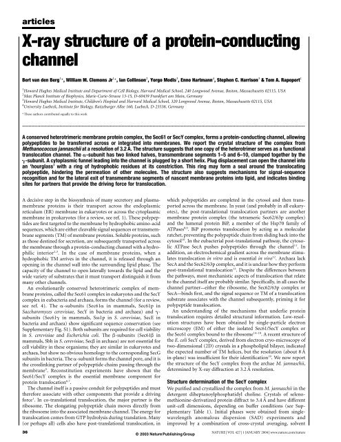
articlesX-<strong>ray</strong> <strong>structure</strong> <strong>of</strong> a <strong>protein</strong>-<strong>conducting</strong><strong>channel</strong>Bert van den Berg 1 *, William M. Clemons Jr 1 *, Ian Collinson 2 , Yorgo Modis 3 , Enno Hartmann 4 , Stephen C. Harrison 3 & Tom A. Rapoport 11 Howard Hughes Medical Institute and Department <strong>of</strong> Cell Biology, Harvard Medical School, 240 Longwood Avenue, Boston, Massachusetts 02115, USA2 Max Planck Institute <strong>of</strong> Biophysics, Marie-Curie-Strasse 13-15, D-60439 Frankfurt am Main, Germany3 Howard Hughes Medical Institute, Children’s Hospital and Harvard Medical School, 320 Longwood Avenue, Boston, Massachusetts 02115, USA4 University Luebeck, Institute for Biology, Ratzeburger Allee 160, Luebeck, D-23538, Germany* <strong>The</strong>se authors contributed equally to this work...........................................................................................................................................................................................................................A conserved heterotrimeric membrane <strong>protein</strong> complex, the Sec61 or SecY complex, forms a <strong>protein</strong>-<strong>conducting</strong> <strong>channel</strong>, allowingpolypeptides to be transferred across or integrated into membranes. We report the crystal <strong>structure</strong> <strong>of</strong> the complex fromMethanococcus jannaschii at a resolution <strong>of</strong> 3.2 Å. <strong>The</strong> <strong>structure</strong> suggests that one copy <strong>of</strong> the heterotrimer serves as a functionaltranslocation <strong>channel</strong>. <strong>The</strong> a-subunit has two linked halves, transmembrane segments 1–5 and 6–10, clamped together by theg-subunit. A cytoplasmic funnel leading into the <strong>channel</strong> is plugged by a short helix. Plug displacement can open the <strong>channel</strong> intoan ‘hourglass’ with a ring <strong>of</strong> hydrophobic residues at its constriction. This ring may form a seal around the translocatingpolypeptide, hindering the permeation <strong>of</strong> other molecules. <strong>The</strong> <strong>structure</strong> also suggests mechanisms for signal-sequencerecognition and for the lateral exit <strong>of</strong> transmembrane segments <strong>of</strong> nascent membrane <strong>protein</strong>s into lipid, and indicates bindingsites for partners that provide the driving force for translocation.A decisive step in the biosynthesis <strong>of</strong> many secretory and plasmamembrane<strong>protein</strong>s is their transport across the endoplasmicreticulum (ER) membrane in eukaryotes or across the cytoplasmicmembrane in prokaryotes (for a review, see ref. 1). <strong>The</strong>se polypeptidesare first targeted to the membrane by hydrophobic amino-acidsequences, which are either cleavable signal sequences or transmembranesegments (TM) <strong>of</strong> membrane <strong>protein</strong>s. Soluble <strong>protein</strong>s, suchas those destined for secretion, are subsequently transported acrossthe membrane through a <strong>protein</strong>-<strong>conducting</strong> <strong>channel</strong> with a hydrophilicinterior 2,3 . In the case <strong>of</strong> membrane <strong>protein</strong>s, when ahydrophobic TM arrives in the <strong>channel</strong>, it is released through anopening in the <strong>channel</strong> wall into the surrounding lipid phase. <strong>The</strong>capacity <strong>of</strong> the <strong>channel</strong> to open laterally towards the lipid and thewide variety <strong>of</strong> substrates that it must transport distinguish it frommany other <strong>channel</strong>s.An evolutionarily conserved heterotrimeric complex <strong>of</strong> membrane<strong>protein</strong>s, called the Sec61 complex in eukaryotes and the SecYcomplex in eubacteria and archaea, forms the <strong>channel</strong> (for a review,see ref. 4). <strong>The</strong> a-subunits (Sec61a in mammals, Sec61p inSaccharomyces cerevisiae, SecY in bacteria and archaea) and g-subunits (Sec61g in mammals, Sss1p in S. cerevisiae, SecE inbacteria and archaea) show significant sequence conservation (seeSupplementary Fig. S1). Both subunits are required for cell viabilityin S. cerevisiae and Escherichia coli. <strong>The</strong> b-subunits (Sec61b inmammals, Sbh in S. cerevisiae, Secb in archaea) are not essential forcell viability in these organisms; they are similar in eukaryotes andarchaea, but show no obvious homology to the corresponding SecGsubunits in bacteria. <strong>The</strong> a-subunit forms the <strong>channel</strong> pore, and it isthe crosslinking partner <strong>of</strong> polypeptide chains passing through themembrane 5 . Reconstitution experiments have shown that theSec61/SecY complex is the essential membrane component for<strong>protein</strong> translocation 6,7 .<strong>The</strong> <strong>channel</strong> itself is a passive conduit for polypeptides and musttherefore associate with other components that provide a drivingforce 1 . In co-translational translocation, the major partner is theribosome. <strong>The</strong> elongating polypeptide chain moves directly fromthe ribosome into the associated membrane <strong>channel</strong>. <strong>The</strong> energy fortranslocation comes from GTP hydrolysis during translation. Many(or perhaps all) cells also have post-translational translocation, in36© 2003 Nature Publishing Groupwhich polypeptides are completed in the cytosol and then transportedacross the membrane. In yeast (and probably in all eukaryotes),the post-translational translocation partners are anothermembrane <strong>protein</strong> complex (the tetrameric Sec62/63p complex)and the lumenal <strong>protein</strong> BiP, a member <strong>of</strong> the Hsp70 family <strong>of</strong>ATPases 8,9 . BiP promotes translocation by acting as a molecularratchet, preventing the polypeptide chain from sliding back into thecytosol 10 . In the eubacterial post-translational pathway, the cytosolicATPase SecA pushes polypeptides through the <strong>channel</strong> 11 . Inaddition, an electrochemical gradient across the membrane stimulatestranslocation in vitro and is essential in vivo 12 . Archaea lackSecA and the Sec62/63p complex, and it is unclear how they performpost-translational translocation 13 . Despite the differences betweenthe pathways, most mechanistic aspects <strong>of</strong> translocation that relateto the <strong>channel</strong> itself are probably similar. Specifically, in all cases the<strong>channel</strong> partner—either the ribosome, the Sec62/63p complex orSecA—binds first, and the signal sequence or TM <strong>of</strong> a translocationsubstrate associates with the <strong>channel</strong> subsequently, priming it forpolypeptide translocation.An understanding <strong>of</strong> the mechanisms that underlie <strong>protein</strong>translocation requires detailed structural information. Low-resolution<strong>structure</strong>s have been obtained by single-particle electronmicroscopy (EM) <strong>of</strong> either the isolated Sec61/SecY complex orthe Sec61 complex bound to the ribosome 14–18 . A recent <strong>structure</strong> <strong>of</strong>the E. coli SecY complex, derived from electron cryo-microscopy <strong>of</strong>two-dimensional (2D) crystals in a phospholipid bilayer, indicatedthe expected number <strong>of</strong> TM helices, but the resolution (about 8 Åin-plane) was insufficient for their identification 19 . We now reportthe <strong>structure</strong> <strong>of</strong> the SecY complex from the archae M. jannaschii,determined by X-<strong>ray</strong> diffraction at 3.2 Å resolution.Structure determination <strong>of</strong> the SecY complexWe purified and crystallized the complex from M. jannaschii in thedetergent diheptanoylphosphatidyl choline. Crystals <strong>of</strong> selenomethionine-derivatized<strong>protein</strong> diffract to 3.4 Å and have differentunit-cell dimensions, depending on buffer conditions (see SupplementaryTable 1). Initial phases were obtained from singlewavelengthanomalous dispersion (SAD) experiments andimproved by a combination <strong>of</strong> cross-crystal averaging, solventNATURE | VOL 427 | 1 JANUARY 2004 | www.nature.com/nature
articlesflattening and histogram matching. After obtaining an initialmodel, we generated point mutants in the a-subunit aimed atstrengthening crystal contacts. One double mutant (Lys422Arg andVal423Thr), called Y1, gave crystals that diffracted to 3.2 Å. <strong>The</strong>reare subtle differences between the mutant and wild-type <strong>structure</strong>s(see Supplementary Fig. S2), and we have used the latter to generatethe figures below. <strong>The</strong> final wild-type model includes all residues,with the exception <strong>of</strong> some at the termini (a-subunit 434–436;b-subunit 1–20, 53; g-subunit 67–73). Examples <strong>of</strong> the fit into theexperimental electron density map are shown in Supplementary Fig.S3, and a summary <strong>of</strong> the quality <strong>of</strong> the <strong>structure</strong>s is given in Table 1.Architecture <strong>of</strong> the SecY complexGeneral characteristics<strong>The</strong> crystal <strong>structure</strong> contains a single copy <strong>of</strong> the SecY complexwith one polypeptide chain <strong>of</strong> each <strong>of</strong> the three subunits. Figure 1ashows a stereo view from the cytoplasm (‘top view’) and Fig. 1bshows the complex from the side (for more details, see SupplementaryFig. S4). As expected (see ref. 4), the a-subunit contains tenTMs with amino and carboxy termini in the cytosol, and the b- andg-subunits have one TM each, with their N termini in the cytosol.Viewed from the top, the SecY complex has an approximatelyrectangular shape (Fig. 1a). <strong>The</strong> a-subunit is open on one side(the ‘front’) and is surrounded on the remaining three sides by thetwo smaller subunits. <strong>The</strong> side view shows that only the cytoplasmicdomains protrude significantly beyond the phospholipid-headgroupregion <strong>of</strong> the membrane (Fig. 1b).<strong>The</strong> a-subunitThis subunit is divided into two halves, TM1–5 and TM6–10(Fig. 1c), which are connected at the back <strong>of</strong> the molecule by anexternal loop between TM5 and TM6. Each half consists <strong>of</strong> threeouter and two inner TMs (Fig. 1d) related by pseudo-symmetrythrough a two-fold rotation axis in the plane <strong>of</strong> the membrane(Fig. 1d), and they have similar folds (Supplementary Fig. S5). <strong>The</strong>second half is essentially an inverted version <strong>of</strong> the first half, andpairs <strong>of</strong> helices in the two parts are topologically related (forexample, TM2 and TM7). A similar pseudo-symmetry that is notobvious from the primary sequence has also been observed in the<strong>structure</strong>s <strong>of</strong> several other membrane <strong>protein</strong>s (for example, refs20, 21).Many <strong>of</strong> the TMs in the a-subunit are not perpendicular to theplane <strong>of</strong> the membrane (for example, TM2, TM5 and TM7), andTable 1 Crystallographic statisticsData set SeMet (X25)* Y1 mutant (8BM)*.............................................................................................................................................................................Resolution (Å) 3.5 3.2Unique reflections 14,439 (1,291) 18,118 (1,439)I/Ij 30.9 (2.74) 26.9 (2.24)Completeness (%) 98.4 (89.6) 93.2 (76.1)R sym † 0.08 (0.67) 0.05 (0.44)Phasing to 3.8 ÅFOM (from SOLVE) 0.29 (0.19)Map correlation‡ 0.24Mean phase difference (8)§ 75.1 (77.6)R cryst k 0.254 (0.383) 0.242 (0.414)R free { 0.334 (0.463) 0.287 (0.442)r.m.s. deviation bond length (Å) 0.008 0.008r.m.s. deviation bond angles (8) 1.3 1.23Mean B-factor 122.4 97.8.............................................................................................................................................................................*Values in parentheses refer to data in the highest-resolution shell (3.63 Å to 3.50 Å and 3.31 Å to3.20 Å in the wild-type and mutant data sets, respectively). SeMet, selenomethionine.†R sym ¼ S hklS ijI iðhklÞ 2 IðhklÞj=S hklS ijI iðhklÞj; where I(hkl) is the average intensity. All reflectionswere used.‡<strong>The</strong> map correlation coefficient is the correlation between a synthetic electron density mapcalculated on the basis <strong>of</strong> the final model and the map corresponding to the experimental set <strong>of</strong>phases, averaged over all grid points.§Mean phase difference between initial phases from SOLVE and phases calculated from the finalrefined wild-type model. <strong>The</strong> phase difference is defined as the average difference betweenexperimental phases and phases calculated from the final refined model.kR cryst ¼ S hklkF obsj 2 kjF calck=S hkljF obsj{R free is the same as R cryst for a selected subset (10%) <strong>of</strong> the reflections that was not included inprior refinement calculations.some do not span the entire membrane (for example, TM9 andTM10). In addition, several loops have a complex secondary<strong>structure</strong>, and some are tucked back into the membrane (forexample, the loop between TM7 and TM8). <strong>The</strong> most strikingfeature <strong>of</strong> the a-subunit is the segment following TM1 (Fig. 1e). Inmost organisms, it begins with the sequence PFXG (F is a conservedhydrophobic amino acid). It then continues as a long loop that runsalong the external side <strong>of</strong> the molecule before leading back into thecentre <strong>of</strong> the a-subunit and ending in a short, distorted helix, calledTM2a. This helix extends to a point about halfway through themembrane (Fig. 1e). It is not particularly hydrophobic and waspredicted to be in the external aqueous phase 22 . TM2a is followedby a segment resembling a b-hairpin loop centred on Gly 68(Supplementary Fig. S3a). This loop ends in the middle <strong>of</strong> themembrane at the completely conserved Gly 74 <strong>of</strong> the sequenceGFXP. A sharp turn leads into TM2b, which extends from the centre<strong>of</strong> the molecule outwards at a ,308 angle with respect to the plane <strong>of</strong>the membrane.<strong>The</strong> b-subunitThis polypeptide begins with a disordered cytoplasmic segmentfollowed by a loop that crosses over the N terminus <strong>of</strong> the a-subunit(Fig. 1a). <strong>The</strong> TM is almost perpendicular to the plane <strong>of</strong> themembrane and comes close to the C terminus <strong>of</strong> the g-subunit atthe external side <strong>of</strong> the membrane (Fig. 1b). <strong>The</strong> b-subunit makesonly limited contact with the a-subunit, which may explain why it isnot essential for the function <strong>of</strong> the complex.<strong>The</strong> g-subunitThis subunit consists <strong>of</strong> two helices. <strong>The</strong> N-terminal helix lies on thecytoplasmic surface <strong>of</strong> the membrane (Fig. 1a, b). In agreement withpredictions and crosslinking studies 23,24 , this helix is amphipathicwith the hydrophobic surface pointing towards the membrane,contacting the C-terminal part <strong>of</strong> the a-subunit. <strong>The</strong> helix isfollowed by a short b-strand that forms a sheet with a segment <strong>of</strong>the b-hairpin between TM6 and TM7 <strong>of</strong> the a-subunit (Fig. 1a).<strong>The</strong> TM <strong>of</strong> the g-subunit is a long, curved helix that crosses themembrane at a ,358 angle with respect to the plane <strong>of</strong> themembrane (Fig. 1b). One side <strong>of</strong> the helix makes limited contactswith TM1, TM5, TM6 and TM10 <strong>of</strong> the a-subunit, and thus clampstogether the two halves <strong>of</strong> the a-subunit.Comparison with the EM <strong>structure</strong> <strong>of</strong> the E. coli SecY complexFunctional interpretations <strong>of</strong> the X-<strong>ray</strong> <strong>structure</strong> must be based onthe large body <strong>of</strong> experimental work carried out on systems fromE. coli, mammals and S. cerevisiae; no functional data are availablefor archaeal SecY complexes, but the X-<strong>ray</strong> <strong>structure</strong> <strong>of</strong> theM. jannaschii complex is probably representative <strong>of</strong> all species.<strong>The</strong> amino-acid sequences <strong>of</strong> the M. jannaschii subunits are quitesimilar to those in eukaryotes and eubacteria (,50% similarity)(Supplementary Fig. S1). Fitting the X-<strong>ray</strong> <strong>structure</strong> into theelectron density map <strong>of</strong> the 2D crystal <strong>structure</strong> <strong>of</strong> the E. coli SecYcomplex, determined by EM 19 , shows that the TM segments <strong>of</strong> theSecY complexes from M. jannaschii and E. coli are arranged innearly identical ways (Fig. 2a). We can identify all TMs in the E. colia-subunit and observe that many features are conserved, includingthe position <strong>of</strong> the unusual segment following TM1 (Fig. 2a). <strong>The</strong>external loops between TM7 and TM8 and between TM5 and TM6are shorter in eubacteria, causing small shifts in some helices, butthe changes are confined largely to lipid-facing parts <strong>of</strong> the molecule,which have few conserved residues (Supplementary Fig. S6).<strong>The</strong> agreement between the X-<strong>ray</strong> and EM models also shows thatthe architecture is the same whether in detergent solution or in aphospholipid bilayer. Moreover, as the a-subunit in the 2D crystal ispart <strong>of</strong> a dimer 19 (Fig. 2a, b), oligomerization does not grosslychange the conformation <strong>of</strong> the SecY complex.<strong>The</strong> small subunits <strong>of</strong> the E. coli complex in the EM <strong>structure</strong> canNATURE | VOL 427 | 1 JANUARY 2004 | www.nature.com/nature 37© 2003 Nature Publishing Group
articlesalso be identified from a comparison with the M. jannaschiicomplex. <strong>The</strong> bacterial b-subunit (SecG) comprises two TMs, thesecond <strong>of</strong> which has the same position and orientation as the TM <strong>of</strong>Secb <strong>of</strong> M. jannaschii 25 (Fig. 2a), suggesting that they may haveanalogous functions. <strong>The</strong> N-terminal TM <strong>of</strong> the bacterial b-subunit(SecG) has no correspondence in archaea and eukaryotes. <strong>The</strong>g-subunit (SecE) <strong>of</strong> E. coli has two non-essential TMs at its Nterminus 23 , which correspond to the two helices that are far apartfrom the others in the 2D crystal <strong>structure</strong> <strong>of</strong> the E. coli <strong>protein</strong>(Fig. 2a). <strong>The</strong>se helices are in approximately the same location as theN terminus <strong>of</strong> the M. jannaschii g-subunit in the X-<strong>ray</strong> <strong>structure</strong>(Fig. 2b).Translocation pore and plugExperiments in different systems have shown that the SecY/Sec61pcomplex forms oligomers during translocation, but as we will arguelater, our <strong>structure</strong> suggests that a single copy <strong>of</strong> the SecY/Sec61pcomplex serves as a functional <strong>channel</strong>. A large, funnel-like cavitywith a diameter <strong>of</strong> 20–25 Å at the cytoplasmic side <strong>of</strong> the SecYcomplex could serve as a <strong>channel</strong> entrance (Fig. 3a, b). It containsmany conserved residues (Supplementary Fig. S6), suggesting that ithas an important function. <strong>The</strong> funnel tapers to a close in themiddle <strong>of</strong> the membrane (Fig. 3b), indicating that the <strong>structure</strong>corresponds to a closed <strong>channel</strong>, impermeable to polypeptides oreven small molecules.How might the <strong>channel</strong> open and how could it recognize signals?A large body <strong>of</strong> data in the literature allows us to propose specificmodels for these and other properties. TM2a, which we call the‘plug’, blocks the bottom <strong>of</strong> the funnel about halfway across themembrane, and separates the cytoplasmic side from the externalaqueous space (Fig. 3a). We propose that the <strong>channel</strong> opens forpolypeptide translocation by displacement <strong>of</strong> the plug. Consistentwith this hypothesis, a cysteine introduced into the plug at residue67 <strong>of</strong> the E. coli a-subunit (SecY; corresponding to M. jannaschiiThr 61) can form a disulphide bridge in vivo with a cysteineintroduced at residue 120 <strong>of</strong> the E. coli g-subunit (SecE;M. jannaschii residue 64) 26 . <strong>The</strong>se residues are more than 20 Åapart in the closed state <strong>of</strong> the <strong>channel</strong> (Fig. 3a). Disulphide bridgeformation results in a dominant-negative phenotype, as would beexpected if the <strong>channel</strong> were locked into a permanently open state.Another combination <strong>of</strong> cysteines (E. coli residue 64 in the a-subunit(SecY) and 124 in the g-subunit (SecE)) is also lethal, whereascombinations <strong>of</strong> neighbouring residues are not 26 , suggesting thatthe helical <strong>structure</strong>s <strong>of</strong> the plug and the g-subunit are maintainedduring movement. Given that the TM <strong>of</strong> the g-subunit is acontinuous helix with one hydrophobic side, we assume that itremains stationary while the plug moves as a rigid body into a cavityon the external side <strong>of</strong> the <strong>channel</strong> next to the C terminus <strong>of</strong> theg-subunit (Fig. 3b, c). This displacement requires a translation <strong>of</strong>,22 Å towards the back <strong>of</strong> the molecule, as well as a shift <strong>of</strong> about12 Å towards the external side <strong>of</strong> the membrane. <strong>The</strong> hinges for themotion could be the Gly residues at positions 49 and 68. Althoughnot universally conserved, all species have small residues in thisregion that could serve this function. Movement <strong>of</strong> the plug wouldopen the pore (Fig. 3b, c), resulting in an hourglass-shaped <strong>channel</strong>with aqueous funnels that taper to a constriction in the middle <strong>of</strong>the membrane (Fig. 3b).<strong>The</strong> funnel-like cavities on both sides <strong>of</strong> the constriction wouldcreate an aqueous <strong>channel</strong> across the membrane (Fig. 4a, b). Thisfeature is consistent with fluorescence life-time measurements 3 ,which suggest that a translocating polypeptide is in an aqueousenvironment. <strong>The</strong> walls <strong>of</strong> both funnel-like cavities contain hydro-Figure 1 General architecture <strong>of</strong> the SecY complex. a, Stereo view from the cytosol (top).<strong>The</strong> a-subunit is coloured blue to red from the N to the C terminus with the TM segmentsnumbered; the b-subunit is shown in pink and the g-subunit in magenta. b, View from theback, with the phospholipid head group and hydrocarbon regions <strong>of</strong> the membraneshown in blue and grey in the background. Cytosolic loops probably involved in ribosomeand SecA binding are indicated. c, Top view with the N- and C-terminal halves <strong>of</strong> thea-subunit in blue and red, respectively. d, Top view sliced through the middle <strong>of</strong> themembrane. <strong>The</strong> solid lines connect the TMs from the N to the C terminus in the two halves;the dotted arrow shows the axis <strong>of</strong> internal symmetry. e, Slab views from the front andback, with the foreground removed and TM1 (dark blue), TM2a and TM2b (sky blue)highlighted.38© 2003 Nature Publishing GroupNATURE | VOL 427 | 1 JANUARY 2004 | www.nature.com/nature
articlesFigure 2 Comparison with the 2D crystal <strong>structure</strong> <strong>of</strong> the E. coli SecY complex. a, X-<strong>ray</strong><strong>structure</strong> <strong>of</strong> the M. jannaschii SecY complex (coloured as in Fig. 1a), visually docked intothe electron density map <strong>of</strong> the E. coli SecY complex, determined by cryo-electronmicroscopy <strong>of</strong> 2D crystals 19 . Shown is a 5 Å slab, viewed from the top, with the mapcontoured at 1 j. <strong>The</strong> M. jannaschii TMs are numbered. TMs <strong>of</strong> the E. coli complex withno correspondence in M. jannaschii were fitted into the density as grey cylinders. <strong>The</strong>diamond symbol indicates the axis <strong>of</strong> two-fold symmetry in the E. coli complex.b, Modelled dimer <strong>of</strong> the SecY complex, in the same orientation as in a, with TM2b andTM7 coloured as in Fig. 1a. TM2a (plug) is shown in dark green. A cysteine introduced atthe position indicated by a red sphere results in efficient crosslinks (X) between two g-subunits.philic residues (Fig. 4a, b), but there are few charges (not shown).<strong>The</strong> absence <strong>of</strong> charges is particularly remarkable for the cytoplasmicfunnel; its interior is uncharged, but its outer rim in thecytoplasm contains a large number <strong>of</strong> both positive and negativeresidues.<strong>The</strong> pore ring<strong>The</strong> nature <strong>of</strong> the constriction suggests a mechanism by which themembrane barrier can be maintained during <strong>protein</strong> translocation.At its narrowest point, the <strong>channel</strong> is lined by a ‘pore ring’ <strong>of</strong> sixhydrophobic residues (Ile 75, Val 79, Ile 170, Ile 174, Ile 260 andLeu 406) (Fig. 3b, c). In E. coli, the corresponding residues are allisoleucines, and this amino acid is frequently found as pore residuesin other species (Supplementary Fig. S1). <strong>The</strong> opening formed bythe ring is about 5–8 Å. <strong>The</strong> hydrophobic residues may form agasket-like seal around a translocating polypeptide, and bulkyisoleucines may be particularly suitable seal residues. Differentamino acids must pass through the pore ring, and the requiredflexibility may be provided largely by lateral shifts <strong>of</strong> the helices towhich the pore residues are attached (see below). Although the sealis not likely to be perfect, it would significantly hinder the passage <strong>of</strong>small molecules during translocation. This model is different fromprevious proposals, in which the ribosome–<strong>channel</strong> junction andthe binding <strong>of</strong> the lumenal <strong>protein</strong> BiP provide the seal in cotranslationaltranslocation in mammals 3,27 . Our model explains howthe membrane barrier can be maintained in both co- and posttranslationaltranslocation pathways, and why a gap between theribosome and the <strong>channel</strong>, seen in EM <strong>structure</strong>s <strong>of</strong> the eukaryoticribosome–<strong>channel</strong> complex 15–18 , may not compromise the membranebarrier.<strong>The</strong> hourglass shape <strong>of</strong> the open <strong>channel</strong> would minimize itsinteractions with a translocating polypeptide chain. Contacts wouldbe confined largely to the constriction formed by the pore residues.Because the constriction consists <strong>of</strong> only a single layer <strong>of</strong> hydrophobicside chains (Fig. 3b), interactions with a polypeptide at itscentre would probably be weak, even when hydrophobic aminoacids are passing through. <strong>The</strong> lack <strong>of</strong> charges in the cavities oneither side <strong>of</strong> the constriction may also help to minimize interactionsbetween the <strong>channel</strong> and the translocating chain.<strong>The</strong> diameter <strong>of</strong> the pore ring seen in the X-<strong>ray</strong> <strong>structure</strong> might besufficient to accommodate an extended polypeptide chain, but witha little expansion it could allow passage <strong>of</strong> an a-helix. An increase inpore size could result from shifts in the helices that line the <strong>channel</strong>,perhaps facilitated by rearrangement <strong>of</strong> the loop between TM4 andTM5, in which the conserved Gly residues <strong>of</strong> a Gly-Ile-Gly-Ser-Gly-Ile-Gly sequence could serve as hinges. TM10 could also move,facilitated by conserved Gly residues in the membrane-embeddedloop between TM9 and TM10, related by pseudo-symmetry to the4/5 loop (Supplementary Fig. S5). <strong>The</strong>se shifts could allow a variablepore width, and might explain how even bulky residues attached tothe side chains <strong>of</strong> amino acids in vitro 28 or a disulphide-bondedpolypeptide loop <strong>of</strong> 13 residues in a secretory <strong>protein</strong> 29 can betransferred through the <strong>channel</strong>. <strong>The</strong> pore size would effectivelyprevent the passage <strong>of</strong> folded domains, consistent with experimentaldata 30,31 . Unless the pores <strong>of</strong> several copies <strong>of</strong> Sec61p/SecYcomplexes fuse into a larger pore (see below), the <strong>structure</strong> seemsFigure 3 <strong>The</strong> <strong>channel</strong> pore. a, View from the top with TM2a (plug) coloured in dark green.b, View from the side with the front half <strong>of</strong> the model cut away. <strong>The</strong> modelled plugmovement towards the g-subunit (magenta) is indicated. <strong>The</strong> hydrophobic pore ring isshown by the side chains coloured in gold. c, Top view with the plug modelled in its openposition. TM2b and TM7 are coloured as in Fig. 1a. <strong>The</strong> star indicates the region whereintroduced cysteines result in crosslinking between TM2a and the TM <strong>of</strong> the g-subunit.NATURE | VOL 427 | 1 JANUARY 2004 | www.nature.com/nature 39© 2003 Nature Publishing Group
articlesto be inconsistent with the claim that the open <strong>channel</strong> has adiameter <strong>of</strong> at least 40 Å (ref. 32).Signal-sequence binding and lateral gatingOne trigger for <strong>channel</strong> opening is binding <strong>of</strong> the signal sequence <strong>of</strong>a translocation substrate 3,33 . At the beginning <strong>of</strong> translocation <strong>of</strong> asecretory <strong>protein</strong>, the polypeptide inserts into the <strong>channel</strong> as a loop,with the signal sequence intercalated into the walls <strong>of</strong> the <strong>channel</strong>and the following, mature region located in the aqueous pore.Photocrosslinking experiments in a yeast in vitro system have shownthat the hydrophobic region <strong>of</strong> a bound signal sequence forms ahelix <strong>of</strong> approximately two turns, contacted on opposite sides byTM2 and TM7 (ref. 34). <strong>The</strong> part <strong>of</strong> the signal sequence precedingthe hydrophobic core contacts TM8 (ref. 34). Each residue <strong>of</strong> thesignal sequence can also be crosslinked to phospholipid molecules,indicating that the binding site is located at the interface between<strong>channel</strong> and lipid 34 . <strong>The</strong> X-<strong>ray</strong> <strong>structure</strong> <strong>of</strong> the SecY complexenables us to rationalize these results. <strong>The</strong> signal-sequence intercalationsite is located between TM2b and TM7 at the front <strong>of</strong>the cytoplasmic funnel (Figs 5a, b); it is not close to the TM <strong>of</strong> theg-subunit, as previously proposed 34 . <strong>The</strong> top <strong>of</strong> TM2b interactswith TM8, explaining why TM8 can be crosslinked to the Nterminus <strong>of</strong> the bound signal sequence 34 . In addition, the weakcrosslinks <strong>of</strong> the signal sequence to TM2a (designated as TM1 inref. 34) are also consistent with the <strong>structure</strong>. <strong>The</strong> residues formingthe signal-sequence-binding site are well conserved (SupplementaryFig. S6). TM2b and TM7 are in opposite halves <strong>of</strong> the a-subunit,and intercalation <strong>of</strong> a signal sequence between them would requirethe front <strong>of</strong> the a-subunit to open. This could result from a small(,158) hinge motion between TM5 and TM6 at the back <strong>of</strong> themolecule (Fig. 5), which would create a pore with dimensions <strong>of</strong>15–20 Å front to back and 10–15 Å side to side, sufficient to allowloop insertion <strong>of</strong> a translocating polypeptide chain. As TM2b andTM7 cross at an angle <strong>of</strong> about 708, the signal sequence could not bein contact with both TMs for more than two turns, consistent withthe minimal requirement for 6–7 consecutive hydrophobic residuesin a functional signal sequence. Small variations in the hingeopening could permit a range <strong>of</strong> separations and relative orientations<strong>of</strong> TM2b and TM7, providing the flexibility needed to insertdifferent signal sequences. Intercalation between TM2b and TM7 atthe open front <strong>of</strong> the a-subunit would also explain why the signalsequence is exposed to lipid 34 .<strong>The</strong> location <strong>of</strong> the TM2b–TM7 intercalation site is consistentwith a role for the signal sequence as a trigger for <strong>channel</strong> opening3,33 , because the signal sequence would have access to its bindingsite in the closed <strong>channel</strong> (Fig. 5b). We propose that the boundsignal sequence destabilizes the interactions <strong>of</strong> the plug that keep itin the centre <strong>of</strong> the pore, opening the <strong>channel</strong>. Insertion into thepore <strong>of</strong> the polypeptide region distal to the signal sequence could fixthe <strong>channel</strong> in the open state by preventing the plug from returningto its closed-state position. <strong>The</strong> plug could return and the porewould close only after the polypeptide chain had left the <strong>channel</strong>.<strong>The</strong> TMs <strong>of</strong> nascent membrane <strong>protein</strong>s must exit laterally fromthe <strong>channel</strong> into the lipid phase. <strong>The</strong> X-<strong>ray</strong> <strong>structure</strong> shows that thefront is the only place where the complex could open towards lipid;all other directions are blocked by the small subunits or obstructedby segments <strong>of</strong> the a-subunit. <strong>The</strong> lateral gate would be formed inthe cytoplasmic half <strong>of</strong> the membrane by the interface betweenTM2b and TM8 and between TM2b and TM7, and in the externalhalf by the interface between TM3 and TM7 (Fig. 5a, b). Openingthe gate would require breaking <strong>of</strong> hydrophobic interactions and <strong>of</strong>hydrogen bonds between the conserved Gln 86 in TM2b and Thr 337in TM8, and between Asn 268 in TM7, Glu 122 in TM3 and Thr 80in TM2b (the last two are conserved; see Supplementary Fig. S1).Whereas signal sequences contain relatively short hydrophobicsegments and may stay intercalated into the lateral gate until theyare cleaved by signal peptidase, longer and more hydrophobic TMsmay partition into lipid 35 .Figure 4 Distribution <strong>of</strong> polar residues. Accessible surface representations <strong>of</strong> the <strong>channel</strong>with the plug in its open-state position and polar amino-acid residues(S,T,C,H,D,E,N,Q,R,K) shown in red. a, b, Internal surface <strong>of</strong> the cytoplasmic and externalfunnels, revealed by cutting the complex into halves. <strong>The</strong> two views are related by 1808rotation. c, d, External surface <strong>of</strong> the complex shown from the front and back. <strong>The</strong> linesindicate the borders <strong>of</strong> the hydrophobic region <strong>of</strong> the membrane (M).40© 2003 Nature Publishing GroupSignal-sequence suppressor mutationsSupport for the postulated mechanism <strong>of</strong> pore opening comes fromthe location <strong>of</strong> signal-sequence suppressor (prl) mutations. Geneticstudies in E. coli have shown that certain mutations in the SecYcomplex allow secretory <strong>protein</strong>s with defective or even deletedsignal sequences to be transported 36,37 . Most <strong>of</strong> the prl mutations arelocated in the centre <strong>of</strong> the <strong>channel</strong>, particularly on the internal side<strong>of</strong> TM7 and in the plug (Fig. 6). Inspection <strong>of</strong> specific mutationsindicates that at least some <strong>of</strong> them could destabilize the closed state<strong>of</strong> the <strong>channel</strong>. For example, the mutations Phe64Cys and Asn65Tyr(prlA300 and prlA8914) in the E. coli a-subunit (SecY) correspondto changes in residues within the plug (Phe 58 and Trp 59) that faceTM7. Other prl mutations change hydrophobic residues in the porering to hydrophilic residues (for example, Ile408Asn (M. jannaschiiLeu 406) in prlA4). <strong>The</strong>se mutations may stabilize the open state <strong>of</strong>the <strong>channel</strong>, in which the residues <strong>of</strong> the pore are in an aqueousenvironment, or they may facilitate widening <strong>of</strong> the pore duringinitiation <strong>of</strong> translocation. <strong>The</strong> prlG3 mutation Ser120Phe in theE. coli g-subunit (SecE; M. jannaschii Gly 64) may also stabilize theopen state <strong>of</strong> the <strong>channel</strong>, as this is the same residue that can becrosslinked to the plug. In general, our analysis supports the ideathat prl mutations mimic the effect <strong>of</strong> signal-sequence binding—that is, they destabilize the closed state <strong>of</strong> the <strong>channel</strong> or stabilize theopen state.Binding <strong>of</strong> cytosolic <strong>channel</strong> partnersFor translocation, the <strong>channel</strong> must associate with a partner, whichcan be—depending on the mode <strong>of</strong> translocation—the ribosome,SecA or the Sec62/63p complex. <strong>The</strong> ribosome must bind predominantlyto the cytosolic domains in the C-terminal half <strong>of</strong> the a-subunit—that is, to the loops between TM6 and TM7 and betweenNATURE | VOL 427 | 1 JANUARY 2004 | www.nature.com/nature
articlesTM8 and TM9, and to the C-terminal tail (Fig. 1b). Proteolysisexperiments with the mammalian Sec61p complex have shown thatthe loop between TM8 and TM9 and the C-terminal tail arerequired for high-affinity ribosome binding 38 . <strong>The</strong> tip <strong>of</strong> the 8/9loop contains several conserved residues, including the universallyconserved Arg 360 and Arg 372, which are prime candidates forinteraction with nucleic acid in the large ribosomal subunit 39 . <strong>The</strong>cytosolic loops in the N-terminal half are much shorter, hardlyprotruding beyond the phopholipid-head-group region (Fig. 1b),and would not be expected to make strong contacts. Binding <strong>of</strong> theribosome to only one half <strong>of</strong> the a-subunit would not prevent therelative movement <strong>of</strong> the two halves, which the <strong>structure</strong> suggests isrequired for signal-sequence intercalation. <strong>The</strong> protrusion <strong>of</strong> thecytosolic loops in the C-terminal half could place the bindingplatform for the ribosome 10–20 Å away from the plane <strong>of</strong> themembrane. A gap <strong>of</strong> this size has been seen by EM <strong>of</strong> ribosome–<strong>channel</strong> complexes 15–18 .In eubacteria, the same cytosolic domains that bind the ribosomeprobably interact with SecA. <strong>The</strong> loop between TM8 and TM9 isparticularly important, and mutagenesis <strong>of</strong> residues correspondingto Arg 360 and a few residues following it results in translocationdefects (for a review, see ref. 40). Mutations in the C-terminal tail,although not abolishing the interaction with SecA, prevent aconformational change in SecA that is required for translocation 40 .Both the overlap <strong>of</strong> the interaction sites and size considerationssuggest that the ribosome and SecA cannot bind at the same time.<strong>The</strong> narrow pore in the SecY complex does not explain observationsthat suggest a deep insertion <strong>of</strong> SecA into the <strong>channel</strong> 11,41 .Binding <strong>of</strong> a <strong>channel</strong> partner probably causes conformationalchanges in the Sec61p/SecY complex. This would be consistent withthe observation that Sec61p <strong>channel</strong>s with bound ribosomes havean increased conductance for small molecules compared with thosewithout a <strong>channel</strong> partner or those containing a translocatingpolypeptide 2,42 . Binding <strong>of</strong> a <strong>channel</strong> partner followed by intercalation<strong>of</strong> a signal sequence may be required to open the <strong>channel</strong>. In thecase <strong>of</strong> a prl mutant in E.coli, SecA binding would be sufficient, and asignal sequence would not be needed.Oligomerization <strong>of</strong> the Sec61/SecY complex<strong>The</strong> translocating <strong>channel</strong> is likely to be an oligomer <strong>of</strong> the Sec61/SecY complex, containing between two and four copies 15–19,43–45 , andit has been assumed that a large pore forms at the interface <strong>of</strong> severalSec61/SecY complexes. However, a side view shows that simpleassociation <strong>of</strong> several copies <strong>of</strong> the complex could not form ahydrophilic pore in the membrane (Fig. 4c, d). A hydrophilicpore could be generated by multiple complexes only if theyassociated at their front surfaces and opened to fuse the a-subunitsinto a larger <strong>channel</strong>. <strong>The</strong> enlarged pore would be lined by the sameresidues as in a single complex, but would have a lateral exit site ateach <strong>of</strong> the interfaces between a-subunits. Although we cannotexclude this possibility, we consider it unlikely. In fact, for thebacterial post-translational translocation system, experimental evidencesuggests that a dimer with a back-to-back association <strong>of</strong> thecomplexes is the basic functional unit. This is the arrangement seenin the 2D crystal <strong>structure</strong> 19 (Fig. 2). A cavity between the twomonomers, previously thought to be a potential pore 19 , is entirelyhydrophobic and is probably filled with lipids. A functional backto-backassociation <strong>of</strong> two SecY complexes is supported by severaldifferent experiments. Cysteine crosslinking shows that two g-subunitsare close to one another during translocation 46 , with the mostefficient crosslinking occurring at residue 106 <strong>of</strong> the E. coli g-subunit(SecE; M. jannaschii Ile 50), which is indeed at the interfacebetween two g-subunits in the 2D crystals (Fig. 2b). Crosslinkingalso shows that the N and C termini <strong>of</strong> two a-subunits are inproximity, and that the crosslinked dimer has translocationactivity 47 . A tandem molecule, in which the C terminus <strong>of</strong> the firsta-subunit is linked with the N terminus <strong>of</strong> the second, is als<strong>of</strong>unctional 45 . Because the lateral exit sides in the back-to-back dimerface different directions and there is no connection between the twopores (Fig. 2b), we conclude that one copy <strong>of</strong> the SecY complex issufficient to serve as an active pore. In support <strong>of</strong> this proposal, adetergent-solubilized translocation intermediate contains just onecopy <strong>of</strong> SecY complex associated with one SecA and one translocationsubstrate molecule 45,48 . <strong>The</strong> active translocation <strong>channel</strong> mayalso be contained in a tetramer <strong>of</strong> SecY complexes 43 , which couldFigure 5 Signal-sequence-binding site and lateral gate. a, b, Views from the top (a) andthe front (b), with faces <strong>of</strong> the helices that form the signal-sequence-binding site and thelateral gate through which TMs <strong>of</strong> nascent membrane <strong>protein</strong>s exit the <strong>channel</strong> into lipidhighlighted in colours as in Fig. 1a. <strong>The</strong> plug is coloured in green. <strong>The</strong> hydrophobic core <strong>of</strong>the signal sequence probably forms a helix, modelled as a magenta cylinder, whichintercalates between TM2b and TM7 above the plug. Intercalation requires opening thefront surface, as indicated by the broken arrows, with the hinge for the motion being theloop between TM5 and TM6 at the back <strong>of</strong> the molecule (5/6 hinge). A solid arrow pointingto the magenta circle in the top view indicates schematically how a TM <strong>of</strong> a nascentmembrane <strong>protein</strong> would exit the <strong>channel</strong> into lipid.NATURE | VOL 427 | 1 JANUARY 2004 | www.nature.com/nature 41© 2003 Nature Publishing Group
articlesFigure 6 Signal-sequence suppressor (prl) mutations. a, Stereo view from the top with theCb atoms <strong>of</strong> the residues corresponding to E. coli prl mutations in the a-subunit (SecY)and g-subunit (SecE) shown as pink and purple spheres, respectively. TM2a, TM2b andTM7 are highlighted in colour. b, View from the front.form by the association <strong>of</strong> two dimers side by side, similar to theirarrangement in the 2D crystal lattice 19 . Crosslinks between the TM<strong>of</strong> the E. coli g-subunit (SecE) and TM2 and TM7 <strong>of</strong> the a-subunit(SecY) have been observed 49 , but they do not fit with either front-t<strong>of</strong>rontor back-to-back orientation <strong>of</strong> the monomers.Ribosome-associated Sec61p <strong>channel</strong>s contain three or fourcopies <strong>of</strong> the complex 15–18 . In view <strong>of</strong> their sequence similarity(Supplementary Fig. S1), it seems likely that the co-translationaleukaryotic and the post-translational bacterial complexes functionsimilarly. We therefore favour a model in which two dimers, with aback-to-back arrangement <strong>of</strong> the complexes, associate side by sidebeneath the ribosome. <strong>The</strong> front sides <strong>of</strong> all complexes would thenface outwards, and only one complex at any given time would formthe active pore and contain a translocating polypeptide. As theribosome is asymmetric and would make different contacts with thefour copies <strong>of</strong> the Sec61p/SecY complex, the monomers may havedifferent conformations. Our model implies that the appearance <strong>of</strong>a pore in low-resolution EM <strong>structure</strong>s <strong>of</strong> ribosome–<strong>channel</strong>Figure 7 Different stages <strong>of</strong> translocation <strong>of</strong> a secretory <strong>protein</strong>. See main text fordescription.42© 2003 Nature Publishing Groupcomplexes is simply an indentation between the subunits ratherthan a <strong>channel</strong>. <strong>The</strong> recent higher-resolution EM <strong>structure</strong>s do nothave a pore, and the observed central indentation is <strong>of</strong>fset from theexit site <strong>of</strong> the nascent chain from the ribosome 17,18 .Oligomers <strong>of</strong> the Sec61p complex may also be required for posttranslationaltranslocation in S. cerevisiae 14 , but there is no evidencethat the signal sequence intercalates between TMs <strong>of</strong> differenta-subunits 50 . Thus, in all three translocation pathways, a singlecopy <strong>of</strong> the Sec61p/SecY complex in an oligomeric assembly mayform the translocation pore.If the active pore is formed by a monomer, what is the role <strong>of</strong>oligomerization? Allosteric interactions between the monomers,regulation <strong>of</strong> the binding <strong>of</strong> partners and display <strong>of</strong> sites forrecruitment <strong>of</strong> other factors (for example, signal peptidase, oligosaccharidetransferase, TRAM or TRAP) are possible answers.Nonetheless, the issue remains an important puzzle for future work.A model for <strong>protein</strong> translocation<strong>The</strong> X-<strong>ray</strong> <strong>structure</strong> allows us to propose the following refinedmodel for the translocation <strong>of</strong> secretory <strong>protein</strong>s. Initially, the<strong>channel</strong> is closed because the plug blocks the pore (Fig. 7, stage1). Next, a <strong>channel</strong> partner binds; depending on the mode <strong>of</strong>translocation and the organism, this can be a ribosome, theSec62/63p complex or SecA (Fig. 7, stage 2 indicates the situationwith a ribosome). Although part <strong>of</strong> an oligomer, only one copy <strong>of</strong>the SecY/Sec61p complex forms the active pore. <strong>The</strong> closed state <strong>of</strong>the <strong>channel</strong> may be destabilized by a conformational change, butbinding <strong>of</strong> the partner alone is insufficient to completely open the<strong>channel</strong>. In the next step, the substrate inserts as a loop into the<strong>channel</strong>, with its signal sequence intercalated between TM2b andTM7, and with its mature region in the pore (Fig. 7, stage 3).Insertion requires a hinge motion to separate TM2b and TM7, anddisplacement <strong>of</strong> the plug to its open-state position close to theg-subunit. <strong>The</strong> mature region <strong>of</strong> the polypeptide chain is thentransported through the pore, and the signal sequence is cleaved atsome point during translocation (Fig. 7, stage 4). While thepolypeptide chain is moving from an aqueous cytoplasmic cavityto an external one, the pore ring forms a seal around the chain,hindering the permeation <strong>of</strong> other molecules. Finally, when thepolypeptide has passed through, the plug returns to its closed-stateposition (Fig. 7, stage 5). Membrane <strong>protein</strong> biosynthesis, althoughless well understood, might occur in a similar way, except that TMscould move from the lateral gate all the way into lipid, and cytosolicdomains would form by emerging through the gap between ribosomeand <strong>channel</strong>. Several <strong>of</strong> these points differ from conventionalNATURE | VOL 427 | 1 JANUARY 2004 | www.nature.com/nature
articlesmodels, but the <strong>structure</strong> now provides the basis for experimentaltesting.AMethodsProtein preparationWe cloned SecY complexes from ten different species into the pBAD22 vector (seeSupplementary Information) under control <strong>of</strong> the arabinose promoter. <strong>The</strong> plasmidscontained the genes for the g-, a- andb-subunits in this order, each with a separatetranslation-initiation site. <strong>The</strong> genes were amplified by polymerase chain reaction (PCR)from genomic DNA, and digested with NcoI/SalI, SalI/XbaI andXbaI/KpnI, respectively.Four-component ligation with NcoI/KpnI-digested pBAD22 resulted in the intergenicsequence AGGAGGAGCATC between the 5 0 restriction site and the initiation codon <strong>of</strong> thea- andb-subunits. <strong>The</strong> g-subunit contained at its N terminus a hexa-histidine tagfollowed by a thrombin cleavage site, resulting after cleavage in the N-terminal sequenceGly-Ser instead <strong>of</strong> Met. <strong>The</strong> following procedure was used for the SecY complex fromM. jannaschii, which gave the highest expression levels and was the most stable in anumber <strong>of</strong> detergents. Initially, we crystallized the SecYE dimer, but the crystals neverdiffracted beyond ,6.5 Å. By consulting sequence databases, we discovered archaealpolypeptides with sequence similarity to the b-subunits in eukaryotes. We co-expressedthe b-subunit from M. jannaschii and found that it associated with SecYE. <strong>The</strong> trimericcomplex turned out to be significantly more stable than the dimer. For its purification,cells <strong>of</strong> E. coli strain C43 (DE3) were induced with 0.2% arabinose for 3 h at 37 8C or12–16 h at room temperature, and lysed on ice in TSG buffer (20 mM Tris, 300 mM NaCl,10% glycerol, pH 7.8) using a micr<strong>of</strong>luidizer. Membranes were collected by centrifugationfor 40 min at 40,000 r.p.m., and solubilized in 1.5% (w/v) 1,2-diheptanoyl-sn-glycero-3-phosphocholine (DHPC; Avanti Polar Lipids) in TSG buffer. After centrifugation for30 min at 40,000 r.p.m., the extract was loaded onto a 15–20 ml Ni column (metalchelatingSepharose, Amersham Pharmacia Biotech). <strong>The</strong> column was washed with 20volumes <strong>of</strong> 0.2% DHPC in TSG buffer in the presence <strong>of</strong> 30 mM imidazole. <strong>The</strong> SecYcomplex was eluted with 300 mM imidazole, exchanged into PBS (pH 7.4) containing 10%glycerol and 0.1% DHPC, and treated with bovine thrombin at 5–10 U mg 21 <strong>protein</strong> for16 h at 30 8C. After adding imidazole to 30 mM, the solution was passed through a small(2 ml) Ni column. Further purification was carried out by gel filtration on Superdex-200 inbuffer A (10 mM HEPES, pH 7.5, 100 mM NaCl, 10% glycerol, 0.1% DHPC), followed byion-exchange chromatography on Mono-S (Pharmacia) using a gradient from 100 mM to400 mM NaCl in buffer A. <strong>The</strong> b-subunit was present at sub-stoichiometric levels relativeto the YE subunits; obtaining crystals depended critically on removing contaminatingSecYE complexes from the trimeric complex, which was achieved by the gel-filtration andion-exchange steps. <strong>The</strong> complex was concentrated to 10–15 mg ml 21 with a CentriprepPlus-20 concentrator (Amicon; molecular mass cut<strong>of</strong>f, 30 kDa) and dialysed overnight at4 8C.Selenomethionine-substituted SecY complex from M. jannaschii was expressed inwild-type C43 (DE3) cells by inhibition <strong>of</strong> the methionine biosynthesis pathway. <strong>The</strong> cellswere grown in M9 minimal medium with 0.2% glucose and 5% (v/v) glycerol as carbonsources, and induced with 1% arabinose. Purification was as described for the native<strong>protein</strong>, with all buffers supplemented with 0.5 mM EDTA and 0.5 mM Tris-(2-carboxyethyl)phosphine to avoid oxidation.CrystallizationCrystallization <strong>of</strong> the M. jannaschii trimeric SecY complex was performed by hangingdropvapour diffusion at 4 8C, mixing 1 ml each <strong>of</strong> <strong>protein</strong> (5–10 mg ml 21 ) and reservoirsolutions. Initial crystallization conditions were found using a broad screen. Afteroptimization, the best crystals were obtained with 50–55% PEG400, 50 mM glycine buffer,pH 9.0–9.5. <strong>The</strong>y appeared overnight and grew to maximum dimensions <strong>of</strong>150 £ 150 £ 400 mm within a week. <strong>The</strong>y belong to space group P2 1 2 1 2 and diffract to3.4 Å. Selenomethionine-substituted crystals <strong>of</strong> the SecY complex were obtained with,45% PEG400.After obtaining an initial model, several double mutants were made in which residuesinvolved in crystal contacts between the C terminus <strong>of</strong> one monomer and the 5/6 loop <strong>of</strong>another were changed. One <strong>of</strong> them (Lys422Arg/Val423Thr), called Y1, crystallized in thinplates at 35% PEG400.Crystals were flash frozen in liquid nitrogen directly from the drop. We discovered thaton gradually increasing the PEG concentration from 35–40% to 52%, the unit cells shranksubstantially (Supplementary Table 1). A similar reduction in cell dimensions wasobserved on soaking certain heavy-atom derivatives such as K 2 PtCl 4 into the crystals. <strong>The</strong>different crystal forms were useful for cross-crystal averaging <strong>of</strong> electron density maps.Data collection, <strong>structure</strong> determination and refinementDiffraction data were collected at 100 K on beamlines at either the National SynchrotronLight Source at Brookhaven National Labs (X25) or the Advanced Photon Source atArgonne National Labs (8BM, 14BM-C, 19ID) (see Table 1). <strong>The</strong> data were indexed andscaled with HKL2000. As the crystals were radiation sensitive, we collected highlyredundant single-wavelength anomalous dispersion (SAD) data sets at the selenium peakwavelength. Thirteen out <strong>of</strong> 15 selenium sites were located using SOLVE. <strong>The</strong> seleniumsites in other crystal forms were found by molecular replacement using AMORE. Aftersolvent flattening with CNS, electron density maps were generated with useful phases to,4Å. An initial model was built with the program O for the a-helical TM segments, usingselenium sites and large amino-acid side chains to determine the registry. <strong>The</strong> model wasimproved by an iterative process, using cross-crystal averaging, solvent flattening andhistogram matching with a modified version <strong>of</strong> DMMULTI, and model building.Molecular replacement with the improved model was used in AMORE to obtain electrondensity maps for the native SecY complex and the Y1 mutant, which were then included inthe averaging. Torsion angle refinement with hydrogen bond restraints and B-factorrefinement <strong>of</strong> the model were performed with the Y1 data set using CNS, resulting in anR free <strong>of</strong> 28.7%. This model was used in the subsequent refinement <strong>of</strong> the wild-type crystalform, giving an R free <strong>of</strong> 33.4%. <strong>The</strong> following residues in the best-refined <strong>structure</strong> <strong>of</strong> thea-subunit were poorly defined, presumably because <strong>of</strong> conformational flexibility: 18–20,48–55, 288–292 and 301–314. Figures shown in the paper were rendered with RIBBONSand POV-Ray (http://www.pov<strong>ray</strong>.org/), except for Fig. 4, which was generated withRaster3D and SPOCK, and Supplementary Fig. S4, which was made using Molscript.Received 14 October; accepted 19 November 2003; doi:10.1038/nature02218.Published online 3 December 2003.1. Matlack, K. E. S., Mothes, W. & Rapoport, T. A. Protein translocation—tunnel vision. Cell 92, 381–390(1998).2. Simon, S. M. & Blobel, G. A <strong>protein</strong>-<strong>conducting</strong> <strong>channel</strong> in the endoplasmic reticulum. Cell 65,371–380 (1991).3. Crowley, K. S., Liao, S. R., Worrell, V. E., Reinhart, G. D. & Johnson, A. E. Secretory <strong>protein</strong>s movethrough the endoplasmic reticulum membrane via an aqueous, gated pore. Cell 78, 461–471 (1994).4. Rapoport, T. A., Jungnickel, B. & Kutay, U. Protein transport across the eukaryotic endoplasmicreticulum and bacterial inner membranes. Annu. Rev. Biochem. 65, 271–303 (1996).5. Mothes, W., Prehn, S. & Rapoport, T. A. Systematic probing <strong>of</strong> the environment <strong>of</strong> a translocatingsecretory <strong>protein</strong> during translocation through the ER membrane. EMBO J. 13, 3937–3982 (1994).6. Brundage, L., Hendrick, J. P., Schiebel, E., Driessen, A. J. M. & Wickner, W. <strong>The</strong> purified E. coli integralmembrane <strong>protein</strong> SecY/E is sufficient for reconstitution <strong>of</strong> SecA-dependent precursor <strong>protein</strong>translocation. Cell 62, 649–657 (1990).7. Gorlich, D. & Rapoport, T. A. Protein translocation into proteoliposomes reconstituted from purifiedcomponents <strong>of</strong> the endoplasmic reticulum membrane. Cell 75, 615–630 (1993).8. Deshaies, R. J., Sanders, S. L., Feldheim, D. A. & Schekman, R. Assembly <strong>of</strong> yeast Sec <strong>protein</strong>s involvedin translocation into the endoplasmic reticulum into a membrane-bound multisubunit complex.Nature 349, 806–808 (1991).9. Panzner, S., Dreier, L., Hartmann, E., Kostka, S. & Rapoport, T. A. Posttranslational <strong>protein</strong> transportin yeast reconstituted with a purified complex <strong>of</strong> Sec <strong>protein</strong>s and Kar2p. Cell 81, 561–570 (1995).10. Matlack, K. E., Misselwitz, B., Plath, K. & Rapoport, T. A. BiP acts as a molecular ratchet duringposttranslational transport <strong>of</strong> prepro-a-factor across the ER membrane. Cell 97, 553–564 (1999).11. Economou, A. & Wickner, W. SecA promotes pre<strong>protein</strong> translocation by undergoing ATP-drivencycles <strong>of</strong> membrane insertion and deinsertion. Cell 78, 835–843 (1994).12. Schiebel, E., Driessen, A. J. M., Hartl, F.-U. & Wickner, W. DmH þ and ATP function at different steps <strong>of</strong>the catalytic cycle <strong>of</strong> pre<strong>protein</strong> translocase. Cell 64, 927–939 (1991).13. Irihimovitch, V. & Eichler, J. Post-translational secretion <strong>of</strong> fusion <strong>protein</strong>s in the halophilic archaeaHal<strong>of</strong>erax volcanii.. J. Biol. Chem. 278, 12881–12887 (2003).14. Hanein, D. et al. Oligomeric rings <strong>of</strong> the Sec61p complex induced by ligands required for <strong>protein</strong>translocation. Cell 87, 721–732 (1996).15. Beckmann, R. et al. Alignment <strong>of</strong> conduits for the nascent polypeptide chain in the ribosome-Sec61complex. Science 19, 2123–2126 (1997).16. Menetret, J. et al. <strong>The</strong> <strong>structure</strong> <strong>of</strong> ribosome-<strong>channel</strong> complexes engaged in <strong>protein</strong> translocation. Mol.Cell 6, 1219–1232 (2000).17. Beckmann, R. et al. Architecture <strong>of</strong> the <strong>protein</strong>-<strong>conducting</strong> <strong>channel</strong> associated with the translating80S ribosome. Cell 107, 361–372 (2001).18. Morgan, D. G., Menetret, J. F., Neuh<strong>of</strong>, A., Rapoport, T. A. & Akey, C. W. Structure <strong>of</strong> the mammalianribosome-<strong>channel</strong> complex at 17Å resolution. J. Mol. Biol. 324, 871–886 (2002).19. Breyton, C., Haase, W., Rapoport, T. A., Kuhlbrandt, W. & Collinson, I. Three-dimensional <strong>structure</strong><strong>of</strong> the bacterial <strong>protein</strong>-translocation complex SecYEG. Nature 418, 662–665 (2002).20. Murata, K. et al. Structural determinants <strong>of</strong> water permeation through aquaporin-1. Nature 407,599–605 (2000).21. Dutzler, R., Campbell, E. B., Cadene, M., Chait, B. T. & MacKinnon, R. X-<strong>ray</strong> <strong>structure</strong> <strong>of</strong> a ClCchloride <strong>channel</strong> at 3.0 A reveals the molecular basis <strong>of</strong> anion selectivity. Nature 415, 287–294 (2002).22. Flower, A. M., Osborne, R. S. & Silhavy, T. J. <strong>The</strong> allele-specific synthetic lethality <strong>of</strong> prlA-prlG doublemutants predicts interactive domains <strong>of</strong> SecY and SecE. EMBO J. 14, 884–893 (1995).23. Murphy, C. K. & Beckwith, J. Residues essential for the function <strong>of</strong> SecE, a membrane component <strong>of</strong>the Escherichia coli secretion apparatus, are located in a conserved cytoplasmic region. Proc. Natl Acad.Sci. USA 91, 2557–2561 (1994).24. Satoh, Y., Mori, H. & Ito, K. Nearest neighbor analysis <strong>of</strong> the SecYEG complex. 2. Identification <strong>of</strong> aSecY-SecE cytosolic interface. Biochemistry 42, 7442–7447 (2003).25. Nishiyama, K., Suzuki, T. & Tokuda, H. Inversion <strong>of</strong> the membrane topology <strong>of</strong> SecG coupled withSecA-dependent pre<strong>protein</strong> translocation. Cell 85, 71–81 (1996).26. Harris, C. R. & Silhavy, T. J. Mapping an interface <strong>of</strong> SecY (PrlA) and SecE (PrlG) by using syntheticphenotypes and in vivo cross-linking. J. Bacteriol. 181, 3438–3444 (1999).27. Hamman, B. D., Hendershot, L. M. & Johnson, A. E. BiP maintains the permeability barrier <strong>of</strong> the ERmembrane by sealing the lumenal end <strong>of</strong> the translocon pore before and early in translocation. Cell 92,747–758 (1998).28. Kurzchalia, T. V. et al. tRNA-mediated labelling <strong>of</strong> <strong>protein</strong>s with biotin. A nonradioactive method forthe detection <strong>of</strong> cell-free translation products. Eur. J. Biochem. 172, 663–668 (1988).29. Tani, K., Tokuda, H. & Mizushima, S. Translocation <strong>of</strong> proOmpA possessing an intramoleculardisulfide bridge into membrane vesicles <strong>of</strong> Escherichia coli. Effect <strong>of</strong> membrane energization. J. Biol.Chem. 265, 17341–17347 (1990).30. Mingarro, I., Nilsson, I., Whitley, P. & von Heijne, G. Different conformations <strong>of</strong> nascent polypeptidesduring translocation across the ER membrane. BMC Cell Biol. 1, 3 (2000).31. Kowarik, M., Kung, S., Martoglio, B. & Helenius, A. Protein folding during cotranslationaltranslocation in the endoplasmic reticulum. Mol. Cell 10, 769–778 (2002).32. Hamman, B. D., Chen, J. C., Johnson, E. E. & Johnson, A. E. <strong>The</strong> aqueous pore through the transloconhas a diameter <strong>of</strong> 40–60Å during cotranslational <strong>protein</strong> translocation at the ER membrane. Cell 89,535–544 (1997).33. Jungnickel, B. & Rapoport, T. A. A posttargeting signal sequence recognition event in the endoplasmicreticulum membrane. Cell 82, 261–270 (1995).NATURE | VOL 427 | 1 JANUARY 2004 | www.nature.com/nature 43© 2003 Nature Publishing Group
articles34. Plath, K., Mothes, W., Wilkinson, B. M., Stirling, C. J. & Rapoport, T. A. Signal sequence recognitionin posttranslational <strong>protein</strong> transport across the yeast ER membrane. Cell 94, 795–807 (1998).35. Heinrich, S. U., Mothes, W., Brunner, J. & Rapoport, T. A. <strong>The</strong> Sec61p complex mediates theintegration <strong>of</strong> a membrane <strong>protein</strong> by allowing lipid partitioning <strong>of</strong> the transmembrane domain. Cell102, 233–244 (2000).36. Bieker, K. L., Phillips, G. J. & Silhavy, T. J. <strong>The</strong> sec and prl genes <strong>of</strong> Escherichia coli.. J. Bioenerg.Biomembr. 22, 291–310 (1990).37. Derman, A. I., Puziss, J. W., Bassford, P. J. & Beckwith, J. A signal sequence is not required for <strong>protein</strong>export in prlA mutants <strong>of</strong> Escherichia coli. EMBO J. 12, 879–888 (1993).38. Raden, D., Song, W. & Gilmore, R. Role <strong>of</strong> the cytoplasmic segments <strong>of</strong> Sec61a in the ribosomebindingand translocation-promoting activities <strong>of</strong> the Sec61 complex. J. Cell Biol. 150, 53–64 (2000).39. Prinz, A., Behrens, C., Rapoport, T. A., Hartmann, E. & Kalies, K. U. Evolutionarily conserved binding<strong>of</strong> ribosomes to the translocation <strong>channel</strong> via the large ribosomal RNA. EMBO J. 19, 1900–1906(2000).40. Mori, H. & Ito, K. <strong>The</strong> Sec <strong>protein</strong>-translocation pathway. Trends Microbiol. 9, 494–500 (2001).41. Kim, Y. J., Rajapandi, T. & Oliver, D. SecA <strong>protein</strong> is exposed to the periplasmic surface <strong>of</strong> the E.coliinner membrane in its active state. Cell 78, 845–853 (1994).42. Heritage, D. & Wonderlin, W. F. Translocon pores in the endoplasmic reticulum are permeable to aneutral, polar molecule. J. Biol. Chem. 276, 22655–22662 (2001).43. Manting, E. H., van Der Does, C., Remigy, H., Engel, A. & Driessen, A. J. SecYEG assembles into atetramer to form the active <strong>protein</strong> translocation <strong>channel</strong>. EMBO J. 19, 852–861 (2000).44. Mori, H. et al. Fluorescence resonance energy transfer analysis <strong>of</strong> <strong>protein</strong> translocase. SecYE from<strong>The</strong>rmus thermophilus HB8 forms a constitutive oligomer in membranes. J. Biol. Chem. 278,14257–14264 (2003).45. Duong, F. Binding, activation and dissociation <strong>of</strong> the dimeric SecA ATPase at the dimeric SecYEGtranslocase. EMBO J. 22, 4375–4384 (2003).46. Kaufmann, A., Manting, E. H., Veenendaal, A. K., Driessen, A. J. & van der Does, C. Cysteine-directedcross-linking demonstrates that helix 3 <strong>of</strong> SecE is close to helix 2 <strong>of</strong> SecYand helix 3 <strong>of</strong> a neighboringSecE. Biochemistry 38, 9115–9125 (1999).47. van der Sluis, E. O., Nouwen, N. & Driessen, A. J. SecY–SecYand SecY–SecG contacts revealed by sitespecificcrosslinking. FEBS Lett. 527, 159–165 (2002).48. Yahr, T. L. & Wickner, W. T. Evaluating the oligomeric state <strong>of</strong> SecYEG in pre<strong>protein</strong> translocase.EMBO J. 19, 4393–4401 (2000).49. Veenendaal, A. K., van der Does, C. & Driessen, A. J. Mapping the sites <strong>of</strong> interaction between SecYandSecE by cysteine scanning mutagenesis. J. Biol. Chem. 276, 32559–32566 (2001).50. Plath, K., Wilkinson, B. M., Stirling, C. J. & Rapoport, T. A. Interactions between Sec-complex andprepro-a-factor during posttranslational <strong>protein</strong> transport into the ER. Mol. Biol. Cell (in the press).Supplementary Information accompanies the paper on www.nature.com/nature.Acknowledgements We thank R. MacKinnon for advice and suggestions <strong>of</strong> reagents; F. Duong forclones; J. Walker for C43 cells; C. Vonrhein, T. Terwilliger and K. Cowtan for help with s<strong>of</strong>tware;M. Becker, L. Berman and S. LaMarra for support at beamline X25 at the National SynchrotronLight Source (Brookhaven National <strong>Laboratory</strong>, supported by the US Department <strong>of</strong> Energy,Division <strong>of</strong> Materials Sciences and Division <strong>of</strong> Chemical Sciences); A. Joachimiak, S. Ginell andR. Alkire for help at beamline 19ID at the Advanced Photon Source (supported by the USDepartment <strong>of</strong> Energy, Office <strong>of</strong> Science, Office <strong>of</strong> Basic Energy Sciences); and C. Ogata andM. Capel for help at beamline 8BM at the Advanced Photon Source (Northeastern CollaborativeAccess Team supported by an award from the National Center for Research Resources at theNational Institutes <strong>of</strong> Health). We thank C. Akey, V. Ramakrishnan and particularly K. Matlackfor critical reading <strong>of</strong> the manuscript. This work was supported by a fellowship from the DamonRunyon Cancer Research Foundation to W.M.C., and by fellowships from the Human FrontierScience Program Organization to I.C. and Y.M. E.H. was supported by grants from the DeutscheForschungsgemeinschaft and Fonds der Chemischen Industrie. T.A.R and S.C.H. are HowardHughes Medical Institute Investigators.Competing interests statement <strong>The</strong> authors declare that they have no competing financialinterests.Correspondence and requests for materials should be addressed to T.A.R.(tom_rapoport@hms.harvard.edu). Coordinates for the native and mutant SecY complexes havebeen deposited in the Protein Data Bank, accession codes 1RHZ and 1RH5, respectively.44© 2003 Nature Publishing GroupNATURE | VOL 427 | 1 JANUARY 2004 | www.nature.com/nature