In vivo angiogenic activity of urokinase: role - Journal of Cell ...
In vivo angiogenic activity of urokinase: role - Journal of Cell ...
In vivo angiogenic activity of urokinase: role - Journal of Cell ...

You also want an ePaper? Increase the reach of your titles
YUMPU automatically turns print PDFs into web optimized ePapers that Google loves.
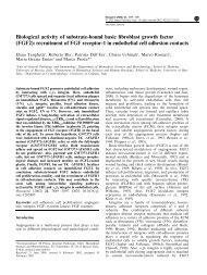
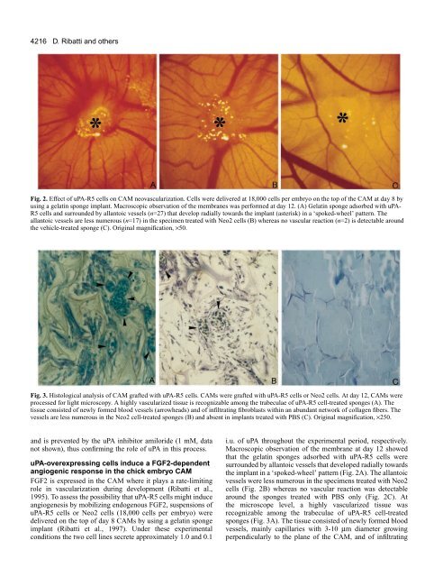
4216D. Ribatti and othersFig. 2. Effect <strong>of</strong> uPA-R5 cells on CAM neovascularization. <strong>Cell</strong>s were delivered at 18,000 cells per embryo on the top <strong>of</strong> the CAM at day 8 byusing a gelatin sponge implant. Macroscopic observation <strong>of</strong> the membranes was performed at day 12. (A) Gelatin sponge adsorbed with uPA-R5 cells and surrounded by allantoic vessels (n=27) that develop radially towards the implant (asterisk) in a ‘spoked-wheel’ pattern. Theallantoic vessels are less numerous (n=17) in the specimen treated with Neo2 cells (B) whereas no vascular reaction (n=2) is detectable aroundthe vehicle-treated sponge (C). Original magnification, ×50.Fig. 3. Histological analysis <strong>of</strong> CAM grafted with uPA-R5 cells. CAMs were grafted with uPA-R5 cells or Neo2 cells. At day 12, CAMs wereprocessed for light microscopy. A highly vascularized tissue is recognizable among the trabeculae <strong>of</strong> uPA-R5 cell-treated sponges (A). Thetissue consisted <strong>of</strong> newly formed blood vessels (arrowheads) and <strong>of</strong> infiltrating fibroblasts within an abundant network <strong>of</strong> collagen fibers. Thevessels are less numerous in the Neo2 cell-treated sponges (B) and absent in implants treated with PBS (C). Original magnification, ×250.and is prevented by the uPA inhibitor amiloride (1 mM, datanot shown), thus confirming the <strong>role</strong> <strong>of</strong> uPA in this process.uPA-overexpressing cells induce a FGF2-dependent<strong>angiogenic</strong> response in the chick embryo CAMFGF2 is expressed in the CAM where it plays a rate-limiting<strong>role</strong> in vascularization during development (Ribatti et al.,1995). To assess the possibility that uPA-R5 cells might induceangiogenesis by mobilizing endogenous FGF2, suspensions <strong>of</strong>uPA-R5 cells or Neo2 cells (18,000 cells per embryo) weredelivered on the top <strong>of</strong> day 8 CAMs by using a gelatin spongeimplant (Ribatti et al., 1997). Under these experimentalconditions the two cell lines secrete approximately 1.0 and 0.1i.u. <strong>of</strong> uPA throughout the experimental period, respectively.Macroscopic observation <strong>of</strong> the membrane at day 12 showedthat the gelatin sponges adsorbed with uPA-R5 cells weresurrounded by allantoic vessels that developed radially towardsthe implant in a ‘spoked-wheel’ pattern (Fig. 2A). The allantoicvessels were less numerous in the specimens treated with Neo2cells (Fig. 2B) whereas no vascular reaction was detectablearound the sponges treated with PBS only (Fig. 2C). Atthe microscope level, a highly vascularized tissue wasrecognizable among the trabeculae <strong>of</strong> uPA-R5 cell-treatedsponges (Fig. 3A). The tissue consisted <strong>of</strong> newly formed bloodvessels, mainly capillaries with 3-10 µm diameter growingperpendicularly to the plane <strong>of</strong> the CAM, and <strong>of</strong> infiltrating