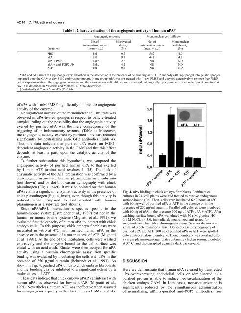
4218D. Ribatti and othersTable 4. Characterization <strong>of</strong> the <strong>angiogenic</strong> <strong>activity</strong> <strong>of</strong> human uPA*Angiogenic responseMononuclear cell infiltrateNo. <strong>of</strong> Microvessel No. <strong>of</strong> Mononuclearintersection points density intersection points cell densityTreatment (mean ± s.d.) (%) (mean ± s.d.) (%)PBS 1±1 0.7 5±2 4.9uPA 12±2 9.7 4±2 4.2uPA + PMSF 4±1‡ 2.8 ND NDuPA + anti-FGF2 Ab 5±1‡ 4.2 ND NDATF 1±1 0.7 ND ND*uPA and ATF (both at 1 µg/sponge) were absorbed in the absence or in the presence <strong>of</strong> neutralizing anti-FGF2 antibody (400 ng/sponge) into gelatin spongesimplanted onto the CAM at day 8 (10 embryos per group). <strong>In</strong> one group, uPA was pre-treated with 1 mM PMSF and dialyzed extensively to remove free PMSFbefore experimentation. The <strong>angiogenic</strong> response and the mononuclear cell infiltrate were assessed histologically by a planimetric method <strong>of</strong> ‘point counting’ atday 12 as described in Materials and Methods. ND: not determined.‡Statistically different from uPA (P
Angiogenic <strong>activity</strong> <strong>of</strong> uPA4219implicating endogenous FGF2 in mediating the <strong>angiogenic</strong><strong>activity</strong> <strong>of</strong> uPA.FGF2 is characterized by the capacity to interact withHSPGs present on the cell surface and in the ECM and thebiological implications <strong>of</strong> this interaction are manifold (for adiscussion <strong>of</strong> this point see Rusnati and Presta, 1996). Amongthem, ECM may act as a physiological reservoir forextracellular FGF2. <strong>In</strong>deed, FGF2 has been found associatedwith ECM in vitro and in <strong>vivo</strong> (Vlodavsky et al., 1987;Folkman et al., 1988; DiMario et al., 1989; Rogelj et al., 1989;Hageman et al., 1991). <strong>In</strong> vitro studies have shown that variousenzymes, including heparanase, thrombin, collagenases, andplasmin release ECM-bound FGF2 by degrading theglycosaminoglycan chain or the core protein <strong>of</strong> HSPGs(Saksela and Rifkin, 1990; Falcone et al., 1993; Whitelock etal., 1996). Our data show that uPA-overexpressing endothelialuPA-R5 cells, but not mock-transfected Neo2 cells, cause themobilization <strong>of</strong> ECM-bound 125 I-FGF2 in a plasminogendependentmanner, thus confirming the capacity <strong>of</strong> uPA/plasmin to facilitate the release <strong>of</strong> the immobilized growthfactor.Several experimental evidences suggest that the balancebetween storage and release <strong>of</strong> <strong>angiogenic</strong> FGF2 in ECM, aswell as the integrity <strong>of</strong> the matrix, may regulate the biologicaleffects <strong>of</strong> this growth factor on endothelium. For instance,interleukin-1-induced degradation <strong>of</strong> ECM in culturedchondrocytes, with consequent release <strong>of</strong> extracellular FGF2,has been implicated in the neovascularization <strong>of</strong> the synovia <strong>of</strong>arthritic patients (Tamura et al., 1996). Also, uPA productionand FGF2 release by macrophage derived-foam cells have beenimplicated in the development <strong>of</strong> the atherosclerotic lesion(Falcone et al., 1993). Finally, FGF2 mobilization from injuredcorneal epithelial basement membrane by increased plasmin<strong>activity</strong> (Salonen et al., 1987) has been suggested to have a <strong>role</strong>in corneal neovascularization (Folkman et al., 1988).Even though these data strongly suggest that uPA canfacilitate angiogenesis by increasing the bioavailability <strong>of</strong>ECM-stored FGF2, no direct in <strong>vivo</strong> experimental evidenceswere available to support this hypothesis. To this respect,neovascularization <strong>of</strong> the CAM <strong>of</strong> the chicken embryorepresents an unique experimental system to assess thishypothesis. <strong>In</strong>deed, FGF2 protein is present in the CAM whereit plays a limiting <strong>role</strong> in blood vessel growth duringdevelopment (Ribatti et al., 1995). Accordingly, chorionicepithelial cells and endothelial cells <strong>of</strong> the CAM mesodermexpress FGF2 mRNA (Ribatti et al., 1998). Moreover, thepresence <strong>of</strong> significant amounts <strong>of</strong> FGF2 in the chorioallantoicfluid, that parallel the levels <strong>of</strong> FGF2 present in the CAM,demonstrates that FGF2 is released by these cells andaccumulates in the extracellular environment (Ribatti et al.,1995).By utilizing this experimental system, we have shown thatneutralizing anti-FGF2 antibodies reduce significantly the<strong>angiogenic</strong> <strong>activity</strong> exerted by uPA-R5 cells and purifiedhuman uPA, thus implicating extracellular endogenous FGF2in the growth <strong>of</strong> newly formed blood vessels stimulated byuPA. However, the incapacity <strong>of</strong> anti-FGF2 antibody to fullysuppress the <strong>angiogenic</strong> ability <strong>of</strong> purified uPA and uPA-R5cells, even when tested under experimental conditions thatfully inhibit the <strong>angiogenic</strong> <strong>activity</strong> <strong>of</strong> exogenous recombinantFGF2, suggests that more factors besides FGF2 might beimplicated in protease-triggered CAM neovascularization.<strong>In</strong>deed, a variety <strong>of</strong> <strong>angiogenic</strong> growth factors, includingdifferent members <strong>of</strong> the FGF family, some is<strong>of</strong>orms <strong>of</strong> VEGFand placenta growth factor, HGF, and interleukin-8 share thecapacity to interact with HSPGs <strong>of</strong> the ECM (Rusnati andPresta, 1996) and exert an <strong>angiogenic</strong> response in the CAM(Oh et al., 1997; Ziche et al., 1997). This hypothesis issupported by the observation that uPA-R5 cells and Neo2 cellsretain an <strong>angiogenic</strong> potential apparently related to their levels<strong>of</strong> uPA expression also in the presence <strong>of</strong> neutralizing anti-FGF2 antibodies (see Table 1). It is also interesting to note thatboth purified human uPA and uPA-R5 cells exert an <strong>angiogenic</strong>response in the CAM that is less potent than that exerted byexogenous recombinant FGF2 (see Table 1 and 3), suggestingthat the levels <strong>of</strong> endogenous <strong>angiogenic</strong> growth factor(s)available to the protease action may represent a limiting factorin this experimental system.uPA exerts a significant <strong>angiogenic</strong> <strong>activity</strong> when deliveredonto the CAM in a single administration or when continuouslyreleased by uPA-R5 transfectants during the 4 days <strong>of</strong>experimentation. <strong>In</strong> the former experimental conditions, 250 ng<strong>of</strong> uPA (corresponding to approximately 7-8 i.u. <strong>of</strong> uPA<strong>activity</strong>) exert a significant <strong>angiogenic</strong> <strong>activity</strong>. <strong>In</strong> the latterexperimental conditions, as few as 18,000 uPA-R5 cells,releasing approximately 1.0 i.u. <strong>of</strong> uPA <strong>activity</strong> throughout theexperimental period, are sufficient to induce an <strong>angiogenic</strong>response. It must be pointed out that the amount <strong>of</strong> uPAreleased by uPA-R5 transfectants is similar to the amount <strong>of</strong>enzyme released by parental cells when stimulated in vitro byrecombinant FGF2 (Gualandris et al., 1997), thus indicatingthat the levels <strong>of</strong> uPA produced by our transfectants arebiologically significant. Our data are in keeping with previousobservations showing that purified human uPA exerts an<strong>angiogenic</strong> response in the rabbit cornea when administered atdoses ranging from 10 to 500 ng per implant (see Berman etal., 1982; Fibbi et al., 1998). Even though an accuratequantitative comparison among the various experimentalsystems cannot be performed due to their biologicalheterogeneity and the different enzymatic potency <strong>of</strong> humanuPA with plasminogen from different sources (human uPAbeing tested in rabbit or chick experimental models), the bulk<strong>of</strong> data indicate that uPA can elicit a significant <strong>angiogenic</strong>stimulus in the absence <strong>of</strong> a detectable inflammatory response(see Table 4, and Fibbi et al., 1998).Recent observations have shown that uPA/uPAR interactioninduces a pro-<strong>angiogenic</strong> phenotype in cultured endothelialcells that can be triggered also by the uPAR-binding ATF(Fibbi et al., 1998). However, the inability <strong>of</strong> ATF to induceangiogenesis in the CAM indicates that the proteolytic <strong>activity</strong><strong>of</strong> uPA is <strong>of</strong> pivotal importance in mediating its <strong>angiogenic</strong>capacity in <strong>vivo</strong>. This is confirmed by the reduced capacity <strong>of</strong>the PMSF-uPA complex to induce CAM neovascularization.Accordingly, irreversible inhibition <strong>of</strong> uPA by the active siteinhibitor Phe-Ala-Arg-chloromethyl ketone prevents its<strong>angiogenic</strong> <strong>activity</strong> in the rabbit cornea (Berman et al., 1982).Also, the capacity <strong>of</strong> plasmin (Iruela-Arispe et al., 1995)and thrombin (Tsopanoglou et al., 1993) to induceneovascularization in the CAM further support the notion <strong>of</strong>ECM proteolysis as a trigger for new blood vessel growth.Nevertheless, our observations do not rule out the possibilitythat uPAR occupancy may affect endothelial cell behavior also