DDU(Vascular) - Australasian Society for Ultrasound in Medicine
DDU(Vascular) - Australasian Society for Ultrasound in Medicine
DDU(Vascular) - Australasian Society for Ultrasound in Medicine
- No tags were found...
Create successful ePaper yourself
Turn your PDF publications into a flip-book with our unique Google optimized e-Paper software.
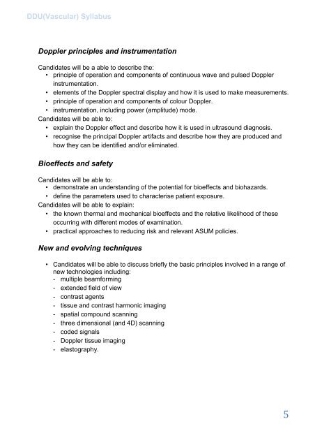
<strong>DDU</strong>(<strong>Vascular</strong>) SyllabusDoppler pr<strong>in</strong>ciples and <strong>in</strong>strumentationCandidates will be a able to describe the:• pr<strong>in</strong>ciple of operation and components of cont<strong>in</strong>uous wave and pulsed Doppler<strong>in</strong>strumentation.• elements of the Doppler spectral display and how it is used to make measurements.• pr<strong>in</strong>ciple of operation and components of colour Doppler.• <strong>in</strong>strumentation, <strong>in</strong>clud<strong>in</strong>g power (amplitude) mode.Candidates will be able to:• expla<strong>in</strong> the Doppler effect and describe how it is used <strong>in</strong> ultrasound diagnosis.• recognise the pr<strong>in</strong>cipal Doppler artifacts and describe how they are produced andhow they can be identified and/or elim<strong>in</strong>ated.Bioeffects and safetyCandidates will be able to:• demonstrate an understand<strong>in</strong>g of the potential <strong>for</strong> bioeffects and biohazards.• def<strong>in</strong>e the parameters used to characterise patient exposure.Candidates will be able to expla<strong>in</strong>:• the known thermal and mechanical bioeffects and the relative likelihood of theseoccurr<strong>in</strong>g with different modes of exam<strong>in</strong>ation.• practical approaches to reduc<strong>in</strong>g risk and relevant ASUM policies.New and evolv<strong>in</strong>g techniques• Candidates will be able to discuss briefly the basic pr<strong>in</strong>ciples <strong>in</strong>volved <strong>in</strong> a range ofnew technologies <strong>in</strong>clud<strong>in</strong>g:- multiple beam<strong>for</strong>m<strong>in</strong>g- extended field of view- contrast agents- tissue and contrast harmonic imag<strong>in</strong>g- spatial compound scann<strong>in</strong>g- three dimensional (and 4D) scann<strong>in</strong>g- coded signals- Doppler tissue imag<strong>in</strong>g- elastography.5


