FreeSurfer
FreeSurfer
FreeSurfer
- No tags were found...

You also want an ePaper? Increase the reach of your titles
YUMPU automatically turns print PDFs into web optimized ePapers that Google loves.
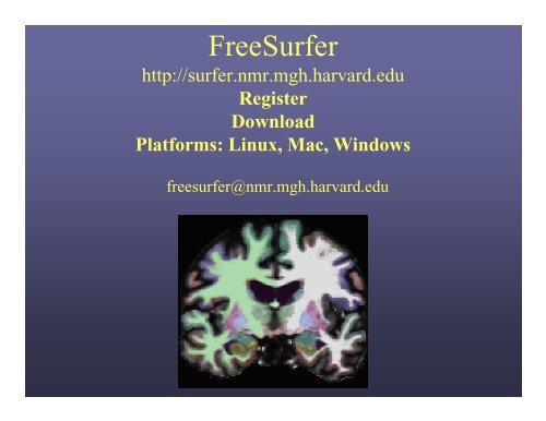
<strong>FreeSurfer</strong>http://surfer.nmr.mgh.harvard.eduRegisterDownloadPlatforms: Linux, Mac, Windowsfreesurfer@nmr.mgh.harvard.edu
Analyzing the Individual Subjectin <strong>FreeSurfer</strong>…What happens?How do I do that?Now What?
Analyzing the Individual Subjectin <strong>FreeSurfer</strong>…What happens?How do I do that?Now What?
Surface and Volume AnalysisCortical Reconstructionand Automatic LabelingInflation and FunctionalMappingAutomatic SubcorticalGray Matter LabelingSurface FlatteningSurface-based IntersubjectAlignment and StatisticsAutomatic Gyral WhiteMatter Labeling
Cortical Surface Reconstruction<strong>FreeSurfer</strong> creates computerizedmodels of the brain from MRI data.Input:T1-weighted (MPRAGE,SPGR)1mm 3 resolution(.dcm)Output:Segmented & parcellated conformedvolume(.mgz)
MR Anatomy CaveatsSurfaces are only as good as your scan.• Dependent on data quality– Contrast to noise– Signal to noise– Voxel resolution• MR Artifacts– MR susceptibility– MR distortions• Variations in MR tissue parameters across regions of the brain arealtered in different populations
Suggested MorphometrySequenceshttp://www.nmr.mgh.harvard.edu/~andre/
Analyzing the Individual Subjectin <strong>FreeSurfer</strong>…Wait…what happens?How do I do that?Now What?
What Happens During CorticalSurface Reconstruction?• Finds wm/gray boundary – white surface• Finds pial/CSF boundary – pial surface• Subcortical Segmentation• Cortical Parcellation• Generates surface-based cross-subjectregistration
What Happens During CorticalSurface Reconstruction?• Finds wm/gray boundary – white surfaceone sliceentire brain
What Happens During CorticalSurface Reconstruction?• Finds pial/CSF boundary – pial surfaceone sliceentire brain
Surface Model• Mesh of triangles gives a measurablesize• Allows us to measure Area, Curv.,Thickness (distance b/w vertices)• Vertex = point of 6 triangles• Triangles/Faces ~ 150,000 per hemi• 1:1 correspondence of vertices• XYZ at each vertex
-finalsurfs-autorecon2Cortical Thickness
What Happens During CorticalSurface Reconstruction?• Subcortical SegmentationWhite MatterCortexLateral VentricleThalamusCaudatePallidumHippocampusPutamenAmygdala
What Happens During CorticalSurface Reconstruction?• Cortical ParcellationPrecentral GyrusPostcentral GyrusSuperior Temporal Gyrus
What Happens During CorticalSurface Reconstruction?• Finds white/gray boundary – wm surface• Finds pial/CSF boundary – pial surface• Subcortical Segmentation• Cortical Parcellation• Generates surface-based cross-subjectregistration
Analyzing the Individual Subjectin <strong>FreeSurfer</strong>…Wait…what really happens?How do I do that?Now What?
Volumetric Processing Stages (subjid/mri):1. Motion Cor, Avg, Conform (orig.mgz)2. Non-uniform inorm (nu.mgz)3. Talairach transform computation(talairach/talairach.xfm)4. Intensity Normalization 1 (T1.mgz)5. Skull Strip (brainmask.mgz)Individual Steps6. EM Register (linear volumetric registration)7. CA Intensity Normalization (norm.mgz)8. CA Non-linear Volumetric Registration9. CA Label (Volumetric Labeling) (aseg.mgz)10. Intensity Normalization 2 (T1.mgz)11. White matter segmentation (wm.mgz)12. Edit WM With ASeg13. Fill and cut (filled.mgz)recon-all -helpSurface Processing Stages (subjid/surf):14. Tessellate (?h.orig.nofix)15. Smooth116. Inflate117. QSphere (?h.qsqhere)18. Automatic Topology Fixer (?h.orig)19. Final Surfs (?h.white ?h.pial ?.thickness)20. Smooth2 (?h.smoothwm)21. Inflate2 (?h.inflated)22. Aseg Statistics (stats/aseg.stats)23. Cortical Ribbon Mask (?h.ribbon.mgz)24. Spherical Morph25. Spherical Registration (?h.sphere.reg)26. Map average curvature to subject27. Cortical Parcellation (Labeling)28. Cortical Parcellation Statistics29. Cortical Parcellation mapped to Aseg30. White Matter Parcellation (wmparc.mgz)Note: ?h.orig means lh.orig or rh.orig
Processing Stream Orderhttp://surfer.nmr.mgh.harvard.edu/fswiki/ReconAllDevTable
How to Get StartedUse dicoms from scanner as input to CorticalReconstruction command:recon-all -all -i -s must do for each subject.Come back in 30 hours …Check your results – accurate to the tissueboundaries?
Reconstruction Stagesrecon-all is broken into three stages– autorecon1– autorecon2– autorecon3these 3 stages are equivalent to -all
Output of Cortical Surface Reconstruction
<strong>FreeSurfer</strong> Directory TreeEach data set has its own unique SubjectId (eg, bert)bertSubject IDbem stats src mri scripts surf tmp label trash
MGZ File Format001.mgz• mgz = compressed MGH file• Can store 4D (like NIFTI)• cols, rows, slices, frames• Generic: volumes and surfaces• Eg, Typical Anatomical Scan Volume: 256 x 256 x 128 x 1• <strong>FreeSurfer</strong> can read/write:NIFTI, Analyze, MINCCareful with NIFTI! (has32k column limit; surfacescould be more)• <strong>FreeSurfer</strong> can read:DICOM, SiemensIMA, AFNI
-autorecon1Volumetric Processing Stages (subjid/mri):1. Motion Cor, Avg, Conform (orig.mgz)2. Non-uniform inorm (nu.mgz)3. Talairach transform computation(talairach/talairach.xfm)4. Intensity Normalization 1 (T1.mgz)5. Skull Strip (brainmask.mgz)6. EM Register (linear volumetric registration)7. CA Intensity Normalization (norm.mgz)8. CA Non-linear Volumetric Registration9. CA Label (Volumetric Labeling) (aseg.mgz)10. Intensity Normalization 2 (T1.mgz)11. White matter segmentation (wm.mgz)12. Edit WM With ASeg13. Fill and cut (filled.mgz)recon-all -helpSurface Processing Stages (subjid/surf):14. Tessellate (?h.orig.nofix)15. Smooth116. Inflate117. QSphere (?h.qsqhere)18. Automatic Topology Fixer (?h.orig)19. Final Surfs (?h.white ?h.pial ?.thickness)20. Smooth2 (?h.smoothwm)21. Inflate2 (?h.inflated)22. Aseg Statistics (stats/aseg.stats)23. Cortical Ribbon Mask (?h.ribbon.mgz)24. Spherical Morph25. Spherical Registration (?h.sphere.reg)26. Map average curvature to subject27. Cortical Parcellation (Labeling)28. Cortical Parcellation Statistics29. Cortical Parcellation mapped to Aseg30. White Matter Parcellation (wmparc.mgz)
-motioncorMotion Correction and Averaging-autorecon1001.mgz+ rawavg.mgz002.mgzmriDoes not change native resolution.mri_robust_templateorigrawavg.mgz001.mgz 002.mgz
-motioncorConform-autorecon1rawavg.mgzorig.mgzChanges to 256^3, 1mm^3All volumes will be conformed to“anatomical space”.orig Volumemri_convert -conform
-talairachTalairach Transform-autorecon1• Computes 12 DOF transform matrix• Does NOT resample• MNI305 template (compute talairach from this)• Used to report Talairach coords in papers• helps with skull stripmritransformstalairach_avitalairach.xfm
-nuintensitycor-autorecon1Non-Uniform Intensity Correction• Uses MNI tool• Removes B1 bias fieldmri_nu_correct.mninu Volume
Intensity Bias• Left side of the image much brighter than right side• Worse with many coil elements• Makes gray/white segmentation difficult
-normalizationIntensity Normalization-autorecon1• Presegmentation (T1.mgz)– All WM = 110 intensity– Pre- and Post-SkullStripmri_normalizeT1 Volume
-skullstripSkull Strip-autorecon1• Removes all non-brain– Skull, Eyes, Neck, Dura• brainmask.mgzmri_watershed, mri_gcutOrig VolumeBrainmask Volume
<strong>FreeSurfer</strong> Directory TreeEach data set has its own unique SubjectId (eg, bert)bertSubject IDbem stats srcmri scripts surf tmp label trashorig T1 brainmask wmaseg aparc+aseg wmparc
Volumetric Processing Stages (subjid/mri):1. Motion Cor, Avg, Conform (orig.mgz)2. Non-uniform inorm (nu.mgz)3. Talairach transform computation(talairach/talairach.xfm)4. Intensity Normalization 1 (T1.mgz)5. Skull Strip (brainmask.mgz)-autorecon26. EM Register (linear volumetric registration)7. CA Intensity Normalization (norm.mgz)8. CA Non-linear Volumetric Registration9. CA Label (Volumetric Labeling) (aseg.mgz)10. Intensity Normalization 2 (T1.mgz)11. White matter segmentation (wm.mgz)12. Edit WM With ASeg13. Fill and cut (filled.mgz)recon-all -helpSurface Processing Stages (subjid/surf):14. Tessellate (?h.orig.nofix)15. Smooth116. Inflate117. QSphere (?h.qsqhere)18. Automatic Topology Fixer (?h.orig)19. Final Surfs (?h.white ?h.pial ?.thickness)20. Smooth2 (?h.smoothwm)21. Inflate2 (?h.inflated)22. Aseg Statistics (stats/aseg.stats)23. Cortical Ribbon Mask (?h.ribbon.mgz)24. Spherical Morph25. Spherical Registration (?h.sphere.reg)26. Map average curvature to subject27. Cortical Parcellation (Labeling)28. Cortical Parcellation Statistics29. Cortical Parcellation mapped to Aseg30. White Matter Parcellation (wmparc.mgz)Note: lh processed completely first, then rh.
-subcortsegAutomatic Volume Labeling-autorecon2• Label subcorticalstructures and wm/gm•Determine volumes ofsubcortical structuressteps 6-9, 22ASeg Volume•Used to fill insubcortical structuresfor later stepsAtlas: RB_all_2008-03-26
-subcortseg-autorecon2Volume-based Labeling% voxels in label10864WMGMlVThCaPuPaHpAm200 20 40 60 80 100 120intensityLabeling is determined by location andintensity.
Markov Random Field: MotivationWhat is the probability that cortical graymatter occurs inferior to hippocampus?
Segmentation: MRFPreliminary SegmentationFinal Segmentation
Why we use atlas + intensity +spatial location + geometric info +neighboring voxels + other info…Problems with Affine (12 DOF) Registration• ROIs need to be individualized.Why not just register to an ROI Atlas?12 DOF(Affine)ICBM AtlasCan’t segment on intensityaloneMarkov Random Field: Motivation% voxels in label10864WMGMlVThCaPuPaHpAm200 20 40 60 80 100 120intensityWhat is the probability that cortical graymatter occurs inferior to hippocampus?
Validation of Volume Labeling *Manual labeling done by Center for Morphometric Analysis(CMA)*Thanks to Drs Larry Seidman and Jill Goldstein for providing this data.
-segmentationWhite Matter Segmentation-autorecon2• Separates white matter from everything else• “Fills in” subcortical structures• Cerebellum removed, brain stem still theremri_segmentmri_edit_wm_with_asegmri_pretesswm Volume
-fillFill and Cut (Subcortical Mass)• Fills in any voids-autorecon2• Removes any islands• Removes brain stem• Separates hemispheres (each hemi has different value)• filled.mgz = “Subcortical Mass”mri_fillWM VolumeFilled Volume
<strong>FreeSurfer</strong> Directory TreeEach data set has its own unique SubjectId (eg, bert)bertSubject IDbem stats srcmri scripts surf tmp label trashorig T1 brainmask wm aseg aparc+aseg wmparc
-tessellationTessellation-autorecon2orig surfacesurf/lh.origsurf/rh.orig• Mosaic of triangles (“tessellation”)• Errors: Donut holes, handles- Subsequently fixed by the automatictopology fixer
-inflate-autorecon2Inflation: VisualizationGyriSulciDale and Sereno, 1993; Dale et al., Dale et al., 1999; Fischl et al., 1999;Fischl et al., 2000; Fischl et al., 2001
-fix-autorecon2Automatic Topology FixerFornixhippocampusoptic nervePallidum andPutamenCorticalDefectsVentriclesand Caudate•Holes• Handles• Automatically Fixed
-finalsurfsWhite Matter Surface-autorecon2• Nudge orig surface• Follow T1 intensity gradients• Smoothness constraint• Vertex Identity stays constant
-finalsurfsPial Surface-autorecon2• Nudge white surface• Follow T1 intensity gradients• Vertex Identity Stays
-finalsurfs-autorecon2Optimal Surface PlacementGray-White BoundaryOuter Cortical Surface
Cortical Thickness• Distance between whiteand pial surfaces• One value per vertex• Surface-based moreaccurate than volumebasedpial surfacewhite surfacelh.thickness, rh.thickness
• Red regions are thinner• Yellow regions are thickerThickness Maps
Curvature (Radial)• Circle tangent to surfaceat each vertex• Curvature measure is1/radius of circle• One value per vertex• Signed (sulcus/gyrus)• Actually use gaussiancurvaturelh.curv, rh.curv
<strong>FreeSurfer</strong> Directory TreeEach data set has its own unique SubjectId (eg, bert)bertSubject IDbem stats src mri scripts surf tmp labellh.white lh.curvlh.thickness lh.pial
Volumetric Processing Stages (subjid/mri):1. Motion Cor, Avg, Conform (orig.mgz)2. Non-uniform inorm (nu.mgz)3. Talairach transform computation(talairach/talairach.xfm)4. Intensity Normalization 1 (T1.mgz)5. Skull Strip (brainmask.mgz)-autorecon36. EM Register (linear volumetric registration)7. CA Intensity Normalization (norm.mgz)8. CA Non-linear Volumetric Registration9. CA Label (Volumetric Labeling) (aseg.mgz)10. Intensity Normalization 2 (T1.mgz)11. White matter segmentation (wm.mgz)12. Edit WM With ASeg13. Fill and cut (filled.mgz)recon-all -helpSurface Processing Stages (subjid/surf):14. Tessellate (?h.orig.nofix)15. Smooth116. Inflate117. QSphere (?h.qsqhere)18. Automatic Topology Fixer (?h.orig)19. Final Surfs (?h.white ?h.pial ?.thickness)20. Smooth2 (?h.smoothwm)21. Inflate2 (?h.inflated)22. Aseg Statistics (stats/aseg.stats)23. Cortical Ribbon Mask (?h.ribbon.mgz)24. Spherical Morph25. Spherical Registration (?h.sphere.reg)26. Map average curvature to subject27. Cortical Parcellation (Labeling)28. Cortical Parcellation Statistics29. Cortical Parcellation mapped to Aseg30. White Matter Parcellation (wmparc.mgz)Note: lh processed completely first, then rh.
Surface-Based Spherical Coord System
“Spherical” RegistrationInflated Surface (Sulcal Map)Spherical InflationAtlas (Target)High-DimensionalRegistration toSpherical TemplateIndividual SubjectTemplate: average.curvature.filled.buckner.40.tiff
-sphere-surfregSpherical InflationRegistration to Atlas-autorecon3Individual SubjectAtlas (Target)
-cortparcCortical ParcellationSpherical Templatebased on ManualParcellation-autorecon3Map to IndividualThru Spherical RegFine-tune based onindividual anatomyAtlases: curvature.buckner40filled.desikan_killiany; destrieux.simple
Non-Cortical Areas of SurfaceAmygdala, Putamen, Hippocampus, Caudate, Ventricles, CC?h.cortex.label
Output of Cortical Surface Reconstruction
Non-Cortical Areas of SurfaceAmygdala, Putamen, Hippocampus, Caudate, Ventricles, CC?h.cortex.label
The “Unknown” Label
-cortparc2Cortical Parcellation: 2-autorecon3ManualAutomaticAtlases: curvature.buckner40filled.desikan_killiany, atlas_2005_simpleThanks to Christophe Destrieux for this slide.
-aparc2asegAparc+Aseg-autorecon3There is also aparc.a2005s+aseg.mgz for Destrieux atlas
-wmparcWhite Matter Parcellation-autorecon3Nearest Cortical Labelto point in White MatterSalat DH, Greve DN, Pacheco JL, Quinn BT, Helmer KG, Buckner RL,Fischl B. Regional white matter volume differences in nondemented agingand Alzheimer’s disease. Neuroimage. 2008 Nov 5.
<strong>FreeSurfer</strong> Directory TreeEach data set has its own unique SubjectId (eg, bert)bertSubject IDbem stats src mri scripts surf tmp labellh.aparc.annot rh.aparc.annot
<strong>FreeSurfer</strong> Directory TreeEach data set has its own unique SubjectId (eg, bert)bertSubject IDbem stats srcmri scripts surf tmp labelorig T1 brainmask wm aseg aparc+aseg wmparc
<strong>FreeSurfer</strong> Directory TreeDirectories used often are in green.bertSubject IDbem stats srcmri scripts surf tmp label trashaseg.stats lh.aparc.stats rh.aparc.stats wmparc.stats
Why use <strong>FreeSurfer</strong>?What happens?How do I do that?Now What?
Using <strong>FreeSurfer</strong>With <strong>FreeSurfer</strong>, certain variables must be setin order to use it correctly:FREESURFER_HOMEtell Operating System where <strong>FreeSurfer</strong> isSUBJECTS_DIRtell <strong>FreeSurfer</strong> where data is
Internal <strong>FreeSurfer</strong>Stablesource /usr/local/freesurfer/nmr-std-env(nmrenv)Devsource /usr/local/freesurfer/nmr-dev-envMailing listmartinos-tech@yahoogroups.com
Freezing <strong>FreeSurfer</strong>rsync -a /usr/local/freesurfer/nmr-std-env /some/location/source /some/location/SetUp<strong>FreeSurfer</strong>.csh(change variables)
Using <strong>FreeSurfer</strong>With <strong>FreeSurfer</strong>, certain variables must be setin order to use it correctly:FREESURFER_HOMEtell Operating System where <strong>FreeSurfer</strong> isSUBJECTS_DIRtell <strong>FreeSurfer</strong> where data is
Set-up Environmental VariablesSubject ID$SUBJECTS_DIRbertfred sara margaret …Before running <strong>FreeSurfer</strong>, mustset $FREESURFER_HOME and$SUBJECTS_DIR
Starting the Reconstruction ProcessBefore running <strong>FreeSurfer</strong>, must set$FREESURFER_HOME and $SUBJECTS_DIRrecon-all \-i /path/to/your/raw/data1 \-i /path/to/your/raw/data2 \-all -s subject_id• This will create the subject directory ‘subject_id’ in your$SUBJECTS_DIR and convert your 2 raw acquisitions to mgz and usethem as input for the ‘-all’command.
Alternative: Add Your Data• cd $SUBJECTS_DIR• mkdir –p bert/mri/orig• mri_convert rawdata.nii bert/mri/orig/001.mgz• mri_convert rawdata.nii bert/mri/orig/002.mgz• recon-all –all –s bertbertbem label src mri scripts surf tmp labelorig001.mgz 002.mgz
<strong>FreeSurfer</strong> Output• Volumes• Surfaces• Surface Overlays• ROI Summaries
Volumesorig.mgzT1.mgz brainmask.mgz wm.mgz filled.mgzSubcortical Massaseg.mgzaparc+aseg.mgz•$SUBJECTS_DIR/bert/mri• All “Conformed” 256^3, 1mm• File format: .mgzVolume Viewer:tkmedit
Surfaces.orig .white .pial.inflated sphere, sphere.reg flat•$SUBJECTS_DIR/bert/surf•Number/Identity of vertices stays the same (except flat)•XYZ Location Changes•Flattening not done as part of standard reconstructionSurface Viewer:tksurfer
Surface Overlayslh.sulc on inflated lh.curv on inflated lh.thickness on inflatedlh.sulc on piallh.curv on inflatedfMRI on flatlh.aparc.annot on inflated• Value for each vertex• Color indicates value• Color: gray,red/green, heat, color table• Rendered on any surface• fMRI/Stat Maps too
Other <strong>FreeSurfer</strong> File FormatsUnique to <strong>FreeSurfer</strong>•Surface: lh.white, lh.pial, lh.orig• Curv: lh.curv, lh.sulc, lh.thickness• Annotation: lh.aparc.annot• Label: lh.pericalcarine.label
Why use <strong>FreeSurfer</strong>?What happens?How do I do that?Now What?
Quality Check Your Recon• Do your surfaces follow gm/wm borders?• Does the subcortical segmentation followintensity boundaries?
Troubleshootingrecon-all fails• check recon-all.log• try to rerun step that failed• look at volume from last successful step• examine data quality to see what might cause error• if it fails again, attempt to run modified version of commandif possible• search <strong>FreeSurfer</strong> mailing list for other instances of thisproblem:http://www.mail-archive.com/freesurfer@nmr.mgh.harvard.edu/• email the mailing list if still need help
<strong>FreeSurfer</strong> Directory TreeEach data set has its own unique SubjectId (eg, bert)bertSubject IDbem stats srcmri scripts surf tmp labelrecon-all.logrecon-all-status.log
Bug Reporting• Report version currently using– see top of recon-all.log– more $FREESURFER_HOME/build-stamp.txt• command line tried to run• attach recon-all.log• Output in terminal window if appropriate• Operating System
ROI Summaries:$SUBJECTS_DIR/subjid/statsaseg.stats – volume summaries?h.aparc.stats – desikan/killiany atlas summaries?h.aparc.a2005s.stats – destrieux atlas summarieswmparc.stats – volume summaries_______________________________________________aseg:• volumes of subcortical structures (mm3)aparc:• thickness of cortical parcellation structures (mm)• total white matter volume (mm3)• number of vertices in cortex• surface area of cortex (mm2)
Aseg Stats FileIndex SegId NVoxels Volume_mm3 StructName Mean StdDev Min Max Range1 2 255076 255076.0 Left-Cerebral-White-Matter 101.5872 7.9167 34.0000 148.0000 114.00002 3 266265 266265.0 Left-Cerebral-Cortex 75.3682 9.4016 28.0000 152.0000 124.00003 4 5855 5855.0 Left-Lateral-Ventricle 37.7920 10.9705 20.0000 88.0000 68.00004 5 245 245.0 Left-Inf-Lat-Vent 56.4091 9.5906 26.0000 79.0000 53.00005 7 16357 16357.0 Left-Cerebellum-White-Matter 91.2850 4.8989 49.0000 106.0000 57.00006 8 60367 60367.0 Left-Cerebellum-Cortex 76.3620 9.5724 26.0000 135.0000 109.00007 10 7460 7460.0 Left-Thalamus-Proper 91.3778 7.4668 43.0000 108.0000 65.00008 11 3133 3133.0 Left-Caudate 78.5801 8.2886 42.0000 107.0000 65.00009 12 5521 5521.0 Left-Putamen 86.9680 5.5752 66.0000 106.0000 40.000010 13 1816 1816.0 Left-Pallidum 97.7162 3.4302 79.0000 106.0000 27.000011 14 852 852.0 3rd-Ventricle 41.9007 11.8230 22.0000 69.0000 47.000012 15 1820 1820.0 4th-Ventricle 39.7053 10.6407 20.0000 76.0000 56.000013 16 25647 25647.0 Brain-Stem 85.2103 8.2819 38.0000 106.0000 68.000014 17 4467 4467.0 Left-Hippocampus 77.6346 7.5845 45.0000 107.0000 62.000015 18 1668 1668.0 Left-Amygdala 74.5104 5.8320 50.0000 94.0000 44.000016 24 1595 1595.0 CSF 52.1348 11.6113 29.0000 87.0000 58.0000Index: nth Segmentation in stats fileSegId: index into lookup tableNVoxels: number of Voxels/Vertices in segmentationStructName: Name of structure from LUTMean/StdDev/Min/Max/Range: intensity across ROIEg: aseg.stats, wmparc.statscreated by mri_segstats
Getting Stats into TableFormat• Organize stats files from a group of subjects into a singletext fileasegstats2table \--subjects 001 002 003 \--meas volume \--tablefile asegstats.txt Command name List subjects to include vol or mean intensity of struct text output fileasegstats.txt created in SUBJECTS_DIR
Import Text File into SpreadsheetFile > OpenSelect asegstats.txtChoose delimited by Space
Cortical, Gray, White, Intracranial VolumesAlso in aseg.stats header:# Measure lhCortex, lhCortexVol, Left hemisphere cortical gray matter volume, 192176.447567, mm^3# Measure rhCortex, rhCortexVol, Right hemisphere cortical gray matter volume, 194153.9526, mm^3# Measure Cortex, CortexVol, Total cortical gray matter volume, 386330.400185, mm^3# Measure lhCorticalWhiteMatter, lhCorticalWhiteMatterVol, Left hemisphere cortical white mattervolume, 217372.890625, mm^3# Measure rhCorticalWhiteMatter, rhCorticalWhiteMatterVol, Right hemisphere cortical white mattervolume, 219048.187500, mm^3# Measure CorticalWhiteMatter, CorticalWhiteMatterVol, Total cortical white matter volume,436421.078125, mm^3# Measure SubCortGray, SubCortGrayVol, Subcortical gray matter volume, 182006.000000, mm^3# Measure TotalGray, TotalGrayVol, Total gray matter volume, 568336.400185, mm^3# Measure SupraTentorial, SupraTentorialVol, Supratentorial volume, 939646.861571, mm^3# Measure IntraCranialVol, ICV, Intracranial Volume, 1495162.656130, mm^3lhCortex, rhCortex, Cortex – surface-based measure of cortical gray matter volumelhCorticalWhiteMatter, … – surface-based measure of cortical white matter volumeSubCortGray – volume-based measure of subcortical gray matterTotalGray – Cortex + Subcortical graySupraTentorial – Cortex + CorticalWhiteMatter + Subcortical grayIntraCranialVol – estimated Total Intracranial vol (eTIV)http://surfer.nm.mgh.harvard.edu/fswiki/eTIV
Parcellation Stats FileStructNameNumVert SurfArea GrayVol ThickAvg ThickStd MeanCurv GausCurv FoldInd CurvIndunknown 10863 7151 13207 1.776 1.629 0.121 0.107 383 50.8bankssts 1222 830 2290 2.711 0.559 0.112 0.027 10 1.3caudalanteriorcingulate 830 585 1459 2.474 0.569 0.128 0.020 10 0.7caudalmiddlefrontal 2509 1658 4979 2.653 0.567 0.125 0.035 27 3.5corpuscallosum 2124 1340 569 0.489 0.631 0.151 0.110 87 8.0cuneus 2737 1706 3086 1.741 0.509 0.162 0.065 52 8.0entorhinal 495 330 1685 3.150 0.753 0.149 0.187 15 1.5fusiform 3878 2638 7887 2.627 0.724 0.137 0.046 57 6.7StructName: Name of structure/ROINumVert: number of vertices in ROISurfArea: Surface area in mm2GrayVol: volume of gray matterThickAvg/ThickStd – average and stddev of thickness in ROIMeanCurv – mean Gaussian curvatureGausCurv – Gaussian curvatureFoldInd – folding indexCurvInd – curvature indexEg, lh.aparc.stats, lh.a2005s.aparc.statscreated by mris_anatomical_stats
Also in ?h.aparc.stats header:Parcellation Stats# Measure Cortex, NumVert, Number of Vertices, 129292, unitless# Measure Cortex, SurfArea, Surface Area, 84209.5, mm^2# Measure Cortex, MeanThickness, Mean Thickness, 2.29807, mmTotal Number of Vertices – same for white and pialTotal Surface Area – white surfaceTotal Mean Thickness – between white and pial
Getting Stats into TableFormataparcstats2table \ Command name--subjects 001 002 003 \ List subjects to include--hemi rh \ Which hemisphere--meas volume \ volume, thickness, or area“Gray Vol”, “Avg Thick”, or “Surf Area” of structure--tablefile rh.aparc.vol.txt text output filerh.aparc.vol.txt created in SUBJECTS_DIR
The Endastevens@nmr.mgh.harvard.edu
Manifold SurgeryBEFOREAFTER