Staging of Rectal Cancer: MRI versus Endoscopic ... - Imedex
Staging of Rectal Cancer: MRI versus Endoscopic ... - Imedex
Staging of Rectal Cancer: MRI versus Endoscopic ... - Imedex
- No tags were found...

You also want an ePaper? Increase the reach of your titles
YUMPU automatically turns print PDFs into web optimized ePapers that Google loves.
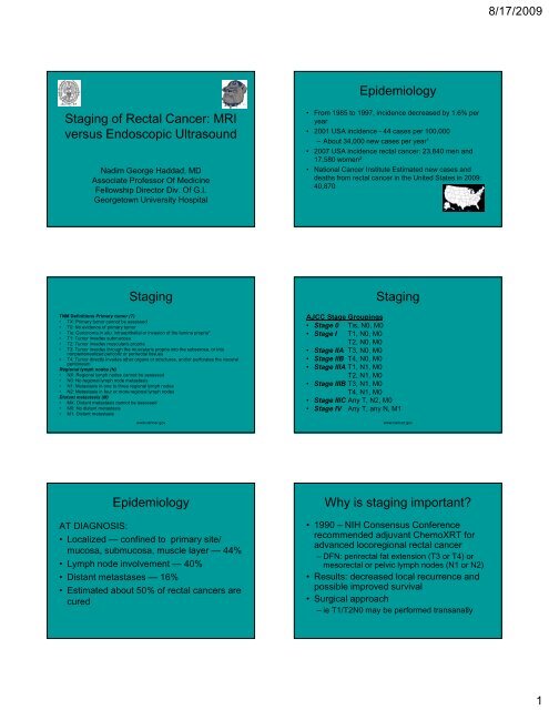
8/17/2009Epidemiology<strong>Staging</strong> <strong>of</strong> <strong>Rectal</strong> <strong>Cancer</strong>: <strong>MRI</strong><strong>versus</strong> <strong>Endoscopic</strong> UltrasoundNadim George Haddad, MDAssociate Pr<strong>of</strong>essor Of MedicineFellowship Director Div. Of G.I.Georgetown University Hospital• From 1985 to 1997, incidence decreased by 1.6% peryear• 2001 USA incidence - 44 cases per 100,000– About 34,000 new cases per year 1• 2007 USA incidence id rectal cancer: 23,840 men and17,580 women 2• National <strong>Cancer</strong> Institute Estimated new cases anddeaths from rectal cancer in the United States in 2009:40,870<strong>Staging</strong>TNM Definitions Primary tumor (T)• TX: Primary tumor cannot be assessed• T0: No evidence <strong>of</strong> primary tumor• Tis: Carcinoma in situ: intraepithelial or invasion <strong>of</strong> the lamina propria*• T1: Tumor invades submucosa• T2: Tumor invades muscularis propria• T3: Tumor invades through the muscularis propria into the subserosa, or intononperitonealized pericolic or perirectal tissues• T4: Tumor directly invades other organs or structures, and/or perforates the visceralperitoneumRegional lymph nodes (N)• NX: Regional lymph nodes cannot be assessed• N0: No regional lymph node metastasis• N1: Metastasis in one to three regional lymph nodes• N2: Metastasis in four or more regional lymph nodesDistant metastasis (M)• MX: Distant metastasis cannot be assessed• M0: No distant metastasis• M1: Distant metastasiswww.cancer.gov<strong>Staging</strong>AJCC Stage Groupings• Stage 0 Tis, N0, M0• Stage I T1, N0, M0T2, N0, M0• Stage IIA T3, N0, M0• Stage IIB T4, N0, M0• Stage IIIA T1, N1, M0T2, N1, M0• Stage IIIB T3, N1, M0T4, N1, M0• Stage IIIC Any T, N2, M0• Stage IV Any T, any N, M1www.cancer.govEpidemiologyAT DIAGNOSIS:• Localized — confined to primary site/mucosa, submucosa, muscle layer — 44%• Lymph node involvement — 40%• Distant metastases — 16%• Estimated about 50% <strong>of</strong> rectal cancers arecuredWhy is staging important?• 1990 – NIH Consensus Conferencerecommended adjuvant ChemoXRT foradvanced locoregional rectal cancer– DFN: perirectal fat extension (T3 or T4) ormesorectal or pelvic lymph nodes (N1 or N2)• Results: decreased local recurrence andpossible improved survival• Surgical approach– ie T1/T2N0 may be performed transanally1
8/17/2009<strong>Staging</strong> Modalities<strong>Staging</strong>•CT Scan•<strong>MRI</strong>•EUS• DRE• <strong>MRI</strong>– Mandatory in UK, Denmark, Norway, Sweden– Circumferential resection margins– Extramural venous invasion– 75-85% accuracy– MERCURY• Study confirming diagnostic accuracy/reproducibility• Predicts involvement <strong>of</strong> surgical margin and extramural tumorinvasion– Taylor et al. AJR Dec 2008• Systematic evaluation <strong>of</strong> <strong>MRI</strong> findings<strong>Staging</strong>Normal anatomy <strong>of</strong> the mesorectum• <strong>MRI</strong>– Zhang et el. AJR May 2008• 38 pts with rectal ca underwent <strong>MRI</strong>• T accuracy 92%• 97% <strong>of</strong> resectable cases, sphincter sparing surgeryaccurately chosen• Questions for the radiologist:– Phased array coils v. endorectal coils– Field Strength• Still need bowel prep– Decrease artifact from air-tissue borderIafrate F. et.al. Radiographics;2006;26:701-714©2006 by Radiological Society <strong>of</strong> North AmericaCoronal turbo spin-echo T2-weighted MR image shows the normal anatomy <strong>of</strong> the rectumStage T1 rectal carcinomaIafrate F. et.al. Radiographics;2006;26:701-714Iafrate F. et.al. Radiographics;2006;26:701-714©2006 by Radiological Society <strong>of</strong> North America©2006 by Radiological Society <strong>of</strong> North America2
8/17/2009<strong>Rectal</strong> adenocarcinomaINVOLVEMENT OF MESORECTALFASCIAIafrate F. et.al. Radiographics;2006;26:701-714©2006 by Radiological Society <strong>of</strong> North AmericaINVOLVEMENT OF PUBORECTAL SPHINCTEREXTRAMURAL VEIN INVASIONPREOPERATIVE STAGING OF RECTALCANCER WITH <strong>MRI</strong>Operative characteristics <strong>of</strong> endorectal <strong>MRI</strong> in staging rectal carcinomaDiagnostic accuracy <strong>of</strong> preoperative <strong>MRI</strong> in predicting curative resection <strong>of</strong>rectal cancer Mercury study group. Br Med J 2006LeBlanc JK (2007) Imaging and management <strong>of</strong> rectal cancerNat Clin Pract Gastroenterol Hepatol 4: 665–676 doi:10.1038/ncpgasthep09773
8/17/2009<strong>Staging</strong>Accuracy <strong>of</strong> tumor (T) staging by endoscopic ultrasound in patients with rectalcancer since 1990• Transrectal <strong>Endoscopic</strong> Ultrasound– Improved T staging• 80-85% accuracy (UTD)– Compared to <strong>MRI</strong> with endorectal coils, EUSbetter for discerning T1 v. T2– Nodal <strong>Staging</strong> Accuracy 70-75% (asge)• Sensitivity decreased for LNs < 5mm• FNA may be needed if T1/T2LeBlanc JK (2007) Imaging and management <strong>of</strong> rectal cancerNat Clin Pract Gastroenterol Hepatol 4: 665–676 doi:10.1038/ncpgasthep0977Accuracy <strong>of</strong> node (N) staging by endoscopic ultrasound in patients with rectalcancer since 1990EUSLeBlanc JK (2007) Imaging and management <strong>of</strong> rectal cancerNat Clin Pract Gastroenterol Hepatol 4: 665–676 doi:10.1038/ncpgasthep0977Possible Downfalls:• Does not asess circumferential resectionmargin or extramural venous invasion• Malignant stricturest– Miniprobes• Obstructing Lesion– UnderstagingCase 1<strong>Rectal</strong> CA38 year old male with anemia andunexplained diarrhea.5
8/17/2009Seminal VesiclesT3N1 <strong>Rectal</strong> CAPeri-rectal Lymph Node Case 258 year old male with change <strong>of</strong> bowelhabits and BRBPR. Patient withcolonoscopy which revealed rectal cancer.T3 <strong>Rectal</strong> <strong>Cancer</strong>Tumor and Prostate6
8/17/2009Peri-rectal Lymph Nodes<strong>Rectal</strong> <strong>Cancer</strong>7
8/17/2009Studies – <strong>MRI</strong> v. EUSConclusion• WJG – June 2008– 34 patients: biopsyproven rectal cancerACCURACY:• <strong>MRI</strong> T staging 89.7%• EUS T staging 85.3%• <strong>MRI</strong> N staging 74.5%• EUS N staging 76.5%SENSITIVITY• <strong>MRI</strong> T staging 79.4%• EUS T staging 70.6%• <strong>MRI</strong> N staging 61.8%• EUS N staging 52.9%SPECIFICITY• <strong>MRI</strong> T staging 93.1%• EUS T staging 90.2%• <strong>MRI</strong> N staging 80.9%• EUS N staging 84.31%• EUS is the best method <strong>of</strong> local T1-T2 staging <strong>of</strong> rectal cancer.• EUS/FNA/FNI for local therapy.• The use <strong>of</strong> EUS for staging rectal cancer seems to improve disease-freesurvival because EUS facilitates the appropriate selection <strong>of</strong> patients forneoadjuvant therapy; the use <strong>of</strong> EUS has also increased the number <strong>of</strong>sphincter-preserving surgeries performed in patients with rectal cancer• <strong>MRI</strong> better for localy advanced tumor(T3/T4).• <strong>Rectal</strong> cancer staging with EUS or <strong>MRI</strong> is not accurate after patients havereceived neoadjuvant therapy .• <strong>MRI</strong> is the most useful staging tool for determining the status <strong>of</strong> thecircumferential resection margin, which is important in the assessment <strong>of</strong>the risk <strong>of</strong> local recurrenceBibliography1. Greenlee RT, Hill-Harmon MB, Murray T, Thun M: <strong>Cancer</strong> statistics,2001. CA <strong>Cancer</strong> J Clin 2001; 51:15-36.2. Jemal A, Siegel R, Ward E, et al: <strong>Cancer</strong> statistics, 2007. CA <strong>Cancer</strong> JClin 2007; 57:43-66.3. Wu X; Chen VW; Martin J, Subsite-specific colorectal cancer incidencerates and stage distributions among Asians and Pacific Islanders in theUnited States, 1995 to 1999. <strong>Cancer</strong> Epidemiol Biomarkers Prev. 2004Jul;13(7):1215-22.4. Thoeni RF, Colorectal cancer. Radiologic staging. Radiol Clin North Am1997 Mar;35(2):457-85.5. LeBlanc JK, Imaging and management <strong>of</strong> rectal cancer Nat Clin PractGastroenterol Hepatol 4: 665–676 doi:10.1038/ncpgasthep0977.8