Titanium mesh cranioplasty for patients with large cranial defects ...
Titanium mesh cranioplasty for patients with large cranial defects ...
Titanium mesh cranioplasty for patients with large cranial defects ...
- No tags were found...

You also want an ePaper? Increase the reach of your titles
YUMPU automatically turns print PDFs into web optimized ePapers that Google loves.
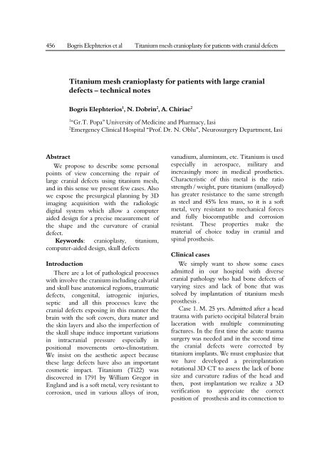
456 Bogris Elephterios et al <strong>Titanium</strong> <strong>mesh</strong> <strong>cranioplasty</strong> <strong>for</strong> <strong>patients</strong> <strong>with</strong> <strong>cranial</strong> <strong>defects</strong><strong>Titanium</strong> <strong>mesh</strong> <strong>cranioplasty</strong> <strong>for</strong> <strong>patients</strong> <strong>with</strong> <strong>large</strong> <strong>cranial</strong><strong>defects</strong> – technical notesBogris Elephterios 1 , N. Dobrin 2 , A. Chiriac 21 “Gr.T. Popa” University of Medicine and Pharmacy, Iasi2 Emergency Clinical Hospital “Prof. Dr. N. Oblu”, Neurosurgery Department, IasiAbstractWe propose to describe some personalpoints of view concerning the repair of<strong>large</strong> <strong>cranial</strong> <strong>defects</strong> using titanium <strong>mesh</strong>,and in this sense we present few cases. Alsowe expose the presurgical planning by 3Dimaging acquisition <strong>with</strong> the radiologicdigital system which allow a computeraided design <strong>for</strong> a precise measurement ofthe shape and the curvature of <strong>cranial</strong>defect.Keywords: <strong>cranioplasty</strong>, titanium,computer-aided design, skull <strong>defects</strong>IntroductionThere are a lot of pathological processes<strong>with</strong> involve the cranium including calvarialand skull base anatomical regions, traumatic<strong>defects</strong>, congenital, iatrogenic injuries,septic and all this processes leave the<strong>cranial</strong> <strong>defects</strong> exposing in this manner thebrain <strong>with</strong> the soft covers, dura mater andthe skin layers and also the imperfection ofthe skull shape induce important variationsin intra<strong>cranial</strong> pressure especially inpositional movements orto-clinostatism.We insist on the aesthetic aspect becausethese <strong>large</strong> <strong>defects</strong> have also an importantcosmetic impact. <strong>Titanium</strong> (Ti22) wasdiscovered in 1791 by William Gregor inEngland and is a soft metal, very resistant tocorrosion, used in various alloys of iron,vanadium, aluminum, etc. <strong>Titanium</strong> is usedespecially in aerospace, military andincreasingly more in medical prosthetics.Characteristic of this metal is the ratiostrength / weight, pure titanium (unalloyed)has greater resistance to the same strengthas steel and 45% less mass, so it is a softmetal, very resistant to mechanical <strong>for</strong>cesand fully biocompatible and corrosionresistant. These properties make thematerial of choice today in <strong>cranial</strong> andspinal prosthesis.Clinical casesWe simply want to show some casesadmitted in our hospital <strong>with</strong> diverse<strong>cranial</strong> pathology who had bone <strong>defects</strong> ofvarying sizes and lack of bone that wassolved by implantation of titanium <strong>mesh</strong>prosthesis .Case 1. M. 25 yrs. Admitted after a headtrauma <strong>with</strong> parieto occipital bilateral brainlaceration <strong>with</strong> multiple comminutingfractures. In the first time the acute traumasurgery was needed and in the second timethe <strong>cranial</strong> <strong>defects</strong> were corrected bytitanium implants. We must emphasize thatwe have developed a preimplantationrotational 3D CT to assess the lack of bonesize and curvature radius of the head andthen, post implantation we realize a 3Dverification to appreciate the correctposition of prosthesis and its connection to
Romanian Neurosurgery (2010) XVII 4: 456 – 460 457the plate bone by titanium screws. Thistechnique was per<strong>for</strong>med <strong>with</strong> SIEMENSAXIOM ARTIS system in a acquisition 3Drotational protocol <strong>with</strong> 60frames /sec on 20seconds that means about 1000 frameswhich need reconstruction in specialsoftware . Figure 1, Figure 2Case 2. F 38 yrs. Admitted after multipleintervention <strong>for</strong> septic problems, subduralempiema and osteitis <strong>with</strong> frontosinusalorigin, presented three <strong>large</strong> <strong>cranial</strong>postoperative <strong>defects</strong>.The correction intervention was madeafter 4 years from the septic episode aftercontrolling the septic problems, imagingrevealed that the risk is practically null.Underwent anterior eradication of septicfoci, 3D exploration highlights accuratelywhether or not there are septic risk. Figures3, 4, 5, 6, 7.Figure 1, 2 3D aspects pre and post implants
458 Bogris Elephterios et al <strong>Titanium</strong> <strong>mesh</strong> <strong>cranioplasty</strong> <strong>for</strong> <strong>patients</strong> <strong>with</strong> <strong>cranial</strong> <strong>defects</strong>Figures 3, 4, 5, 6, 7 first image present thepreoperative aspect the others reveals the correctinsertion of titanium prosthesis <strong>mesh</strong> and screws andalso is the inside view to appreciate the endocraniumDiscussionsThe titanium is considerate the mostcompatible metal and can be easy integratedin the human body. Its biocompatibilitymake it the election material in jointreplacement, dental and craniofacialimplants. Usual the titanium is alloyed <strong>with</strong>aluminium or vanadium.An interesting property of titanium isthe possibility to osseointegrate in this waythere are prosthesis from titanium migrated<strong>with</strong> osteogenic substances (polyvinyl lacticacid) in PLASMA spray.Another property of titanium is thespecial radiolucency so the nonferromagnetic capacity permit the safeexamination in MRI high field, useful <strong>for</strong>long term follow-up of the <strong>patients</strong>.It should be added that very importantthat the properties titanium is compatible<strong>with</strong> MRI exploration (not ferromagnetic)and does not induce any major artifactsexploring the CT or MRI, by providingsuch accurate postoperative imaging(radiolucent). Osseous <strong>defects</strong> of varioussizes and <strong>for</strong>ms can be solved by attachingautologous bone fragments fixed <strong>with</strong>titanium plates and screws. It also showsthat titanium plate are also elastic and hasresistance well calculated to allow easyintraoperative modeling, establishappropriate curvature of the head.
Romanian Neurosurgery (2010) XVII 4: 456 – 460 459Figures 9, 10 <strong>Titanium</strong> dynamic <strong>mesh</strong>prosthesis covers the whole area and isperfectly connected between bone and thebasal region of bifrontal convexity.Figure 8 Some aspects of the devices used duringsurgery sites. The fitting skull comes <strong>with</strong> specialdrills <strong>for</strong> every type of screw sizes and can controlthe insertion depth of boneFigure 9 intraoperative aspectConclusionsIn the neurosurgical pathology there aremany processes that interested the brainand bone shell, <strong>for</strong> various reasons(postsurgical, traumatic, post-replacementprocess), require resection <strong>with</strong> restorationper primam or a subsequent time <strong>for</strong>remaining defect.There are several principles: the implantmust be stable, resistant to daily activitiesand possibly minor injuries, to effectivelyprotect the brain, not skid spontaneous, tobe perfectly biocompatible, does notinterfere <strong>with</strong> voucher skin vasculature andnot least to make a adequate cosmeticcorrection.Now there are a variety of biomaterialsthat meet these goals. Of all the titaniumsite is best suited <strong>for</strong> <strong>cranioplasty</strong> . Surgicalprocedure is relatively simple but requires awell set up infrastructure and obviously adegree of skill. Basically so far I have notreported immediate or delayedcomplications. Is it true that high prices areoften prohibitive, but the cost-effectiveness,ease of implantation process, significantlyreducing operator time and duration ofhospital stay makes it preferable.Figure 10 intraoperative aspectResearch NewsAfter Fraunhofer Institute <strong>for</strong>Manufacturing and Advanced MaterialsIFAM, September 2010, a new titaniumbased material a foam like structure wascreated to replace injured bones. This new<strong>for</strong>m of titanium presentation in medicalpractice will open a lot of possibility, in
460 Bogris Elephterios et al <strong>Titanium</strong> <strong>mesh</strong> <strong>cranioplasty</strong> <strong>for</strong> <strong>patients</strong> <strong>with</strong> <strong>cranial</strong> <strong>defects</strong>bone pathology including vertebraereplacement. The technology involve opencell polyurethane foam saturated <strong>with</strong>titanium powder which make a materialvery close <strong>with</strong> human bone consistency.Interests the authors declare no conflict ofinterests.References1. Carr JC, Fright WR, Beatson RK (1997) Surfaceinterpolation <strong>with</strong> radial basis functions <strong>for</strong> medicalimaging. IEEE Trans Med Imag 16(1):96–1072. Lee M-Y, Chang C-C, Lin C-C et al. (2002) Customimplant design <strong>for</strong> <strong>patients</strong> <strong>with</strong> <strong>cranial</strong> <strong>defects</strong>. IEEEEng Med Biol Mag 21(2): 38–443. Mingzhe Li, Yuhong Liu, Shizhong Su et al. (1999)Multi-point <strong>for</strong>ming: a flexible manufacturing method<strong>for</strong> a 3-d surface sheet. J Mater Process Technol87:277–2804. Cai ZY, Li MZ (2002) Multi-point <strong>for</strong>ming of threedimensionalsheet metal and the control of the <strong>for</strong>mingprocess. Int J Pressure Vessels Piping 79:289–2965. Li MZ, Cai ZY, Sui Z et al. (2002) Multi-point<strong>for</strong>ming technology <strong>for</strong> sheet metal. J Mater ProcessTechnol 129:333–3386. Chen J (2001) The study of multi-point <strong>for</strong>mingCAD and sectional multi-point <strong>for</strong>ming process.Dissertation, Jilin University7. Sanan A, Haines SJ. Repairing holes in the head: ahistory of<strong>cranioplasty</strong>. Neurosurgery 1997;40:588 – 603.8. Miyake H, Ohta T, Tanaka H. A new technique<strong>for</strong><strong>cranioplasty</strong> <strong>with</strong> L-shaped titanium plates andcombinationceramic implants composed ofhydroxyapatite and tricalciumphosphate (Ceratite).Neurosurgery 2000;46:414 – 8.9. Agner C, Dujovny M, Gaviria M. Neurocognitiveassessment be<strong>for</strong>e and after <strong>cranioplasty</strong>. ActaNeurochir (Wien) 2002;144:1033 – 40; discussion 1040.10. Segal DH, Oppenheim JS, Murovic JA.Neurological recoveryafter <strong>cranioplasty</strong>.Neurosurgery1994;34:729 – 31; discussion 731.11. Yoshida K, Furuse M, Izawa A, Iizima N, KuchiwakiH, InaoS. Dynamics of cerebral blood flow andmetabolism in <strong>patients</strong><strong>with</strong> <strong>cranioplasty</strong> as evaluated by133Xe CT and 31P magneticresonance spectroscopy. JNeurol Neurosurg Psychiat 1996;61:166 – 71.12. Blum KS, Schneider SJ, Rosenthal AD. Methylmethacrylate<strong>cranioplasty</strong> in children: long-term results.Pediat Neurosurg1997;26:33 – 5.13. <strong>Titanium</strong> foams replace injured bones - ResearchNews 09-2010-Topic 1 – Fraunhofer-Gesellschaft.Fraunhofer.de. Retrieved on 2010-09-27.14. Winter, Mark (2006). "Chemistry: Periodic table:<strong>Titanium</strong>". WebElements -http://www.webelements.com/webelements/elements/text/Ti/index.html. Retrieved 2006-12-10.