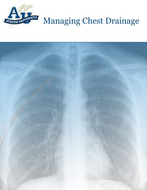
Managing Chest Drainage - Atrium Medical Corporation
Managing Chest Drainage - Atrium Medical Corporation
Managing Chest Drainage - Atrium Medical Corporation

Create successful ePaper yourself
Turn your PDF publications into a flip-book with our unique Google optimized e-Paper software.
Disconnecting The <strong>Chest</strong> <strong>Drainage</strong> Unit . . . . . . . . . . . . . . . . . . . . . . . . . . . . . . . . . . . . . . .29Removing The <strong>Chest</strong> Tube . . . . . . . . . . . . . . . . . . . . . . . . . . . . . . . . . . . . . . . . . . . . . . . . .30Autotransfusion . . . . . . . . . . . . . . . . . . . . . . . . . . . . . . . . . . . . . . . . . . . . . . . . . . . . . . . .31The Future is Now: Mobile <strong>Chest</strong> Drains . . . . . . . . . . . . . . . . . . . . . . . . . . . . . . . . . . . . . . .32Summary . . . . . . . . . . . . . . . . . . . . . . . . . . . . . . . . . . . . . . . . . . . . . . . . . . . . . . . . . . . .34Glossary . . . . . . . . . . . . . . . . . . . . . . . . . . . . . . . . . . . . . . . . . . . . . . . . . . . . . . . . . . . . .35Suggested Readings . . . . . . . . . . . . . . . . . . . . . . . . . . . . . . . . . . . . . . . . . . . . . . . . . . . . .38Classic References . . . . . . . . . . . . . . . . . . . . . . . . . . . . . . . . . . . . . . . . . . . . . . . . . . . . .45Suggested Readings Regarding <strong>Chest</strong> Tube Stripping . . . . . . . . . . . . . . . . . . . . . . . . . . . . . .46iv
Anatomy Of The <strong>Chest</strong>■The ThoraxThe thorax lies between the neck and the abdomen. The walls of the thoracic cavity are made up of the ribs laterally,the sternum anteriorly, and the thoracic vertebrae posteriorly. Internal and external intercostal muscles cover bony thoracicstructures. The dome-shaped, muscular diaphragm forms the lower boundary (sometimes called the floor) ofthe thoracic cavity. See Figure 1 for key anatomical structures of the chest.The thoracic cavity forms a semi-rigid framework that protects the heart, lungs, great vessels, the thymus gland andparts of the trachea and esophagus. In addition, the structure forms an airtight bellows mechanism (which will bedescribed in more detail in the next section), creating a vacuum system that expands the lungs during inspiration.The thorax is divided into three distinct spaces:• The centrally located mediastinum• The right lung cavity• The left lung cavityTrachea<strong>Chest</strong> WallLeft Lung CavityRight LungCavityDiaphragmFigure 1. Anatomy Of The <strong>Chest</strong>■The MediastinumThe mediastinum is a flexible partition in the center of the thoracic cavity. The left and right lung (pleural) cavities arelateral to the mediastinum, the sternum is anterior, and the vertebral column is posterior.The mediastinum contains the heart, covered by the pericardium; the thymus gland; parts of the esophagus and trachea;and a network of nerves and blood vessels.1
■The Lungs And Lung CavitiesThe cone-shaped, spongy, elastic lungs are suspended from the trachea and fill a substantial portion of the thoraciccavity. The left lung is narrower, longer and smaller than the right (because of the position of the heart toward the leftof midline); it is divided into two lobes: the upper and lower lobes. The larger right lung is divided into three lobes:upper, middle and lower.Air is drawn into the thoracic cavity through the upper airway. The trachea divides into two primary bronchi (onebronchus to each lung), which in turn divide many times into smaller and smaller airways that eventually terminate inthe alveoli, where gas exchange takes place across the alveolar-capillary membrane.The boundaries of each airtight lung cavity consist of the chest wall, the diaphragm and the mediastinum. This cavityis lined with a membrane called the parietal pleura. A similar membrane called the pulmonary or visceral pleura coversthe surface of each lung.Figure 2. The membrane lining the chest wall (L) and covering the lungs (R)A thin film of serous lubricating fluid called pleural fluid separates the parietal and visceral pleural surfaces. This fluidallows the moist pleural membranes to adhere to one another while allowing them to slide smoothly as the lungexpands and recoils during inhalation and exhalation. The amount of fluid produced in 24 hours is about 0.3mL/kg ofbody weight or about 25mL.The lungs have a natural tendency to collapse or recoil. The adherence of the pleurae keeps the lungs pulled up againstthe inside of the chest wall, counterbalancing the natural recoil. This tendency for the lungs to pull away from the chestwall results in a subatmospheric, or negative, pressure in the tiny space between the pleurae. Normally, this intrapleuralpressure is approximately -8cmH 2 O during inspiration and -4cmH 2 O during expiration. This negative pressure keepsthe lungs expanded and allows them to move in tandem with the rib cage and diaphragm during inspiration.2
Respiratory PhysiologyNormal breathing consists of:• Ventilation: the mechanical act of moving air into and out of the lungs• Respiration: gas exchange across the alveolar-capillary membraneDuring normal ventilation, air moves in and out of the thoracic cavity through the trachea by the following process (SeeFigure 3):1. During inspiration, the phrenic nerve stimulates the diaphragm to contract, causing it to move downward.At the same time, the external intercostal muscles may also contract, pulling the chest wall out. Both actionsincrease the size of the thoracic cavity.2. Because of the adherence of the pleurae, as the thoracic cavity enlarges, the lungs expand as well.3. As the volume of the lung increases, the pressure within decreases. (This is according to Boyle's gas law, whichstates there is an inverse relationship between volume and pressure.) This creates a negative intrapulmonarypressure.4. Air naturally moves from areas of higher pressure to areas of lower pressure. Thus, air will be drawn into thelungs through the trachea when intrapulmonary pressure becomes more negative.5. During exhalation, the muscles of the diaphragm and intercostals relax. The chest wall moves in, and the lungvolume decreases through natural elastic recoil.6. As lung volume decreases, intrapulmonary pressure rises in relation to atmospheric pressure (again, byBoyle's gas law).7. Air now flows from the lung out through the trachea.8. The cycle then repeats approximately 25,920 times a day.Figure 3. The mechanics of breathing3
PathophysiologyIf air, fluid, or blood enters the tiny space between the parietal and the visceral pleurae, the negative pressure that keepsthe pleurae adherent and holds the lungs against the chest wall will be disrupted. The lung's natural tendency to recoilwill take over and the lung will collapse. When this occurs, the lung cannot fully expand during inspiration. (See Figure4). Depending on the patient's underlying pulmonary condition and the degree of disruption in the pleural space, thepatient may experience minimal symptoms or significant shortness of breath. In addition, the parietal pleurae are highlyinnervated with sensory nerves, so any change in the pleural space may be very painful as well. Pleuritic pain is characterizedby a sharp, stabbing pain during inspiration as the pleurae move. Patients will involuntarily reduce tidal volumewhile increasing respiratory rate to maintain ventilation while limiting the movement of the pleurae to reduce pain.Typically, air or fluid must be removed from the pleural space before the lung can fully re-expand and normal breathingcan resume. In situations in which the air or fluid accumulation is very small, the patient may be monitored carefullywhile the body naturally absorbs the air or fluid.Figure 4. Air in the pleural spaceTwo common clinical conditions require pleural drainage:• Rupture of the surface of the lung (such as a bleb) or tracheobronchial tree, allowing air and possibly serous orserosanguineous fluid into the pleural space while the chest wall remains intact• External penetration of the chest wall resulting from surgical intervention or trauma (such as a gunshotwound or stabbing), allowing air and blood or serosanguineous fluid from damaged tissues into the pleural space(See Figure 5).Since trauma typically injures both the chest wall and the lung surface,air can enter the pleural space from the atmosphere (through the openingin the chest wall) or the lung. Bleeding may come from the chest wallor the lung itself.Figure 5. Stab wound toleft hemithoraxcourtesy trauma.org4
• Increased respiratory rate and effort• Dyspnea• Pleuritic chest pain (if the patient is able to communicate)• Decreased movement of the affected side of the chest (See Figure 9)• Decreased breath sounds on auscultation of the affected side• Falling blood pressure• Rising pulseFigure 9. Left side of chest is fixed at fullinspiration, characteristic of tension pneumothorax.Courtesy trauma.orgFigure 10. Palpating chest withsubcutaneous emphysema.courtesy trauma.orgTextbooks classically describe breath sounds as being absent, which leadsmany nurses to expect that they will hear nothing on the affected side. In reality,sounds from the unaffected side will be transmitted to the side of the chestwith the pneumothorax. Thus, breath sounds will be diminished or distant, notabsent. Also look for tracheal deviation away from the affected side (however,an artificial airway will make this harder to identify); cool, mottled skin; andsubcutaneous emphysema, a feeling of crackling on palpation of the chest,indicating air has entered the subcutaneous tissues (See Figure 10). If thepatient is receiving volume-controlled, positive-pressure ventilation, themanometer on the ventilator will show higher inspiratory pressures and willbe less likely to return to zero (or baseline, if PEEP is used). If the patient isbeing ventilated with a manual resuscitation bag, the bag will become harderand harder to squeeze to deliver a breath.■HemothoraxAfter thoracic surgery or certain chest injuries, blood may collect in the pleural space. This condition is called a hemothorax.A combination of blood and air is called a hemopneumothorax. These conditions typically occur after there hasbeen an opening in the chest wall, either during surgery or from a penetrating injury. However, in some cases, bloodcan accumulate in the pleural space after blunt chest trauma when, for example, sharp ends of fractured ribs laceratelung tissue (pneumothorax) and blood vessels (hemothorax).Like pneumothorax, hemothorax disrupts the normal negative intrapleural pressure.This allows normal lung recoil to occur, resulting in some degree of lung collapse,depending on how much blood is in the pleural space. Once the lung has collapsed,it does not reexpand until the blood is evacuated from the pleural space (See Figure11).Another collection of fluid in the pleural space occurs when there is a disruption ofthe normal balance between the amount of pleural fluid produced and the amount offluid absorbed. This is called a pleural effusion. This condition is commonly seen inpatients with lung and breast cancer. 3See Clinical Update: Dec 2002 and Dec. 2005.Figure 11. Left hemothorax.Note the compression of the leftlung.courtesy trauma.orgEmpyema (pyothorax) is an accumulation of pus in the pleural space, caused by pneumonia, lung abscess or contaminationof the pleural cavity. Chylothorax is the accumulation of lymphatic fluid in the pleural space.7
Like pneumothorax and hemothorax, these collections of material in the pleural space disrupt the normal negativeintrapleural pressure and interfere with breathing, but none of these fluid collections is likely to result in an accumulationof positive pressure that could threaten the patient the way a tension pneumothorax does (See Figure 12). Withoutcontinuous transfusion, a patient would likely exsanguinate before enough blood would collect in the pleural space toaffect the mediastinum.Figure 12. The CT image on the left is a hemothorax. Note the light color of the right hemithorax where the bloodis collected and the lack of mediastinal shift. The CT image on the right is a tension pneumothorax. Note the blacknessof the left hemithorax where air is trapped under pressure and the shift of the mediastinum to the right side.courtesy trauma.orgHowever, blood, fluid, pus, or lymphatic drainage that has accumulated in the pleural space will still cause an inflammatoryresponse and prevent the lung from full expansion during inspiration and should be removed, particularly if thepatient is symptomatic with shortness of breath and/or pleuritic chest pain. Generally, if the costophrenic angle isobscured on an upright chest radiograph, the collection is large enough to be drained.■Cardiac TamponadeFollowing cardiac surgery or chest trauma, blood can pool in the mediastinalcavity. Blood can collect between the pericardium and the heart, externallycompressing the heart in a condition called cardiac tamponade.Cardiac tamponade, like tension pneumothorax, is life-threatening if notidentified and treated promptly because it reduces the heart's ability toaccept venous return, resulting in significantly decreased cardiac output.Emergency treatment is needle pericardiocentesis. (See Figure 13).An accumulation of blood in the pericardium also provides a medium forbacterial growth, potentially leading to postoperative infection.Figure 13. Emergency management of cardiactamponade is a needle pericardiocentesis,usually followed by chest tube placement.courtesy trauma.org8
To reduce the risk of blood accumulation in the mediastinum, at least one, and more commonly, two chest tubes areused postoperatively to drain the mediastinal cavity to allow blood to leave the chest (See Figure 14).Figure 14. Mediastinal chest tube placementcourtesy trauma.org9
<strong>Chest</strong> <strong>Drainage</strong> SystemsMost patients can tolerate a small amount of air or fluid in the pleural space, particularly if they do not have lung disease.If less than ten percent of the pleural space is occupied by air or fluid, the patient will typically have few respiratorysymptoms and the body can usually reabsorb it without external drainage.In some cases, needle drainage will be performed to vent air from the pleural space or to allow drainage of fluid (typicallypleural effusion) from the chest. Other situations will need chest tube drainage. The decision to place a chest tubeis based on the patient's underlying pulmonary condition as well as the amount of air or fluid in the pleural space.The goals of chest tube drainage are to:• Remove the fluid and/or air as quickly as possible• Prevent drained air and/or fluid from re-entering the chest cavity• Re-expand the lungs and restore normal negative intrapleural pressureA chest tube is typically connected to a chest drain that collects drainage from the pleural space and allows the lung tore-expand. The drain must be designed so that it prevents air or fluid drainage from being pulled back into the chestduring inspiration or when negative pressure is restored in the intrapleural space.The same type of drain is used to collect blood from the mediastinum to reduce the risk of cardiac tamponade followingcardiac surgery or chest trauma. However, during mediastinal drainage, negative pressure within the chest is notas significant a factor as it is during pleural drainage.All chest drainage systems have some common components:• A chest tube inserted into the pleural cavity or mediastinal cavity to allow air and/or fluid to leave the chest• A six-foot length of flexible patient tubing that connects the chest tube to the chest drain system• A drainage system that usually is made up of three compartments: (1) a collection chamber that collects fluiddrainage and allows measurement of drainage volume, (2) a one-way water seal chamber or mechanicalvalve that lets air leave the chest and prevents outside air from getting in, (3) a suction control chamber ormechanical valve that limits the amount of negative pressure that is transmitted to the chest; this featureallows the safe use of suction to facilitate quicker evacuation of air and/or fluid.Early chest drainage systems were made up of a set of one, two or three glass or plastic bottles. Sixteen pieces and17 connections were required to set up a three-bottle system properly. Today, most chest drain systems are self-containedunits made of molded plastic. The principles are the same regardless of the type of system used.10
■<strong>Chest</strong> Tubes3 See Clinical Update: Sep 1998A chest tube (sometimes called a thoracic catheter) is generally about 20 inches long,with four to six eyelets that act as drainage holes on the patient (distal) end and an openingfor connection to the chest drainage system on the proximal end, outside the body.A radiopaque line is added to the length of the tube so it can be seen more easily on achest radiograph. Most manufacturers include a break in this radiopaque line to indicatethe location of the eyelet closest to the skin, so that on the radiograph, it will be easy todetermine the position of the most proximal opening so the tube can be repositioned ifit is not fully inside the chest (See figure 15).There are two basic types of chest tubes:Figure 15. <strong>Chest</strong> tube eyelet,indicated by bluearrow, outside the pleuralspace.courtesy trauma.org• Thoracotomy chest tube, a flexible straight or right-angle tube designed for insertion through a small incisionin the chest, typically after a surgical procedure. Although some doctors prefer silicone, most chest tubes aremade of transparent medical-grade polyvinyl chloride (PVC). Right angle catheters are used most often formediastinal drainage.• Trocar chest tube, in which the chest tube is packaged with a removable, pointed and rigid stylet. This styletallows the chest tube to be placed in the chest through a puncture made by the trocar — the physician usesconsiderable force to push the stylet and chest tube through the chest wall and soft tissue and on into thepleural space. The trocar is then removed, leaving the chest tube in place. This technique is morecommonly used in emergency rooms and other areas outside the operating room, in which chest tubes mayneed to be placed quickly in non-surgical patients. These chest tubes may have only two or three eyelets fordrainage. Because of the force needed to insert trochar chest tubes, they present a higher risk for lung injuryduring insertion than thoracostomy chest tubes.The diameter of the chest tube selected depends on the size of the patient, the type of drainage (air and/or fluid), andthe expected duration of drainage. Typical chest tube diameters are:• 8 to 12 French Infants and young children• 16 to 20 French Children and young adults• 24 to 32 French Most adults• 36 to 40 French Large adultsWith the advent of minimally invasive cardiothoracic surgical techniques, smaller chest tubes are more commonplaceto reduce the amount of tissue trauma and speed postoperative recovery.The Food and Drug Administration (FDA) has approved closed wound drains for postoperative drainage in cardiothoracicsurgical patients. The wound drain is attached to a reservoir bulb for drainage — the same type of reservoir bulbused for abdominal wounds, for example — and not connected to a chest drainage device.11
A traditional chest tube is a hollow catheter with a single lumen. One type of wounddrain has a configuration that changes three times from the patient tip to the proximalend that attaches to the reservoir bulb. The distal end has a multi-lumen, fourchanneldesign. If you look at the lumen at the distal tip of the tube, you'll see a "t" —a PVC core divides the catheter into four separate sections for drainage. (See Figure16).Instead of eyelets found on a traditional chest tube, slits along the wound drain allowfluid into the sections for drainage. In the middle part of the tube, the PVC "t" remains, but the outside of the tube isclosed. This section provides the change-over from the open multi-lumen catheter to the third part of the tube that connectsto a drainage device — a single-lumen catheter.Three main variables affect how well blood and fluid leave the chest through a chest tube: the length of the tube, theamount of negative pressure (suction) applied, and the inner diameter of the tube. A tube's ability to evacuate the chestdepends on the smallest or most restrictive part of the tube. The middle part of the three-part wound drain is mostrestrictive, whereas a traditional chest tube's flow rate through a single lumen is constant through the length of thetube. A tube's stated size is determined by its outer diameter, not the flow area inside. When the inner diameter is factoredin, the 20 Fr chest tube allows for slightly greater flow than the 24 Fr three-part wound drain, and more than 2.5times the flow of a 19 Fr wound drain. Thus, a surgeon who might be using a 24 Fr wound drain to achieve betterdrainage can instead use a smaller chest tube that will disturb less tissue. 3 See Clinical Update: Mar 2004<strong>Chest</strong> DrainFigure 16. Compare wounddrain (L) with chest tube (R).Wound DrainVents positive pressureConstant suction levelConsistent flow rate<strong>Drainage</strong> occurs as long as drain is below the chestWill work even if clinician does not actively maintaindrainCan be used for all cardiothoracic patientsRemains a closed system throughout useClosed system with no ventVariable suction levelVariable flow rate as suction changes<strong>Drainage</strong> stops if reservoir fills (100cc)regardless of drain positionClinician-dependent for proper useCannot be used if patient has an air leakMust be opened periodically to discard drainageTable 1. Characteristics of <strong>Chest</strong> Drains and Wound Drains■Patient TubingA six-foot tube connects the chest tube to the collection chamber of the chest drainage system. The length of this tubingallows the patient to turn and move in bed and to walk without tension on the chest tube. It also minimizes thechance that a deep breath could draw any drainage back up into the chest. Sometimes two chest tubes are attachedto a single patient tube and chest drainage system with a Y-connector.12
Reusable <strong>Chest</strong> <strong>Drainage</strong> SystemsThe first chest drainage systems were made up of a series of one to three interconnected reusable glass bottles.Although one-piece molded plastic drainage units have largely replaced these systems today, the principles on whichthe bottle systems were based hold true for today's integrated chest drainage systems.■One-Bottle <strong>Chest</strong> <strong>Drainage</strong> SystemThe simplest way to drain the chest is to set up a single bottle with a tube submerged to a depth of 2 centimeters underwater as illustrated in Figure 6. One short tube leads out of the bottle through the plug at the top, allowing air to ventto the atmosphere. The submerged tube is connected to the patient tubing. Placing the distal end of the tube underwater creates a water seal, the most important element in a pleural drainage system. The water seal provides a lowresistance,one-way valve that allows air to leave the chest while preventing outside air from being pulled into the chestduring breathing.Positive pressure exceeding +2cmH 2 O will push air down the tube. The air will bubble through the water and leave thechest drain system through the atmospheric vent.If the tube that is submerged in the water is marked, indicating each centimeter on the tube, the water seal becomesa manometer that can measure intrapleural pressures. Pressure changes in the pleural space that occur with breathingwill be seen as fluctuations in the level of the water within the tube. These fluctuations, called "tidalling," may be asgreat as 5 to 10 cmH 2 O with normal spontaneous breathing. The water level will go up (more negative) during inspiration,and go down (return to baseline) during exhalation. If the patient is receiving positive pressure ventilation, thewater level will go down (more positive) during inspiration, and go back up (return to baseline) during exhalation,reflecting the higher positive pressure in the chest with mechanical ventilation.Tube To AtmosphereTube From PatientWater Seal / CollectionFigure 17. One bottle chest drain systemThe one-bottle setup is a combination water seal and fluid collection bottle. As fluids drain from the chest into the bottle,the level of the initial sterile fluid combined with drainage will rise. Thus, the submerged tube will be deeper than 2centimeters. The higher the fluid level, the more pressure it takes to push air through the fluid as it leaves the chest.Theoretically, the problem could be solved by emptying some of the drainage from the bottle or pulling the tube furtherout of the top of the bottle in an effort to maintain the 2cmH 2 O water seal level. However, in practice, if fluiddrainage is expected, another bottle is added to collect drainage independent of the water seal. This creates a two-bottlechest drainage system.13
■Two-Bottle <strong>Chest</strong> <strong>Drainage</strong> SystemIn a two-bottle chest drainage system, fluid drains from the chest into a dedicated collection bottle. Air from the pleuralspace, continuing through the tubing that connects the two bottles, bubbles through the water seal and exits to theatmosphere, as illustrated in Figure 18.If the collection bottle has volume markings, the amount and rate of fluid drainage can be measured and monitored.More importantly, adding a separate collection bottle allows the water seal to remain at an undisturbed, fixed level,allowing air to leave the pleural space through a system with low resistance to air flow, regardless of the amount offluid drainage.Tube To AtmosphereTube From PatientWater SealCollectionFigure 18. Two bottle chest drain systemBoth the one- and two-bottle chest drainage systems rely on gravity to create a pressure gradient by which air and fluidleave the chest. Keeping the drainage system below the level of the patient's chest enhances gravity drainage; additionalpressure is created when the patient exhales or coughs. However, if the patient has a large air leak into the pleuralspace, gravity drainage may not be sufficient to evacuate the chest, and suction may be required. This also meansthe addition of a third bottle to the system — a suction control bottle.■Three-Bottle <strong>Chest</strong> <strong>Drainage</strong> SystemWhen suction is required to increase the pressure difference between the pleural space and the drainage system, it isimportant to accurately regulate suction levels to avoid patient injury. If suction pressure is too high, complications canoccur such as hematoma formation at the distal end of the catheter and tissue invagination into the catheter eyelets.A third bottle added to the chest drainage system will limit the amount of negative pressure that can be transmitted tothe patient's chest. A suction control bottle has three tubes (see figure 19 on the next page) :1. A long tube positioned so that the upper end is open to the atmosphere through the plug in the topof the bottle while the lower end is submerged under water, usually to a depth of 20 centimeters.2. A short tube connected to the water seal bottle.3. A tube that connects the bottle to the suction source, which can be either a portable pump or a wall vacuumregulator.When the three bottle set-up is used as illustrated in Figure 19, the maximum level of negative pressure that can be14
transmitted to the patient's chest directly corresponds to the depth of submersion of the tube in the suction controlbottle. If the tube is under 20 centimeters of water, the maximum suction level the patient can be subjected to is -20cmH 2 O.Tube To AtmosphereTube To SuctionTube From PatientSuction Control Water Seal CollectionFigure 19. Three bottle chest drain system■Suction Control BottleIf the system is not connected to a vacuum source, the fluid in the suction control bottle's atmospheric vent tube willbe at the same level as the fluid in that bottle and there will be no bubbling. If the system is connected to a vacuumsource set at the same setting as the water level in the suction control bottle (-20cmH 2 O, for example), the water in theatmospheric vent tube will be pulled down 20 centimeters below the surface of the water in the bottle, there will be nobubbling, and the pressure in all three bottles will be -20cmH 2 O.When the vacuum source is set at a level higher than the water level in the suction control bottle, the controlled maximumsuction imposed on the patient is achieved when fluid is no longer present in the atmospheric vent tube andbubbling occurs in the bottle. Air is drawn in through the atmospheric vent. The air bubbles out the bottom of the submergedtube, and then is evacuated from the system through the vacuum source. The key is that the depth of submersionof the tube in the suction control bottle determines the amount of suction imposed on the patient.■Drawbacks of the Three-Bottle SystemThree-bottle reusable systems have many clinical drawbacks. Set-up is time consuming, and because of all the connections,the potential for error or contamination of the sterile system is high. It can be expensive for the hospital toclean, sterilize and track the processing of the system and all of its pieces. Since there are no valves to vent positiveand negative pressure build-up, the patient does not have the advantages of the safety advances made in disposablechest drainage systems over the past twenty years. These problems are solved with the one-piece, integrated disposablechest drain system.15
Disposable <strong>Chest</strong> <strong>Drainage</strong> SystemsThe first one-piece, disposable three-chamber chest drainage unit was introduced in 1967. Today's chest drainage systemsare compact, sterile, and disposable. They offer many safety features, diagnostic capabilities and conveniencesnot found in the traditional three-bottle chest drain system. Figure 20 shows a schematic illustration of the one-piecechest drain system.The chambers of these one-piece disposable units correspond to the bottles in the three-bottle system. Most one-piecedisposable systems include:• A collection chamber into which fluids drain and volume and rate of drainage can be measured• A water seal chamber that uses sterile fluid or a mechanical one-way valve to allow air to leave the patient andprevent air from entering the patient's chest through the chest tube• A suction control chamber that uses either sterile fluid or a mechanical device to control and limit the level ofsuction imposed on the patient.Tube To SuctionTube From PatientSuction Control Water Seal Collection ChamberFigure 20. Conventional three-chambered disposable chest drain system■Collection ChamberAn easy-to-read, well-calibrated collection chamber permits the nurse to record the amount of fluid collecting in thischamber. Most drains allow the nurse to draw a line indicating the level of drainage and write the time on the front ofthe chamber. This allows all clinicians to assess the rate of fluid drainage from the chest.16
■Water Seal Chamber3 See Clinical Update: Dec 1997The water seal chamber is connected to the collection chamber and provides the protection of the one-way valve discussedearlier. The water seal in most disposable drainage units is formed with an asymmetric U-tube rather than anarrow tube submerged underwater as in the traditional bottle systems. The narrow arm (closest to the collectionchamber) is equivalent to the tube; the larger arm serves as the water reservoir. When the fluid reservoir is filled to 2centimeters above the seal in the U-tube, it has the same effect as submerging the tube in the bottle system 2 centimetersbelow the surface of the water.In addition to providing the one-way valve, a U-tube design can also be used to measure pressure. When pressureson both sides of the U-tube are equal, the water level is the same in both arms. However, if the pressures on each armdiffer, fluid moves away from the side of higher pressure toward the side with lower pressure. If the water seal columnon the front of the chest drain is calibrated with markings, the fluid movement acts as a water manometer for measuringintrapleural pressure, providing additional assessment data for the clinician.Some units have an anti-siphoning float valve in the water seal fluid column that prevents the water from beingsiphoned out of the water seal chamber and into the collection chamber during situations that create high negativepressures, such as chest tube stripping. (This practice is not supported by research, but may be seen in the clinicalsetting.)The original design of the float valve at the top of this chamber permitted uncontrolled vacuum levels to accumulate inthe patient's chest with each subsequent stripping of the patient tube (see discussion on chest tube stripping on page24). To eliminate this pressure accumulation (since tubes may still be stripped) manufacturers have added manual highnegative pressure relief valves to chest drain systems that allow filtered atmospheric air to enter the system to preventany accumulation of negative pressure in the patient. However, with manual devices, the clinician must recognize thecondition of high negativity, evidenced by the rise in the water level in the water seal, and depress the relief valve toremedy the situation.In 1983, automatic high negative pressure protection was introduced. Many systems now employ a float ball designat the top of the water seal chamber with a notch that allows fluid to pass through it. A compartment above the ballholds the water that fills the water seal chamber. Thus, no water spills into the collection chamber, and no water is lost,so the one-way valve protection is not put at risk during conditions of high negative intrapleural pressure. The speedat which disposable systems release accumulating negative pressure varies, depending on the manufacturer and a particulardrain's design.■Dry Seal <strong>Chest</strong> Drains3 See Clinical Update: Jun 2000Some chest drains use a mechanical one-way valve in place of a conventional water seal. The mechanical one-wayvalve allows air to escape from the chest and prevents air from entering the chest. An advantage of a mechanical onewayvalve is that it does not require water to operate and it is not position-sensitive the way a water-filled chamber is.A dry seal drain protects from air entering the patient's chest if a drain is knocked over.17
A drawback to any mechanical one-way valve is that it does not provide the same level of patient assessment informationas a conventional water seal; for example, the clinician cannot see changes in the water level reflecting pressurechanges in the chest. For optional air leak detection, a separate air leak monitor can be filled with water. A vacuumindicator on the face of the drain provides visual evidence of negative pressure (vacuum) inside the collectionchamber.■Suction Control Chamber3 See Clinical Update: Mar 2008The suction control chamber is another safety device that protects the patient from excess suction pressure in the pleuralcavity or mediastinum. Suction control mechanisms in one-piece drains are either "wet" or "dry.""Wet" suction control systems regulate suction pressure transmitted to the chest by the height of a column of water inthe suction control chamber. Like the water seal chamber, the wet suction chamber is an asymmetric U-tube manometer.The narrow arm is the atmospheric vent and the large arm is the reservoir. The amount of negative pressure thatis transmitted to the patient's chest is determined by the height of water in this chamber, not the level of vacuum seton the wall (or source) regulator."Dry" suction control systems regulate suction pressure mechanically rather than with a column of water. Some drysuction systems use a screw-type valve that varies the size of the opening to the vacuum source, thereby limiting theamount of negative pressure that can be transmitted to the chest. These valves narrow the opening of the chest drainin order to adjust the level of negative pressure; therefore, the total amount of air thatcan flow out of the chest drain is also limited. Thus, this type of dry suction controlmechanism is impractical for patients with significant pleural air leaks.Two manufacturers use a calibrated, spring-loaded, self-regulating mechanism thatallows suction levels to be adjusted with the simple turn of a dial to the desired level ofsuction, in place of water (See Figure 21). These systems are capable of handling largevolumes of airflow and can also compensate for changes in patient air leaks or fluctuationsin the source vacuum while maintaining a consistent level of negative pressure inthe patient's chest. The screw-type valves cannot compensate for these changes.Figure 21. Dialing indesired suction levelDry suction control mechanisms are quieter and often easier to set up than wet units. But because the dry unit is silentit is not as obvious that the unit is working properly without careful examination of the front of the drain. The sound ofbubbling in wet units provides feedback that the system is working. Proper set-up and monitoring is covered in thenext section.If the tubing leaving the drain from the suction source becomes obstructed or if the source vacuum fails, and the patienthas an active air leak from the pleural space, positive pressure could build up in the pleural cavity, significantly impairingbreathing. This situation could even lead to a tension pneumothorax. To safeguard against this potentially life-threateningcomplication, most chest drain systems have a positive pressure relief valve (PPRV) that vents accumulatedpressure greater than 2cmH 2 O (the depth of the water seal).18
■Double Collection <strong>Chest</strong> DrainsDouble collection chest drains are designed to be connected to two chest tubes. The drain consists of two collectionchambers: a major chamber and a minor chamber. This type of drain is rarely used for chest tubes on both sides ofthe chest at the same time; rather, the tubes are on the same side of the chest. Typically, this drain is used when onetube is placed high in the chest to evacuate air, and one tube is placed low in the chest to drain fluid on the same side.Since the lower tube is likely to drain both fluid and air, it is connected to the major collection chamber. Since the uppertube will mostly evacuate air, it is connected to the minor collection chamber.Double units may also be used in cardiovascular surgery when the surgeon wants to monitor drainage from two mediastinaltube locations separately. The tube(s) placed below the heart are connected to the major chamber and thetube(s) above the heart are connected to the minor chamber. Or, if a pleural tube is required because the parietal pleurawas entered during cardiac surgery (particularly if the internal mammary artery is used for a bypass), the pleuraltube can be connected to the minor chamber since it is placed to evacuate air. The mediastinal tubes, draining fluid,are then connected to the major chamber.■Infant <strong>Chest</strong> <strong>Drainage</strong> SystemsThe most prominent feature of infant chest drainage units is the smaller collection chamber that holds less drainagethan an adult unit. The patient tubing may have a narrower inner diameter compared with adult drains and usually hassmaller connectors to connect the patient tubing to the smaller chest tubes used for infants.■ Closed Wound Reservoirs3 See Clinical Update: Jun 2004Closed wound drainage systems were originally designed to remove fluid from closed surgical sites; now they arebeing used for cardiothoracic surgical patients. Bulb suction reservoirs connect to the wound drain and create suctionto evacuate fluid. It must be a completely closed system; any venting to the atmosphere will disrupt the system's selfgeneratedsuction. In a cardiothoracic patient, a closed system with no vent presents the potential for a catastrophiccomplication: tension pneumothorax. <strong>Chest</strong> drains vent to the atmosphere and have positive pressure relief valves forsafety; wound drains do not. They can only be used after the lung is expanded and air leaks have sealed. However, notall air leaks are immediately apparent, particularly when there is no water seal or air leak indicator. Whenever an air leakis present, a drainage catheter must be attached to an appropriate pleural drainage system to prevent tension pneumothorax.To use a bulb reservoir system, the reservoir is first "activated," creating unmeasured, unregulated suction that is transmittedto the surgical site. When a bulb reservoir is initially compressed and attached to a wound drain, it generatesapproximately -120cmH 2 O suction — far more than the carefully regulated -20cmH 2 O vacuum levels generated by achest drain attached to a thoracic catheter. As the reservoir fills, tissues are exposed to varying levels of decreasingsuction, and the bedside clinician has no way of knowing the level of suction being applied to the pericardial space orpleural cavity. As the reservoir fills, less negative pressure is present to draw fluid into the reservoir by suction, thusthe flow rate of fluid and air leaving the chest will drop.19
If the drain fills (100cc) and is not emptied immediately, the pressures between the surgical site and the reservoir willequalize. A pressure gradient between the patient and any drain (reservoir) is necessary for drainage. When pressuresequalize, drainage stops. Thus, unlike a chest drainage system described earlier, a bulb reservoir system requires regularmaintenance by the nurse to preserve patency. It must be emptied to maintain suction and keep drainage flowing.If pericardial drainage stops because the reservoir is filled, the patient is at risk for cardiac tamponade. Table 1 on page12 compares the characteristics of a chest drain and a wound drain (reservoir) system.Setting Up A <strong>Chest</strong> Drain SystemSetting up a chest drainage system involves inserting the chest tube, settingup the drainage unit, making the proper connections and applying suction, asprescribed.■Thoracostomy3 See Clinical Update: Sep 2003 (part 1), Dec 2003 (part 2), Sept 2007,Dec 2007The procedure for inserting a chest tube is called a thoracostomy. The preciselocation of the chest tube depends on whether the tube is to drain air,fluid or both. There is a difference of opinion among surgeons as to whetherthe incision should be made in the mid-axillary line or the mid-clavicular line.Figure 22. <strong>Chest</strong> tube insertion sitein the R mid-axillary linecourtesy trauma.orgSome avoid the mid-clavicular line because the pectoralis muscle is often very developed and difficult to penetrate, andto avoid a scar in such a prominent location on the front of the chest. These surgeons place the tube in the mid-axillaryline and direct the distal end of the chest tube to the anterior location (See Figure 22).• For most pneumothorax cases, the end of the tube is directed anterior and superior in the pleural space near theapex of the lung. Typically, this will be at the level of the second or third intercostal space.Figure 23. <strong>Chest</strong> tube placement: superior tube evacuates air, inferiortube drains fluid20
• To drain a hemothorax or pleural effusion, the chest tube is directed inferior and posterior in the pleural spacesince gravity will pull fluid toward the base of the lung in a patient who is upright or in semi-Fowler's position.Again, the tube is placed in the mid-axillary line at about the seventh or eighth intercostal space.• Frequently, two or more chest tubes are used, positioned at different locations within the pleural space tofacilitate removal of all air and fluid. Figure 23 illustrates locations for chest tube placement.When a chest tube is placed at the end of a surgical procedure, the open end of the chest tube is passed from the insideof the chest wall out through a small incision, leaving the end of the tube with eyelets for drainage strategically positionedwithin the chest for optimal drainage. A tight fit through the intercostal muscles is preferred to minimize bleedingand to achieve an airtight thoracic cavity closure.In emergency situations, such as with spontaneous or traumatic pneumothorax, the chest tube is inserted directlythrough the skin and chest wall into the pleural space.■Steps for <strong>Chest</strong> Tube Insertion and Drain Setup(The order of steps may be changed based on the patient's condition and the preferences of the clinician inserting thechest tube.)1. Get the chest drain from storage; the chest tube (if not included in the insertion kit); and and the chest tubeinsertion kit (or thoracostomy tray). Hospitals will determine the contents of an instrument tray from sterile processingor choose a disposable insertion kit. A kit may contain a syringe for local anesthetic, a skin antiseptic,sterile gloves, scalpels, hemostat(s), sutures and dressing material – if not, these supplies will need to be addedto the sterile field. Check whether a local anesthetic such as lidocaine is in the kit or tray, or if unit stock is usedinstead.2. Assure that the patient understands the procedure about to be done. Check that a consent form is completedand on the chart.3. Check to see that the insertion site is marked, as required by the Joint Commission's Universal Protocol forPreventing Wrong Site, Wrong Procedure, Wrong Person Surgery TM , according to hospital policy. If a chestradiograph is on the wall, two people should check to make sure the film is properly positioned on the viewbox and verify the right and left sides of the image.4. Set up the chest drain according to the manufacturer's instructionsfor use. This may include adding water to the water seal chamber orair leak indicator and the suction control chamber.5. As long as the procedure is not an emergency, requiring short cutsto save a patient's life, medicate the patient or begin proceduralsedation (with proper orders or by protocol).6. After the skin is cleaned and the local anesthetic injected, a smallskin incision is made over the rib below the selected intercostalspace. Dissection with a hemostat is carried out through the superiorintercostal muscles and then into the pleural space. The catheterwill then be inserted through this tract (See Figure 24). (When a trocaris used, a puncture is made through the intercostal muscles withthe trocar stylet instead of using tissue dissection).Figure 24. Inserting chest tube with clamp.courtesy trauma.org21
7. Once the chest tube is placed, it will be sutured in place (See Figure25). The chest tube is typically clamped to prevent air from enteringthe chest until the tube is secured to the chest and connected to thechest drain system.8. The open end of the chest tube is then attached to the stepped connectoron the end of the patient tubing attached to the collection chamberof the chest drainage system.Figure 25. Sutures used to secure thetube to the chest wall.courtesy trauma.org9. The insertion site is covered with a sterile occlusive dressing. Padsdesigned as tracheostomy dressings — with the slit in the middle— are ideal for positioning around the chest tube itself. The BritishThoracic Society's guidelines recommend using a transparent dressingto allow direct visualization of the insertion site and to reduce the risk of limited movement a bulky dressingcan cause.10. Place the chest drain below the chest tube, either by hanging it on the bed frame or by using a floor stand andplacing it on the floor. Mobile drains should be positioned according to manufacturer's instructions for use.11. If suction is ordered, attach the suction tubing from the chest drain to a vacuum source (typically a wallvacuum regulator). Use connecting tubing if needed. If a wet suction control system is used, slowly increasesource vacuum (suction) until constant gentle bubbling occurs in the suction control chamber. For dry suctioncontrol units, set the dial to the prescribed level of suction, and increase source vacuum until the indicator— a small bellows or float — appears in the indicator window and the vacuum source is set at least -80mmHg.12. Confirm that a radiograph has been ordered to check the position of the chest tube and to evaluate resolutionof the pneumothorax or fluid removal.22
Caring For Patients Requiring <strong>Chest</strong> <strong>Drainage</strong>3See Clinical Update Dec 2007After chest drainage has been initiated, the nurse should perform regular patient assessments. The frequency willdepend on the reason chest drainage is required, the patient's condition and any comorbidities present such as underlyinglung disease.■RespirationsNote the rate, regularity, depth and ease of respirations. Listen for changes in breath sounds, paying particular attentionto the symmetry of sounds. If breath sounds are asymmetrical, double check the chest drainage system to assureit is patent and working properly. Diminished breath sounds on the affected side may indicate re-accumulation of airor fluid in the pleural space.Every hour or two, have the patient take in deep breaths and cough. Explain that this helps keep the lungs expandedand makes breathing easier.Be sure to teach splinting of the thoracic incision if you are caring for a postoperative patient. When the patient coughs,have him or her place a pillow over the incision and squeeze or hug the pillow close to the chest wall during coughing.■Knowledge LevelContinually assess the patient's understanding of the use of the chest tube and the postoperative regimen of care. Ifyour institution provides a patient version of a clinical pathway for bypass surgery, for example, review it with the patientregularly.■Pain ControlSince the parietal pleura is innervated by intercostal nerves and is very sensitive to pain, regular pain assessments arecritical to successful care of the patient requiring chest drainage. Failure to adequately manage incision pain or pleuralpain can lead to hypoventilation, putting the patient at much higher risk for complications such as atelectasis and pneumonia.Also be aware of the risk of hypoventilation associated with opioid analgesics and patient-controlled analgesia.Some surgeons use local nerve blocks for pain management to reduce opioid side effects.■Vital SignsMonitor vital signs regularly. If the patient has mediastinal chest tubes, be sure to listen to the quality of heart tones.Muffled or distant heart tones are one sign of cardiac tamponade.■Patient Position / Movement3 See Clinical Update: Dec 1998, March 2005, June 2007Research shows that patients who get out of bed and walk around postoperatively will have fewer complications and23
shorter lengths of stay. According to the American Hospital Association, in 2004, the average cost of a patient day inan acute care hospital was $1289.87. Even reducing length of stay by one-half day results in significant cost savings.Unfortunately, many patients who need chest drainage are tethered to wall vacuum because it has been assumed thatpulling air and fluid out of the chest rather than using gravity drainage will hasten recovery. In recent years, however,this practice has been examined to see if, indeed, suction is required.One research study examined pulmonary resection patients and compared continuous suction to discontinuing suctionfor gravity drainage on postop day 2. In the gravity drainage water seal group, 67% of air leaks resolved one dayafter wall vacuum was discontinued. In patients who had continuous suction, only 7% of air leaks resolved by postopday 3.A subsequent study compared patients after pulmonary wedge resection. This time, all patients were connected to wallvacuum in the operating room to re-expand the lung at the end of the case, then vacuum was disconnected for transportto the PACU. There, patients were randomized to resume vacuum or to stay on gravity water seal drainage — twodays earlier than in the previous study.The researchers found that the duration of air leaks in the gravity water seal group was about one-half the time of thewall vacuum group. Since many argue that suction is critical for apposition of the pleurae postoperatively, theseresearchers initially used suction on all patients in the operating room. These researchers note that on inspection, bubblingis more vigorous in the water seal chamber when the chest drain is connected to wall vacuum, indicating a greaterflow of air out of the lung. By switching to gravity drainage, airflow is reduced which allows the lung suture line to bemore closely approximated, and speeds healing. They state that routinely using wall vacuum postoperatively is counterproductive.If a chest drain is disconnected from suction, be sure the tube is open to the air. Disconnect the extension tubing usedto reach the vacuum source. Do not clamp this tube. If there is a stopcock on the tubing, it should be in the open position.3 See Clinical Update: Sep 2002Any drain should be kept below the level of the chest tube to facilitate gravity drainage. Most drains have a carry handlethat allows the patient to carry the drain while walking. One manufacturer makes a holder for drains that attachesto the bottom of an IV pole. The drain simply slips into the holder and is automatically held in the proper position.While the patient is in bed, enhance drainage by changing the patient's position regularly and placing him or her inhigh- or semi-Fowler's position to facilitate gravity drainage of pleural fluid.. Coil tubing on the bed, and let it fall in astraight line to the collection chamber of the chest drain. Avoid dependent loops in the patient tubing since they canimpede drainage from the chest.The chest tube should not be clamped during patient movement, ambulation, or during trips to other parts of the hospital.Clamping the chest tube blocks drainage, which could result in a tension pneumothorax or cardiac tamponade.Clamp chest tubes only to:24• Locate an air leak• Simulate chest tube removal (to assess patient's tolerance)• Replace a drain• Connect or disconnect an in-line autotransfusion bag
■<strong>Chest</strong> Tube Site / DressingRegularly assess the chest tube insertion site. Check to see that the dressing is dry and intact, and palpate around thedressing and the insertion site for subcutaneous emphysema that could indicate air escaping from the pleural spaceand into the subcutaneous tissues (See Figure 26).If subcutaneous emphysema is present, take down the dressing and carefullyinspect the site where the chest tube leaves the chest wall. Look for any evidencedrainage eyelets may have pulled out of the pleural space, such as broken sutures.Tube movement can allow air to enter the subcutaneous tissue. If eyelets are visible,the chest tube will need to be repositioned. If no eyelets are visible, re-dressthe site. In both cases, notify the physician.If the dressing is soiled with drainage, change it as necessary. Otherwise, leave thedressing in place and do not change it regularly unless required by hospital policy.There are no research data guiding the decision whether to use petrolatum gauzeunderneath the dry sterile dressing.Figure 26. Subcutaneous emphysemarelated to chest drainagecan also dissect through fascialplanes and into the face.courtesy trauma.orgA semi-conscious or agitated patient may pull the tube out of the chest. If the patient had an air leak from the chesttube, indicated by bubbling in the water seal chamber, cover the site with a sterile dressing. Tape it on only three sides,allowing air to escape through the fourth side, preventing air accumulation and the risk of tension pneumothorax. Staywith the patient while a colleague calls the physician STAT and gets the equipment so a new tube can be placed. If therewas no air leak evident at your last assessment, apply a sterile occlusive dressing and monitor the patient carefully forany signs of respiratory distress. Notify the physician, who will typically order a chest x-ray to see if the lung is expandedand if the patient needs to have a chest tube inserted.■TubingRegularly inspect the drainage tubing for leaks, kinks, fluid-filled dependent loops, or compression or occlusion andtrace the tubing from the chest wall to the collection chamber of the chest drain.Check tubing connections any time a patient returns from a trip off the nursing unit; for example, after going to theradiology department. If the tubing comes apart, clean the ends with an alcohol wipe and reconnect them. Ask thepatient to cough a few times to push any residual air out of the pleural space.Research has shown that chest tube manipulation (stripping or milking) does not enhance bloody drainage from thechest (see Suggested Readings). A current Cochrane review states there is not enough evidence to support the practice.Blood that comes in contact with the pleurae or pericardium becomes defibrinogenated and should not clot; that'swhy this shed blood can be used for autotransfusion. Furthermore, research shows that stripping chest tubes can generatepressures as high as -400cmH 2 O, which can suck lung tissue into the drainage eyelets in the end of the chesttube. Remember, typical suction pressure is -20cmH 2 O.Tube manipulation should be limited to situations in which patients are receiving medication or blood products that willenhance clotting, or when a blood clot or tissue fragment is visible in the tube and poses the risk of tube occlusion.25
Use gentle techniques, such as squeezing hand over hand along the tubing and releasing the tubing between eachsqueeze. Alternatively, small sections of tubing can be fan-folded and squeezed together, then released. Begin at thepatient and work down the tubing to the chest drain. Be particularly careful in patients with fragile lung tissue such asin emphysema. The automatic high negative pressure relief valve on many chest drains will help protect the patientfrom exposure to high negative pressures caused by vigorous manipulation of the chest drainage tubing. However,there is no evidence that supports tubing manipulation during routine patient care.■<strong>Drainage</strong> FluidDepending on hospital policy, samples of drainage fluid may be taken by inserting a needle (20 gauge or smaller)attached to a syringe directly into the patient drainage tube (See Figure 27). Alternatively, on selected chest drain models,samples can be taken directly from the luer-lock needleless access port located on the patient tube.Regularly monitor the volume, rate, color and characteristics of the collecteddrainage. Mark the level, time and date on the face of the collection chamber at regularintervals. The frequency will be determined by the reason the patient has thechest tube and the volume and rate of drainage. Most one-piece chest drainageunits are designed with a write-on surface; the calibrations of the drainage measurementswill vary by manufacturer and type of drain (adult or pediatric/infant).<strong>Drainage</strong> volume from bleeding is usually relatively small. Over 100mL/hour postoperativelyis considered excessive drainage; even bleeding after chest trauma isseldom more than 200 to 300 mL/hr. If drainage is greater, the patient will likelyhave an exploratory thoracotomy. After cardiac surgery, mediastinal drainage is usually less than 300mL in the firsthour, less than 250mL in the second hour, and less than 150mL/hr after that. Always monitor the patient for the unexpectedsituation in which there is significant postoperative bleeding that may require immediate intervention and anurgent trip to the operating room.Be aware that bloody drainage can collect in the pleural space until the patient moves into a more favorable positionfor gravity drainage. If you suddenly see increased drainage, particularly after position change, check the color of thedrainage. If it's dark, it is old drainage; fresh drainage will be more red in color. This type of drainage typically lasts fora few minutes.■Water SealFigure 27. Withdrawing adrainage sample from the patienttubeCheck periodically to see that the water seal is filled to the appropriate level, and that the waterlevel moves as the patient breathes (tidalling). If there is no tidalling, it could mean that:• The tubing is kinked• The tubing is clamped• The patient is lying on the tubing• There is a dependent, fluid-filled loop in the tubing• Lung tissue or adhesions are blocking the catheter eyelets• No air is leaking into the pleural space and the lung has re-expandedFigure 28. Water seal withair leak meter26
When you first apply suction, there should be a little bubbling in the water seal (or the air leak monitor in a dry sealchest drain system) as air is pulled through from the collection chamber. If no other air enters, the bubbling shouldsoon stop. Bubbling continues when air is entering the system. If an air leak is not expected from your patient assessment,there may be a leak in the system – somewhere between the chest tube and the drain itself. To locate the leak,clamp the tubing with a special tubing clamp or rubber-tipped (booted) hemostat. Start by clamping the chest tubewhere it leaves the chest, and work your way down to the collection chamber. Leave the clamp in place no longer thanten seconds while you glance at the water seal chamber. Once the clamp is placed between the air leak and the waterseal, the bubbling should stop.Proceed as follows:1. Clamp the tube where it leaves the dressing. If the bubbling stops, the leak is likely from the lung/pleural space.However, the tube itself may be displaced. If the bubbling is new and unexpected, take down the dressing andexamine to see if a drainage eyelet has moved outside the chest wall as discussed earlier on page 25.2. If the bubbling continues when you place the clamp at the chest wall, place the clamp on the patient side of theconnector between the chest tube and the tubing leading to the chest drain. If bubbling stops, the leak isbetween the patient and the clamp.3. If bubbling continues, move the clamp to the other side of the connector. If bubbling stops, the leak is likelycoming from the connector. Check to see that the tubing is attached tightly on each side of the connector andpush the tubing and connector together as tightly as possible. Then look to see if bubbling stops. If necessary,replace the connector.4. If bubbling persists when you place the clamp on the drain side of the connector, the leak could be coming froma hole or puncture in the patient tubing.5. If bubbling does not stop after you have clamped at intervals all the way down the tubing, the drainage unitmay be cracked and may need to be replaced.■SuctionCheck suction connections and tubing routinely to ensure the tubing is patent and the system is operating properly.Check that the suction control chamber on the drain is set at the level ordered, or according to protocol. Typically, thesuction level on the drain is -15 to -20cmH 2 O for adults; lower levels may be used for children, although there are noresearch studies to guide practice in this area.If the chest drain uses a wet suction control mechanism, pinch the suction tubing closed momentarily to stop bubblingso you can see the water level in this chamber. Adjust the vacuum source (typically a wall regulator) so that thereis gentle, continuous bubbling in the chamber. Bubbling that is too vigorous makes a lot of noise, which could disturbthe patient with the chest tube as well as other patients nearby. Vigorous bubbling will cause faster evaporation andwater may need to be added to maintain the desired level of suction control.Dry suction chest drain systems that use the screw-type valve mechanism to regulate suction levels do not automaticallycompensate for changes in the patient's air leak or changes in vacuum source pressure the way the other dry suctiondrain mechanisms do. Therefore, it is important to check the suction indicator frequently to watch for unintendedchanges in imposed suction.27
28Most one-piece chest drains have a positive pressure relief valve that prevents excess pressure from building up in thesystem. If someone inadvertently steps on the suction tubing, for example, or if equipment should roll over it, pressurewill be vented through this valve, reducing the risk of tension pneumothorax.
Disconnecting The <strong>Chest</strong> <strong>Drainage</strong> UnitThe chest drainage unit is usually disconnected and is typically replaced when the collection chamber is full, when thepatient's condition has healed, or when the unit is cracked or broken.To replace a unit, follow these steps:1. Prepare the new unit, adding water where needed.2. Untape and slightly loosen the connection between the chest tube and the stepped connector so you knowyou can disconnect the two when you are ready to change the drain.3. Ask the patient to perform a Valsalva maneuver to force air out of the pleural space and keep air fromentering while you switch the tubing. If the patient cannot do this, make the switch at the end of exhalation ifthe patient is breathing spontaneously or the end of inspiration of a machine-generated breath.4. Using sterile technique, clamp off the chest tube and quickly disconnect the old drainage tubing fromthe chest tube and replace it with new tubing connected to the new drain. (Some hospitals don't call for aclamp; follow your institution's policy and procedure guide.) Tell the patient to breathe normally when you aredone and take off the clamp. Be sure to keep the clamp in plain sight so you don't forget about it. If you havetrouble getting the connectors apart, take the clamp off, let the patient breathe normally, and start over.5. Dispose of the chest drain unit according to hospital policy and procedure, following standard precautions.In the unlikely event that the drainage unit is accidentally broken, disconnect it from the chest tube and submerge theend of the chest tube a few centimeters below the surface of a bottle of sterile water or saline. This will provide a temporarywater seal to protect the patient while a new drainage unit is being set up.29
Removing The <strong>Chest</strong> Tube3 See Clinical Update: Dec 2000 and Dec 2006The chest tube can be removed when:• <strong>Drainage</strong> diminishes to little or nothing• Any air leak has disappeared• Fluctuations in the water seal chamber stop• The patient is breathing normally without any signs of respiratory distress• Breath sounds are equal and at baseline for the patient• <strong>Chest</strong> radiograph shows the lung is re-expanded and there is no residual air or fluid in the pleural spaceAbout 8 to 12 hours before the chest tube is removed, the physician may order that the chest tube be clamped for severalhours to simulate chest tube removal and assess the patient's response. A chest radiograph may be taken about2 hours after the tube is clamped to verify that the lung has re-expanded and that there is no significant residual air orfluid in the pleural space. However, recent research has shown that clinical assessment identifies respiratory compromisefrom air or fluid, and that a chest radiograph is not needed if the assessment is normal. Monitor the patient's respiratorystatus carefully during this time, and unclamp the tube if the patient develops signs or symptoms of respiratorydistress.The chest tubes are usually removed at the bedside. Prepare for the tube removal by collecting a suture set, petrolatumgauze, 4 x 4 sterile gauze pads and occlusive tape. Any other equipment will be specified by physician preferenceor hospital policy and procedure. Medicate the patient as ordered (see the Suggested Readings for nursing researchabout sensations associated with chest tube removal).Once the dressing is removed and the anchoring (stay) suture is cut, the patient will need to exhale and perform aValsalva maneuver to increase intrathoracic pressure as the tube is pulled out. The tube will be pulled out quickly, andthe skin closure suture pulled tight to close the wound. The British Thoracic Society recommends against purse-stringsutures because they turn a linear wound into a circular wound that is more uncomfortable for the patient and takeslonger to heal. Once the tube is removed, the patient can then breathe normally. The prepared dressing will be placedover the site and should be taped as an occlusive dressing. A chest radiograph may be done shortly after the procedureto assure that the lung remains expanded. Monitor the patient frequently for any signs of respiratory distress aftertube removal, and then at longer intervals if the assessment remains normal.30
AutotransfusionA patient who is bleeding heavily postoperatively or preoperatively, from chest trauma may need to be transfused (SeeFigure 29). Reinfusion of the patient's own blood, called autotransfusion, may be used as an alternative to transfusingbanked blood. The blood is readily available, does not need to be crossmatched, and is easy to collect and rapidly reinfuse.Most chest drain manufacturers have an optional in-line blood recovery bag that can be connected between thedrainage tubing and the collection chamber so that the blood will drain into the bag before it gets to the collection chamber.When enough blood has been collected, disconnect the bag from the patient and the drainage unit, attach filteredblood tubing and administer the blood to the patient.Another option is closed loop, or continuous autotransfusion (ATS). In this method, an infusion pump is used to reinfusethe blood instead of the blood recovery bag. Shed blood that collects in the ATS collection chamber can be givenback to the patient by connecting IV tubing to a port in the bottom of the chamber and using a blood-compatible infusioncontroller to administer the blood to the patient. This can be done hourly or on a continuous basis.The third alternative is the self-filling ATS bag. The self-filling bag can pull blood out of the collection chamber, allowingfor the most rapid autotransfusion blood collection during emergency situations where high volume blood lossoccurs in a short period of time — either postoperatively or during trauma resuscitation. This approach is particularlybeneficial if the amount of blood loss into the drain is unexpected; with the self-filling bag, that blood is no longer wastedin the drain until a drainage bag can be attached to the drainage tubing.Be sure to follow all hospital policies, procedures, and protocols for handling blood, administering anticoagulants,autologous whole blood autotransfusion, pressure blood infusion, disposal, and infection control. Follow the manufacturers'instructions for use, warnings and cautions for anticoagulant medication, transfusion filters, blood infusionsets, blood-compatible infusion pumps, and pressure infusion devices prior to using any blood collection and reinfusionsystem. Manufacturers have limits on the amount of pressure that can be used for pressure infusion; be sure tocheck the instructions for use for the particular bag you are using.Figure 29. Blood transfusion31
The Future Is Now: Mobile <strong>Chest</strong> Drains3 See Clinical Update: Mar 2002 (part 1), June 2002 (part 2), March 2005, June 2007<strong>Chest</strong> drainage technology has followed trends in today's healthcare system. One of the most prominent of thesetrends is the move to reduce patients' lengths of stay in acute care hospitals. Shorter stays mean lower cost of care.This has led to routine fast-track programs for both cardiac and general thoracic surgery patients.Getting patients up and walking is a critical step toward the goal of early discharge. That's difficult to accomplish if thepatient is tethered to a wall suction source or has a relatively large chest drain to carry around. This challenge has ledto the development of mobile chest drains.There are two types of mobile chest drains: one for air alone and one for both fluid and air. Mobile chest drains usedto treat pneumothorax are one-way valves that allow air to leave the chest and not re-enter. The current recommendationsfrom the American College of <strong>Chest</strong> Physicians state that patients without underlying lung disease who have smallpneumothorax and are reliable for follow up may go home with a one-way valve in place. These mobile drains may beused to facilitate ambulation and reduce length of stay in hospitalized patients as well.The first device for mobile chest drainage was the Heimlich valve, which consists of a flattenedPenrose drain housed in a plastic cylinder that acts as a one-way valve. When it wasintroduced in Vietnam in the 1960s, it didn't matter that the device could not contain fluiddrainage. To meet today's needs for a device to manage uncomplicated pneumothorax, onemanufacturer has designed a latex-free, lightweight, portable device that contains a one-wayvalve (so air can leave the chest and not re-enter) and a 30cc fluid reservoir that collectspleural fluid so that standard precautions are maintained (See Figure 30).For postoperative patients who do not have significant fluid drainage, or for those who canbe stepped down to a mobile device, a mini chest drain is now available. One manufacturer'sdevice has a 500cc collection chamber, a mechanical one-way valve in place of a waterseal, an air leak monitor, and a mechanical suction regulator in a device that measures 8.5 inches tall, 5 inches wideand 1.25 inches deep. The drain can be "worn" by the patient with straps that can go over the shoulder or around thewaist to encourage ambulation when suction is not required (See Figure 31).The first study that used mobile, mini chest drains to send patients home with prolonged air leaks after surgery wasreported from Indiana University Hospital in 2005. Previously, these patients remained inthe hospital, tethered to a traditional chest drain.Figure 31. Mini mobile drainsallow for ambulationFigure 30. Closed mobiledrain (L) and Heimlichvalve (R)Over 20 months, 10% (n=50) of patients met criteria to go home; 7.8% (n=36) did gohome with a mini drain. This approach saved 404 days of hospitalization. At $1,289.87(average) per day, the savings over 20 months were about $500 thousand. There wereno significant complications and patient satisfaction was very high.This newly emerging technology is designed to meet the needs of today's healthcare sys-32
SummaryYou have just reviewed the principles of chest drainage and the steps involved in implementing safe, effective care foryour patients. Incorporating this knowledge into your daily practice will help you manage patients with chest tubesmore confidently, and allow you to help choose chest drainage systems and devices that best meet the needs of yourpatients, your hospital and your health care system.34
Suggested ReadingsAbramov D, M Yeshaaiahu, V Tsodikov, et al.: Timing of chest tube removal after coronary artery bypass surgery. JCard Surg 2005;20(2):142-146.Adame N, Jr., BT Horwood, D Caruso, T Wallace, L Velasco: A test to detect chest tube kinking. Acad Emerg Med2006;13(1):114-116.Adrales G, T Huynh, B Broering, et al.: A thoracostomy tube guideline improves management efficiency in traumapatients. J Trauma 2002;52(2):210-214; discussion 214-216.Aguinagalde B, J Zabaleta, M Fuentes, et al.: Percutaneous aspiration versus tube drainage for spontaneouspneumothorax: systematic review and meta-analysis. Eur J Cardiothoracic Surg 2010;37(5):1129-1135.Akrofi M, et al: A randomized comparison of three methods of analgesia for chest drain removal in postcardiac surgicalpatients. Anaesthesia Analgesia 2005;100:205-209Allibone L: Nursing management of chest drains. Nurs Stand 2003;17(22):45-54.Alphonso N, C Tan, M Utley, et al.: A prospective randomized controlled trial of suction versus non-suction to theunder-water seal drains following lung resection. Eur J Cardiothorac Surg 2005;27(3):391-394.Anderson B, Higgins L, Rozmus C: Critical pathways: application to selected patient outcomes following coronaryartery bypass graft. Applied Nursing Research 1999;12(4):168-174.Argall J, J Desmond: Seldinger technique chest drains and complication rate. Emerg Med J 2003;20(2):169-170.Antunes G, Neville E, Duffy J, Ali N: BTS guidelines for the management of malignant pleural effusions. Thorax2003;58(Suppl II):ii29-ii38.Ayed AK: Suction versus water seal after thoracoscopy for primary spontaneous pneumothorax: prospective randomizedstudy. Annals of Thoracic Surgery 2003;75:1593-1596.Aylwin CJ, K Brohi, GD Davies, MS Walsh: Pre-hospital and in-hospital thoracostomy: indications and complications.Ann R Coll Surg Engl 2008;90(1):54-57.Baumann MH: Less is more? <strong>Chest</strong> 2001;120(1):1-3.Baumann MH: What size chest tube? What drainage system is ideal? And other chest tube management options.Current Opinions in Pulmonary Medicine 2003;9:276-281.Baumann MH, Patel PB, Roney CW, Petrini MF: Comparison of function of commercially available pleural drainageunits and catheters. <strong>Chest</strong> 2003;123:1878-1886.Baumann MH, C Strange: The clinician's perspective on pneumothorax management. <strong>Chest</strong> 1997;112(3):822-828.Baumann MH, Strange C, Heffner JE et al.: Management of spontaneous pneumothorax: an American College of<strong>Chest</strong> Physicians Delphi consensus statement. <strong>Chest</strong> 2001;119(2):590-602.Bell RL, P Ovadia, F Abdullah, S Spector, R Rabinovici: <strong>Chest</strong> tube removal: end-inspiration or end-expiration? JTrauma 2001;50(4):674-677.Bjessmo S, S Hylander, J Vedin, D Mohlkert, T Ivert: Comparison of three different chest drainages after coronaryartery bypass surgery--a randomised trial in 150 patients. Eur J Cardiothorac Surg 2007;31(3):372-375.Brasel KJ, RE Stafford, JA Weigelt, JE Tenquist, DC Borgstrom: Treatment of occult pneumothoraces from blunttrauma. J Trauma 1999;46(6):987-990; discussion 990-981.Brinelli A, Monteverde M, Borri A, et.al.: Comparison of water seal and suction after pulmonary lobectomy: aprospec- tive, randomized trial. Annals of Thoracic Surgery 2004;77(6):1932-1937.Broscious SK: Music: an intervention for pain during chest tube removal after open heart surgery. Am J Crit Care1999;8(6):410-415.Bruce EA, RF Howard, LS Franck: <strong>Chest</strong> drain removal pain and its management: a literature review. J Clin Nurs2006;15(2):145-154.Brunelli A, A Sabbatini, F Xiume, MA Refai, M Salati, R Marasco: Alternate suction reduces prolonged air leak afterpulmonary lobectomy: a randomized comparison versus water seal. Ann Thorac Surg 2005;80(3):1052-1055.Butler J, I Sammy, J Desmond: Towards evidence based emergency medicine: best BETs from Manchester RoyalInfirmary. Antibiotics in patients with isolated chest trauma requiring chest drains. Emerg Med J2002;19(6):553-554.Carroll P: A guide to mobile chest drains. RN 2002;65(5):56-60,65.Carroll P: Ask the experts: dry suction chest drainage system. Critical Care Nurse 2003;23(4):73-74.Carroll P: <strong>Chest</strong> drainage made easy. RN 1995;58(12):46-56.
Carroll P: Enhancing the safety of medical suction through innovative technology. RT Magazine 2008;21(2):30, 32-34. Available online at: http://www.rtmagazine.com/issues/articles/2008-02_04.aspCarroll P: Exploring chest drain options. RN 2000;63(10):50-54.Carroll P: Mobile chest drainage: coming soon to a home near you. Home Healthcare Nurse 2002;20(7):434-441Carroll PF: Patients with pleural air leaks. Focus on Critical Care 1987;14(3):48-51.Carroll PL: The principles of vacuum and its use in the hospital environment. 1995. Ohmeda, Inc.; Columbia, MD.Cerfolio RJ, Bass C, Katholi CR: Prospective randomized trial compared suction versus water seal for air leaks. Annalsof Thoracic Surgery 2001;71(5):1613-1617.Cerfolio RJ, Bryant AS, Bass CS, Alexander JR, Bartolucci AA: Fast tracking after Ivor Lewis esophagogastrectomy.<strong>Chest</strong> 2004;126(4):1187-1194.Cerfolio RJ, Bryant AS, Singh S, Bass CS, Bartolucci AA, et al: The management of chest tubes in patients with apneumothorax and an air leak after pulmonary resection. <strong>Chest</strong> 2005; 128(2):816-820.Cerfolio RJ, Pickens A, Bass C, Katholi C: Fast-tracking pulmonary resections. Journal of Thoracic and CardiovascularSurgery 2001; 122(2):318-324.Cerfolio RJ, Price TN, Bryant AS, Sale Bass C, Bartolucci AA: Intracostal sutures decrease the pain of thoracotomy.Annals of Thoracic Surgery 2003;76:407-12Cerfolio RJ: Recent advances in the treatment of air leaks. Curr Opin Pulm Med 2005;11(4):319-323.Cerfolio RJ, AS Bryant: The benefits of continuous and digital air leak assessment after elective pulmonary resection: aprospective study. Ann Thorac Surg 2008;86(2):396-401.Cerfolio RJ, AS Bryant: Results of a prospective algorithm to remove chest tubes after pulmonary resection with highoutput. J Thorac Cardiovasc Surg 2008;135(2):269-273.Cerfolio RJ, AS Bryant, CS Bass, AA Bartolucci: A prospective, double-blinded, randomized trial evaluating the use ofpreemptive analgesia of the skin before thoracotomy. Ann Thorac Surg 2003;76(4):1055-1058.Chambers A, M Scarci: In patients with first-episode primary spontaneous pneumothorax is video-assistedthoracoscopic surgery superior to tube thoracostomy alone in terms of time to resolution of pneumothoraxand incidence of recurrence? Interactive Cardiovascular and Thoracic Surgery 2009;9(6):1003-1008.Chan SS: The role of simple aspiration in the management of primary spontaneous pneumothorax. J Emerg Med2008;34(2):131-138.Chen JS, HH Hsu, KT Tsai, A Yuan, WJ Chen, YC Lee: Salvage for unsuccessful aspiration of primary pneumothorax:thoracoscopic surgery or chest tube drainage? Ann Thorac Surg 2008;85(6):1908-1913.Chen KY, JS Jerng, WY Liao, et al.: Pneumothorax in the ICU: patient outcomes and prognostic factors. <strong>Chest</strong>2002;122(2):678-683.Coughlin AM, C Parchinsky: Go with the flow of chest tube therapy. Nursing 2006;36(3):36-41.Crocker HL, Ruffin RE: Patient-induced complications of a Heimlich flutter valve. <strong>Chest</strong> 1998;113(3), 838-839.Cunnington J: Spontaneous pneumothorax. Clinical Evidence 2003;10:1738-1746.Currie GP, Kennedy A, Paterson E, Watt SJ: A chronic pneumothorax and fitness to fly. Thorax 2007;62:187-189.Davidson JE, HG Colt: Thoracoscopy: nursing implications for optimal patient outcomes. Dimens Crit Care Nurs1997;16(1):20-28.Davies CWH, Gleeson FV, Davies RJO: BTS guidelines for the management of pleural infection. Thorax 2003;58(SupplII):ii18-ii28.Day TG, RR Perring, K Gofton: Is manipulation of mediastinal chest drains useful or harmful after cardiac surgery?Interact Cardiovasc Thorac Surg 2008;7(5):888-890.Demmy TL, C Nwogu, P Solan, S Yendamuri, G Wilding, O DeLeon: <strong>Chest</strong> tube-delivered bupivacaine improves painand decreases opioid use after thoracoscopy. Ann Thorac Surg 2009;87(4):1040-1046; discussion 1046-1047.Duncan CR, RS Erickson, RM Weigel: Effect of chest tube management on drainage after cardiac surgery. Heart &Lung 1987;16(1):1-9.Evans JT, JD Green, PE Carlin, LO Barrett: Meta-analysis of antibiotics in tube thoracostomy. Am Surg 1995;61(3):215-219.Fitzgerald M, CF Mackenzie, S Marasco, R Hoyle, T Kossmann: Pleural decompression and drainage during traumareception and resuscitation. Injury 2008;39(1):9-20.Fox V, Gould D, Davies N, Owen S: Patients' experiences of having an underwater seal chest drain: a replication study.Journal of Clinical Nursing 1999;8:684-692.
Friesner SA, DM Curry, GR Moddeman: Comparison of two pain-management strategies during chest tube removal:relaxation exercise with opioids and opioids alone. Heart Lung 2006;35(4):269-276.Gareeboo S, S Singh: Tube thoracostomy: how to insert a chest drain. Br J Hosp Med (Lond) 2006;67(1):M16-18.Gercekoglu H, NB Aydin, B Dagdeviren, et al.: Effect of timing of chest tube removal on development of pericardialeffusion following cardiac surgery. J Card Surg 2003;18(3):217-224.Godden J, C Hiley: <strong>Managing</strong> the patient with a chest drain: a review. Nurs Stand 1998;12(32):35-39.Golden P: Follow-up chest radiographs after traumatic pneumothorax or hemothorax in the outpatient setting: aretro- spective review. International Journal of Trauma Nursing 1999;5(3):88-94.Goodman MD, NL Huber, JA Johannigman, TA Pritts: Omission of routine chest x-ray after chest tube removal is safein selected trauma patients. American Journal of Surgery 2010;199(2):199-203.Gordon P, Norton JM: <strong>Managing</strong> chest tubes: what is based on research and what is not? Dimensions of Critical CareNursing 1995;14(1):14-16.Gordon P, Norton JM, Merrel R: Refining chest tube management: analysis of the state of practice. Dimensions ofCritical Care Nursing 1995;14(1):6-13.Gordon PA, JM Norton, JM Guerra, ST Perdue: Positioning of chest tubes: effects on pressure and drainage. AmericanJournal Of Critical Care 1997;6(1):33-38.Graham AN, AP Cosgrove, JR Gibbons, JA McGuigan: Randomised clinical trial of chest drainage systems. Thorax1992;47(6):461-462.Gray E: Pain management for patients with chest drains. Nurs Stand 2000;14(23):40-44; quiz 44-46.Gross SB: Current challenges, concepts, and controversies in chest tube management. AACN Clin Issues Crit Care Nurs1993;4(2):260-275.Hagl C, Harringer W, Gohrbandt B, Haverich A: Site of pleural drain insertion and early postoperative pulmonaryfunc- tion following coronary artery bypass grafting with internal mammary artery. <strong>Chest</strong> 1999;115(3):757-761.Halm MA: To strip or not to strip? Physiological effects of chest tube manipulation. Am J Crit Care 2007;16(6):609-612.Hayes DD: Stemming the tide of pleural effusions. Nursing Management 2001;32(12):30-34.Hazelrigg SR, Cetindag IB, Fullerton J: Acute and chronic pain syndromes after thoracic surgery. Surgical Clinics ofNorth America 2002;82(4):849-865.Heimlich HJ: Heimlich valve for chest drainage. Med Instrum 1983;17(1):29-31.Heimlich HJ: Valve drainage of the pleural cavity. Diseases of the <strong>Chest</strong> 1968;53(3):282-287.Henry M, T Arnold, J Harvey: BTS guidelines for the management of spontaneous pneumothorax. Thorax 2003;58Suppl 2:ii39-52.Houston S, Jesurum J: The quick relaxation technique: effect on pain associated with chest tube removal. AppliedNursing Research 1999;12(4):196-205.Huber-Wagner S, Korner M, Ehrt A, Kay MV, Pfeifer K, Mutschler W, Kanz K: Emergency chest tube placement intrauma care – which approach is prefereable? Resuscitation 2007;72:226-233.Hunter J: <strong>Chest</strong> drain removal. Nurs Stand 2008;22(45):35-38.Irwin JP, O-Yurvati A, Peska D: Rapid ambulation post-thoracotomy with the <strong>Atrium</strong> Express Mini-500 system.Available online at: http://www.atriummed.com/PDF/RapidAmbulation.pdfJones PM, Hewer RD, Wofenden HD, Thomas PS: Subcutaneous emphysema associated with chest tube drainage.Respirology 2001;6:87-89.Joshi VS, S Chauhan, U Kiran, AK Bisoi, PM Kapoor: Comparison of analgesic efficacy of fentanyl and sufentanil forchest tube removal after cardiac surgery. Ann Card Anaesth 2007;10(1):42-45.Kaczala GW, Skippen PW: Air medical evacuation in patients with airleak syndromes. Air <strong>Medical</strong> Journal2008;27(2):91-98.Kam AC, M O'Brien, PCA Kam: Pleural drainage systems. Anaesthesia 1993;48:154-161.Kelly AM, M Clooney: Deviation from published guidelines in the management of primary spontaneouspneumothorax in Australia. Intern Med J 2008;38(1):64-67.Khan T, G Chawla, R Daniel, M Swamy, WR Dimitri: Is routine chest X-ray following mediastinal drain removal aftercardiac surgery useful? Eur J Cardiothorac Surg 2008;34(3):542-544.Landay M, Oliver Q, Estera A, Friese R, Boonswang N, DiMaio JM: Lung penetration by thoracostomy tubes: imagingfindings on CT. Journal of Thoracic Imaging 2006;21(3):197-204.
Law S, JP Boey, KF Kwok, KH Wong, KM Chu, J Wong: Pleural drainage after transthoracic esophagectomy: experiencewith a vacuum system. Dis Esophagus 2004;17(1):81-86.Laws D, Neville E, Duffy K: BTS guidelines for the insertion of a chest drain. Thorax 2003;58(Suppl II):ii53-ii59.Leavitt BJ, O'Connor GT, Olmstead EM, et al.: Use of the internal mammary artery graft and in-hospital mortalityand other adverse outcomes associated with coronary artery bypass surgery. Circulation 2001;103:507-512.Lehwaldt D, F Timmins: The need for nurses to have in service education to provide the best care for clients withchest drains. J Nurs Manag 2007;15(2):142-148.Leonard M: A 21-year-old man with pneumothorax, subcutaneous emphysema, and a persistent air leak after chesttube insertion. Journal of Emergency Nursing 2003;29:5:425-426.Leong CS, PN Cascade, EA Kazerooni, SF Bolling, GM Deeb: Bedside chest radiography as part of a postcardiac surgerycritical care pathway: a means of decreasing utilization without adverse clinical impact. Crit Care Med2000;28(2):383-388.Lichtenstein DA, Meziere G, Lascols N, et al: Ultrasound diagnosis of occult pneumothorax. Critical Care Medicine2005;33(6):1231-1238.Light RW: Tension pneumothorax. Intensive Care Medicine 1994;20:468-469.Lim K, Tai S, Chan C, Hsu Y, Hsu W, Lin B, Lee K: Diagnosis of malpositioned chest tubes after emergency tube thoracostomy:is computed tomography more accurate than chest radiograph? Journal of Clinical Imaging2005;29:401-405.Lin YK, CP Chen, WC Tsai, YC Chiao, BY Lin: Cost-effectiveness of clinical pathway in coronary artery bypass surgery. JMed Syst 2011;35(2):203-213.Linden PA, Bueno R, Colson YL, et al: Lung resection in patients with preoperative FEV1 < 35% predicted. <strong>Chest</strong>2005;127(6):1984-1990.Lodi R, A Stefani: A new portable chest drainage device. Ann Thorac Surg 2000;69(4):998-1001.Lois M, M Noppen: Bronchopleural fistulas: an overview of the problem with special focus on endoscopicmanagement. <strong>Chest</strong> 2005;128(6):3955-3965.Luchette FA, Barrie PS, Oswanski MF et al.: Practice management guidelines for prophylactic antibiotic use in tubetho- racostomy for traumatic hemopneumothorax: the EAST practice management guidelines work group.Journal of Trauma, Injury, Infection & Critical Care 2000;48(4):753-757.Luketich JD, M Kiss, J Hershey, et al.: <strong>Chest</strong> tube insertion: a prospective evaluation of pain management. Clin J Pain1998;14(2):152-154.Marshall MB, Deeb ME et al: Suction vs water seal after pulmonary resection: a randomized prospective study. <strong>Chest</strong>2002;121(3):831-835.Martino K, S Merrit, K Boyakye, et al.: Prospective randomized trial of thoracostomy removal algorithms. J Trauma1999;46(3):369-371; discussion 372-363.Maskell NA, Butland RJA: BTS guidelines for the investigation of a unilateral pleural effusion in adults. Thorax2003;58(Suppl II):ii8-ii17.Massarutti D, G Trillo, G Berlot, et al.: Simple thoracostomy in prehospital trauma management is safe and effective:a 2-year experience by helicopter emergency medical crews. Eur J Emerg Med 2006;13(5):276-280.Mattox KL, AC Beall: Autotransfusion use in penetrating trauma. Texas Medicine 1975;71:69-77.Maxwell RA, DJ Campbell, TC Fabian, et al.: Use of presumptive antibiotics following tube thoracostomy for traumatichemopneumothorax in the prevention of empyema and pneumonia--a multi-center trial. J Trauma2004;57(4):742-748; discussion 748-749.McKenna RJ, Jr., A Mahtabifard, A Pickens, D Kusuanco, CB Fuller: Fast-tracking after video-assisted thoracoscopicsurgery lobectomy, segmentectomy, and pneumonectomy. Annals of Thoracic Surgery 2007;84(5):1663-1667;discussion 1667-1668.McMurray TJ, S Bhanumurthy, F Groogan: Low dose propofol for chest tube removal. Anaesthesia 1995;50(6):566.Meyers L, J Jones: Towards evidence based emergency medicine: best BETs from the Manchester Royal Infirmary. BET3: Do patients with a thoracostomy tube placed in the lung fissure need an additional thoracostomy tubeplaced? Emerg Med J 2008;25(8):523-525.Miller KS, SA Sahn: <strong>Chest</strong> tubes. Indications, technique, management and complications. <strong>Chest</strong> 1987;91(2):258-264.Mimnaugh L, Winegar M, Mabrey Y, Davis, JE: Sensations experienced during removal of tubes in postoperativepatients. Applied Nursing Research 1999;12(2):78-85.
Moser C, I Opitz, W Zhai, et al.: Autologous fibrin sealant reduces the incidence of prolonged air leak and duration ofchest tube drainage after lung volume reduction surgery: a prospective randomized blinded study. J ThoracCardiovasc Surg 2008;136(4):843-849.Noppen M, P Alexander, P Driesen, H Slabbynck, A Verstraeten: Manual aspiration versus chest tube drainage in firstepisodes of primary spontaneous pneumothorax: a multicenter, prospective, randomized pilot study. Am JRespir Crit Care Med 2002;165(9):1240-1244.Obney J, Barnes MJ, Lisagor PG, Cohen DJ: Is bigger better for draining the mediastinum and thorax? [abstract] <strong>Chest</strong>2000;118(4):116S.Okur E, Y Arisoy, V Baysungur, et al.: Prophylactic intraoperative pneumoperitoneum decreases pleural spaceproblems after lower lobectomy or bilobectomy of the lung. Thorac Cardiovasc Surg 2009;57(3):160-164.Osinowo O, AL Softah, M Eid Zahrani: Ectopic chest tube insertions: diagnosis and strategies for prevention. Afr J MedMed Sci 2002;31(1):67-70.Ouanes-Besbes L, M Golli, J Knani, et al.: Prediction of recurrent spontaneous pneumothorax: CT scan findings versusmanagement features. Respir Med 2007;101(2):230-236.Owen S, D Gould: Underwater seal chest drains: the patient's experience. J Clin Nurs 1997;6(3):215-225.Pacanowski JP, Waack ML, Daley BJ: Is routine roentgenography needed after closed tube thoracostomy removal?Journal of Trauma, Injury, Infection & Critical Care 2000;48(4):684-688.Pawelczyk K, M Marciniak, G Kacprzak, J Kolodziej: One or two drains after lobectomy? A comparison of bothmethods in the immediate postoperative period. Thoracic and Cardiovascular Surgeon 2007;55(5):313-316.Pierce JD, D Piazza, DC Naftel: Effects of two chest tube clearance protocols on drainage in patients after myocardialrevascularization surgery. Heart Lung 1991;20(2):125-130.Pieters T: The best treatment for the first episode of primary spontaneous pneumothorax: an unanswered question.Eur Respir J 2003;21(4):732; author reply 732-733.Pizano LR, DE Houghton, SM Cohn, MS Frisch, RH Grogan: When should a chest radiograph be obtained after chesttube removal in mechanically ventilated patients? A prospective study. J Trauma 2002;53(6):1073-1077.Powner DJ: A review of "chest tubes" during donor care and after transplantation. Progress in Transplantation2002;12:61-67.Powner DJ, Cline CD, Rodman GH: Effect of chest-tube suction on gas flow through a bronchopleural fistula. CriticalCare Medicine 1985;13(2):99-101. [classic for discussion of physics]Proehl JA: One-way valve. In: JA Proehl (Ed.), Emergency nursing procedures. WB Saunders Company Philadelphia1999. pp.137-139.Puskas JD, Williams WH, Duke PG, et al. Off pump coronary artery bypass grafting provides complete revascularizationwith reduced myocardial injury, transfusion requirements, and length of stay: A prospective, randomizedcompar- ison of two hundred unselected patients undergoing off-pump versus conventional coronary arterybypass grafting. Journal of Thoracic and Cardiovascular Surgery 2003;125:797-808.Putnam, JB: Malignant pleural effusions. Surgical Clinics of North America 2002;82(4):867.Puntillo K, SJ Ley: Appropriately timed analgesics control pain due to chest tube removal. Am J Crit Care2004;13(4):292-301; discussion 302; quiz 303-294.Reed MF, JM Lyons, FA Luchette, JA Neu, JA Howington: Preliminary report of a prospective, randomized trial ofunderwater seal for spontaneous and iatrogenic pneumothorax. J Am Coll Surg 2007;204(1):84-90.Remerand F, Luce V, Badachi Y, Lu Q, Bouhemad B, Rouby J: Incidence of chest tube malposition in the critically ill.Anesthesiology 2007;106:1112-1119.Rice TW, IC Okereke, EH Blackstone: Persistent air-leak following pulmonary resection. <strong>Chest</strong> Surg Clin N Am2002;12(3):529-539.Roch A, Bojan M, Michelet P, et al: Usefulness of ultrasonography in predicting pleural effusions > 500ml in patientsreceiving mechanical ventilation. <strong>Chest</strong> 2005;127(1):224-232.Roman M, Weinstein A, Macaluso S: Primary spontaneous pneumothorax. MedSurg Nursing 2003;12(3):161-16.Rosen DA, JL Morris, KR Rosen, et al.: Analgesia for pediatric thoracostomy tube removal. Anesth Analg2000;90(5):1025-1028.Rusch VW, Capps JS, Tyler ML, Pierson DL: The performance of four pleural drainage systems in an animal model ofbronchopleural fistula. <strong>Chest</strong> 1988;93:859-863. [classic for discussion of physics]Russo L, Wiechmann RJ, Magovern JA et al.: Early chest tube removal after video-assisted thoracoscopic wedgeresection of the lung. Annals of Thoracic Surgery 1998;66:1751-1754
Sahn SA: Malignant pleural effusions. Seminars in Respiratory and Critical Care Medicine 2001;22(6):607-615.Salhanick M, M Corneille, R Higgins, et al.: Autotransfusion of hemothorax blood in trauma patients: is it the same asfresh whole blood? Am J Surg 2011;202(6):817-821; discussion 821-812.Sarniak RM: Randomised clinical trial of chest drainage systems. Thorax 1992;47(12):1086.Sauls J: The use of ice for pain associated with chest tube removal. Pain Manag Nurs 2002;3(2):44-52.Schmelz JO, D Johnson, JM Norton, M Andrews, PA Gordon: Effects of position of chest drainage tube on volumedrained and pressure. American Journal Of Critical Care 1999;8(5):319-323.Schulman CI, SM Cohn, L Blackbourne, et al.: How long should you wait for a chest radiograph after placing a chesttube on water seal? A prospective study. J Trauma 2005;59(1):92-95.Shackcloth MJ, Poullis M, Jackson M, et.al.: Intrapleural instillation of autologous blood in the treatment of prolongedair leak after lobectomy: a prospective randomized controlled trial. Annals of Thoracic Surgery 2006;82:1052-1056.Simpson G: Management of spontaneous pneumothorax. Intern Med J 2004;34(7):442-443; author reply 443-444.Stafford RE, Linn J, Washington L: Incidence and management of occult hemothoraces. American Journal of Surgery2006;192:722-726.Stolz AJ, J Schutzner, R Lischke, J Simonek, P Pafko: Predictors of prolonged air leak following pulmonary lobectomy.Eur J Cardiothorac Surg 2005;27(2):334-336.Sullivan B: Nursing management of patients with a chest drain. Br J Nurs 2008;17(6):388-393.Swain FR, F Martinez, M Gripp, R Razdan, J Gagliardi: Traumatic complications from placement of thoracic cathetersand tubes. Emerg Radiol 2005;12(1-2):11-18.Tang A, T Hooper, R Hasan: A regional survey of chest drains: evidence-based practice? Postgrad Med J1999;75(886):471-474.Tang ATM, Velissaris TJ, Weeden DF: An evidence-based approach to drainage of the pleural cavity: evaluation of bestpractice. Journal of Evaluation in Clinical Practice 2002;8(3):333-340.Tattersall DJ, Traill ZC, Gleeson FV: <strong>Chest</strong> drains: does size matter? Clinical Radiology 2000;55:415-421.Thomson SC, S Wells, M Maxwell: <strong>Chest</strong> tube removal after cardiac surgery. Crit Care Nurse 1997;17(1):34-38.Tomlinson MA, T Treasure: Insertion of a chest drain: how to do it. Br J Hosp Med 1997;58(6):248-252.Tschopp JM, R Rami-Porta, M Noppen, P Astoul: Management of spontaneous pneumothorax: state of the art.European Resp J 2006;28(3):637-650.Vahey MH: Nursing care of the patient with a Heimlich valve. Nurs Clin North Am 1968;3(2):369-372.Van Schil P: Spontaneous pneumothorax: needle aspiration or chest drain? Evidence-based medicine. Acta Chir Belg2002;102(1):14-16.Vricella LA, Trachiotis GD: Heimlich valve in the management of pneumothorax in patients with advanced AIDS. <strong>Chest</strong>2001;120(1):15-18.Vuorisalo S, P Aarnio, J Hannukainen: Comparison between flutter valve drainage bag and underwater seal device forpleural drainage after lung surgery. Scand J Surg 2005;94(1):56-58.Wakai A, RG O'Sullivan, G McCabe: Simple aspiration versus intercostal tube drainage for primary spontaneouspneumothorax in adults. Cochrane Database Syst Rev 2007ePub ahead of print;10.1002/14651858.CD004479.pub2 [doi](1):CD004479.Walker KJ, Millar IL, Fock A: The performance and safety of a pleural drainage unit under hyperbaric conditions.Anaesthesia and Intensive Care 2006; 43:61-67.Wallen M, A Morrison, D Gillies, E O'Riordan, C Bridge, F Stoddart: Mediastinal chest drain clearance for cardiacsurgery. Cochrane Database Syst Rev 2002ePub ahead of print;CD003042 [pii] 10.1002/14651858.CD003042[doi](2):CD003042.Weissberg D: Pneumothorax: Experience with 1,199 patients. <strong>Chest</strong> 2000;117:1279-1285.White PF, Rawal S, Latham P, et al.: Use of continuous local anesthetic infusion for pain management after medianster- notomy. Anesthesiology 2003; 99(4): 918-923.Williams P, Laing R: Tension pneumothorax complicating autologous “blood patch” pleurodesis. Thorax2005;60A:1066-1067.Wisbach GG, MJ Sise, DI Sack, et al.: What is the role of chest X-ray in the initial assessment of stable traumapatients? Journal of Trauma 2007;62(1):74-78; discussion 78-79.Yamagami T, Nakamura T, Iida S, Kate T, Nishimura T: Management of pneumothorax after percutaneous CT-guidedlung biopsy. <strong>Chest</strong> 2002:121:1159-1164.
Zehtabchi S, CL Rios: Management of emergency department patients with primary spontaneous pneumothorax:needle aspiration or tube thoracostomy? Ann Emerg Med 2008;51(1):91-100, 100 e101.Classic ReferencesCapps JS, Tyler ML, Rusch VW, Pierson DJ: Potential of chest drainage units to evacuate broncho-pleural air leaks.<strong>Chest</strong> 1985;88S:57S. [classic for discussion of physics]Carroll PL: The principles of vacuum and its use in the hospital environment. 1995. Ohmeda, Inc.; Columbia, MD. [classicfor discussion of physics]Duncan C, Erickson R: Pressures associated with chest tube stripping. Heart & Lung 1992;11(2):166-171. [classic onchest tube manipulation]Duncan CR, Erickson RS, Weigel RM: Effect of chest tube management on drainage after cardiac surgery. Heart & Lung1987;16(1):1-9. [classic on chest tube manipulation]Gift AG, Bolgiano CS, Cunningham J: Sensations during chest tube removal. Heart & Lung 1991;20(2):131-137. [classicon chest tube removal]Gordon P, Norton JM: <strong>Managing</strong> chest tubes: what is based on research and what is not? Dimensions of Critical CareNursing 1995;14(1):14-16. [early evidence-based practice]Gordon P, Norton JM, Merrel R: Refining chest tube management: analysis of the state of practice. Dimensions ofCritical Care Nursing 1995;14(1):6-13. [early evidence-based practice]Heimlich HJ: Valve drainage of the pleural cavity. Diseases of the <strong>Chest</strong> 1968;53(3):282-287. [first mention of Heimlichvalve in the literature by its inventor]Kam AC, O'Brien M, Kam PCA: Pleural drainage systems. Anaesthesia 1993;48:154-161. [classic for discussion ofphysics]Powner DJ, Cline CD, Rodman GH: Effect of chest-tube suction on gas flow through a bronchopleural fistula. CriticalCare Medicine 1985;13(2):99-101. [classic for discussion of physics]Rusch VW, Capps JS, Tyler ML, Pierson DL: The performance of four pleural drainage systems in an animal model ofbronchopleural fistula. <strong>Chest</strong> 1988;93:859-863. [classic for discussion of physics and gas flow]
Suggested Readings Regarding <strong>Chest</strong> Tube StrippingDuncan C, Erickson R: Pressures associated with chest tube stripping. Heart & Lung 1992;11(2):166-171.Duncan CR, Erickson RS, Weigel RM: Effect of chest tube management on drainage after cardiac surgery. Heart &Lung 1987;16(1):1-9.Gordon P, Norton JM: <strong>Managing</strong> chest tubes: what is based on research and what is not? Dimensions of Critical CareNursing 1995;14(1):14-16.Gordon P, Norton JM, Merrel R: Refining chest tube management: analysis of the state of practice. Dimensions ofCritical Care Nursing 1995;14(1):6-13.Gross, SB: Current challenges, concepts and controversies in chest tube management. AACN Clinical Issues in CriticalCare 1993;4(2):260-275.Isaacson JJ, George LT, Brewer MJ: The effect of chest tube manipulation on mediastinal drainage. Heart & Lung1986;15(6):601-605.Lim-Levy F, Babler SA, DeGroot-Kosolcharoen J et al.: Is milking and stripping chest tubes really necessary? Annals ofThoracic Surgery 1986;42(1):77-80.Oakes LL, Hinds P, Rao B et al.: <strong>Chest</strong> tube stripping in pediatric oncology patients: an experimental study. AmericanJournal of Critical Care 1993;2(4):293-301.Pierce JD, Naftel DC: Effects of two chest tube clearance protocols on drainage in patients after myocardial revascularizationsurgery. Heart & Lung 1991;20(2):125-130.Teplitz L: Update: Are milking and stripping chest tubes really necessary? Focus on Critical Care 1991;18(6):506-511.46
GlossaryAlveoliAutologousAutotransfusionBronchusCardiac TamponadeChylothoraxDiaphragmDyspneaEmphysemaEmpyemaExsanguinationHemopneumothoraxHemothoraxInfusion PumpIntercostalThin-walled, sac-like dilations of the bronchioles, alveolar ducts, and alveolar sacs, acrosswhich gas exchange occurs between alveolar air and the pulmonary capillaries.Originating with the same individual, especially from the tissues or fluids (e.g., autologousblood).Procedure in which blood is collected from a patient and reinfused into that same patient’scirculation. Also known as autologous autotransfusion.One of the larger branches of the trachea, a connecting airway that leads to the lungs.External compression of the heart by fluid in the pericardial sac, eventually limiting fillingcapacity, venous return and cardiac output.Accumulation of milky lymphatic fluid in the pleural space, usually on the left.Musculomembranous partition between the abdominal and thoracic cavities.Shortness of breath; a subjective difficulty or distress in breathing, usually associatedwith disease of the heart or lungs.Increase in the size of the air spaces distal to the terminal bronchioles, with destructivechanges in their walls and reduction in their number.Presence of pus in a pleural cavity. Also called Pyothorax.Excessive loss of blood due to internal or external hemorrhage.Accumulation of air and blood in a pleural cavity.Collection of blood in a pleural cavity, usually the result of traumatic injury.Device that controls the rate of fluid delivered to the patient through a vascular accessdevice.Between the ribs.35
ManometerMediastinal ShiftMediastinumPericardiumPleuraPleural EffusionPleural SpacePneumothoraxPositive PressureRelief Valve (PPRV)PostoperativeAutotransfusionPyothoraxSerosanguineousInstrument that measures liquid or gaseous pressure. The measurement is usually givenin millimeters of mercury (mmHg) or centimeters of water (cmH 2 O).Compression of the central mediastinal cavity toward the opposite lung in response to atension pneumothorax. May lead to collapse of the lung and compression of the large veinsthat return blood back to the heart, decreasing blood pressure and causing extremerespiratory distress.Space between the two lungs that contains the heart and its large vessels, the trachea,esophagus, thymus, lymph node, and other structures and tissues.Membranous sac covering the heart. It has two layers that form a potential cavity knownas pericardial cavity or pericardial sac.Serous membrane enveloping the lungs and lining the walls of the pleural cavity. Parietalpleura: the pleura that lines the different parts of the wall of the pleural cavity. Visceral(pulmonary) pleura: the pleura that covers the lungs.Escape of fluid from the blood vessels or lymphatics into the pleural space.Potential space between the parietal and pulmonary pleurae.Presence of air or gas in the pleural cavity. Closed pneumothorax: Air enters the pleuralspace from an opening in the lung. The chest wall remains intact. Open pneumothorax:An opening in both the chest wall and the lung that allows air to enter the pleural space.Also called a sucking chest wound. Spontaneous pneumothorax: Air enters the pleuralspace without obvious trauma to the lung or chest wall; most common in patients withadvanced emphysema and blebs, or in young, tall men after a growth spurt. Tension pneumothorax:Air is trapped in the pleural space, is trapped, and during exhalation, intrathoracicpressure builds to levels higher than atmospheric pressure. This pressure build-upcompresses the lung and may displace the mediastinum toward the opposite side.A valve on a chest drain that prevents pressure above atmospheric pressure from buildingup in the system.Collection and reinfusion of the patient’s blood shed from the mediastinum, pleural cavityor joint space after surgery.See Empyema.Liquid drainage that contains both serum and blood, usually pink or straw-colored.36
SternumSubcutaneousEmphysemaThoracostomyThoracotomyThoraxTracheaBreastbone.Presence of air in the interstices of the subcutaneous tissue.Creating an opening in the thoracic cavity to drain unwanted air or fluid.Incision into the chest wall.<strong>Chest</strong>; upper part of the trunk between the neck and the abdomen.Windpipe.37