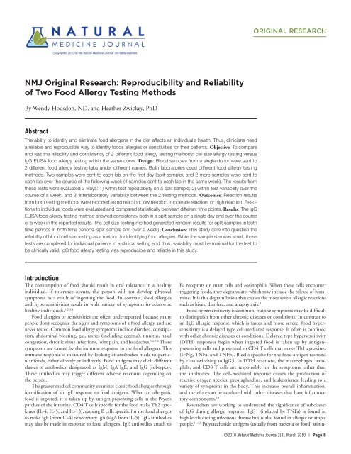
NMJ Original Research: Reproducibility and Reliability of Two Food ...
NMJ Original Research: Reproducibility and Reliability of Two Food ...
NMJ Original Research: Reproducibility and Reliability of Two Food ...

You also want an ePaper? Increase the reach of your titles
YUMPU automatically turns print PDFs into web optimized ePapers that Google loves.
<strong>NMJ</strong> <strong>Original</strong> <strong>Research</strong>: <strong>Reproducibility</strong> <strong>and</strong> <strong>Reliability</strong><br />
<strong>of</strong> <strong>Two</strong> <strong>Food</strong> Allergy Testing Methods<br />
By Wendy Hodsdon, ND, <strong>and</strong> Heather Zwickey, PhD<br />
Abstract<br />
The ability to identify <strong>and</strong> eliminate food allergens in the diet affects an individual’s health. Thus, clinicians need<br />
a reliable <strong>and</strong> reproducible way to identify foods allergies or sensitivities for their patients. Objective: To compare<br />
<strong>and</strong> test the reliability <strong>and</strong> consistency <strong>of</strong> 2 different food allergy testing methods: cell size allergy testing versus<br />
IgG ELISA food allergy testing within the same donor. Design: Blood samples from a single donor were sent to<br />
2 different food allergy testing labs under different names. Both laboratories used different food allergy testing<br />
methods. <strong>Two</strong> samples were sent to each lab on the first day (split sample), <strong>and</strong> 2 more samples were sent to<br />
each lab over the course <strong>of</strong> the following week (4 samples sent to each lab in the same week). The results from<br />
these tests were evaluated 3 ways: 1) within test repeatability on a split sample; 2) within test variability over the<br />
course <strong>of</strong> a week; <strong>and</strong> 3) interlaboratory variability between the 2 testing methods. Outcomes: Reaction results<br />
from both testing methods were reported as no reaction, low reaction, moderate reaction, or high reaction. Reactions<br />
to individual foods were evaluated <strong>and</strong> compared statistically between different time points. Results: The IgG<br />
ELISA food allergy testing method showed consistency both in a split sample on a single day <strong>and</strong> over the course<br />
<strong>of</strong> a week in the reported results. The cell size testing method generated r<strong>and</strong>om results for split samples in both<br />
time periods in both time periods (split sample <strong>and</strong> over a week). Conclusion: This study calls into question the<br />
reliability <strong>of</strong> blood cell size testing as a method for identifying food allergies. While the sample size was small, these<br />
tests are completed for individual patients in a clinical setting <strong>and</strong> thus, variability must be minimal for the test to<br />
be clinically valid. IgG food allergy testing was reproducible <strong>and</strong> reliable in this study.<br />
Introduction<br />
The consumption <strong>of</strong> food should result in oral tolerance in a healthy<br />
individual. If tolerance occurs, the person will not develop physical<br />
symptoms as a result <strong>of</strong> ingesting the food. In contrast, food allergies<br />
<strong>and</strong> hypersensitivities result in wide variety <strong>of</strong> symptoms in otherwise<br />
healthy individuals. 1,2,3,4<br />
<strong>Food</strong> allergies or sensitivities are <strong>of</strong>ten underreported because many<br />
people don’t recognize the signs <strong>and</strong> symptoms <strong>of</strong> a food allergy <strong>and</strong> are<br />
never tested. Common food allergy symptoms include diarrhea, constipation,<br />
abdominal bloating, gas, rashes (including eczema), tinnitus, nasal<br />
congestion, chronic sinus infections, joint pain, <strong>and</strong> headaches. 5,6,7,8 These<br />
symptoms are caused by the immune response to the food allergen. This<br />
immune response is measured by looking at antibodies made to particular<br />
foods, either directly or indirectly. <strong>Food</strong> antigens may elicit different<br />
classes <strong>of</strong> antibodies, designated as IgM, IgA IgE, <strong>and</strong> IgG (subtypes).<br />
These antibodies may trigger different adverse reactions depending on<br />
the person.<br />
The greater medical community examines classic food allergies through<br />
identification <strong>of</strong> an IgE response to food antigens. When an allergenic<br />
food is ingested, it is taken up by antigen-presenting cells in the Peyer’s<br />
patches <strong>of</strong> the intestine. CD4 T cells specific for the food make Th2 cytokines<br />
(IL-4, IL-5, <strong>and</strong> IL-13), causing B cells specific for the food allergen<br />
to make IgE (from IL-4) or secretory IgA (sIgA from IL-5). IgG antibodies<br />
may also be made in response to food allergens. IgE antibodies attach to<br />
ORIGINAL RESEARCH<br />
Fc receptors on mast cells <strong>and</strong> eosinophils. When these cells encounter<br />
triggering foods, they degranulate, which may include the release <strong>of</strong> histamine.<br />
It is this degranulation that causes the more severe allergic reactions<br />
such as hives, diarrhea, <strong>and</strong> anaphylaxis. 9<br />
<strong>Food</strong> hypersensitivity is common, but the symptoms may be difficult<br />
to distinguish from other chronic diseases or conditions. In contrast to<br />
an IgE allergic response which is faster <strong>and</strong> more severe, food hypersensitivity<br />
is a delayed type cell-mediated response. It <strong>of</strong>ten is confused<br />
with other chronic diseases or conditions. Delayed type hypersensitivity<br />
(DTH) responses begin when ingested food is taken up by antigenpresenting<br />
cells <strong>and</strong> presented to CD4 T cells that make Th1 cytokines<br />
(IFNg, TNFa, <strong>and</strong> TNFb). B cells specific for the food antigen respond<br />
by class switching to IgG3. In DTH reactions, the macrophages, basophils,<br />
<strong>and</strong> CD8 T cells are responsible for the symptoms rather than<br />
the antibodies. The cell-mediated response causes the production <strong>of</strong><br />
reactive oxygen species, prostagl<strong>and</strong>ins, <strong>and</strong> leukotrienes, leading to a<br />
variety <strong>of</strong> symptoms in the body. This increases overall inflammation,<br />
<strong>and</strong> therefore can be confused with other diseases that have inflammatory<br />
components. 10<br />
<strong>Research</strong>ers are working to underst<strong>and</strong> the significance <strong>of</strong> subclasses<br />
<strong>of</strong> IgG during allergic response. IgG1 (induced by TNFa) is found in<br />
high levels during infectious disease but is also found in allergic or atopic<br />
people. 11,12 Polysaccharide antigens (usually from bacteria or food) stimu-<br />
©2010 Natural Medicine Journal 2(3), March 2010 | Page 8
late IgG2. 13 IgG3 (induced by IFNg <strong>and</strong> TNFa) is most <strong>of</strong>ten elevated<br />
during infectious disease. IgG3 is at lower levels in people with allergies<br />
<strong>and</strong> higher levels in people with DTH. Elevated IgG4 antibodies<br />
have been found in patients with atopic dermatitis <strong>and</strong> eczema. 14 IgG4<br />
is thought to be related to prolonged antigen exposure. 15 Similar to IgE,<br />
IgG4 requires IL-4 <strong>and</strong> IL-13 for production. 16 Another cytokine, IL-10<br />
may induce IgG4 secretion. 17 IL-10 may also determine whether B cells<br />
continue to produce IgG4 or class switch to IgE. 18 Some allergens do not<br />
induce an IgG response at all. 19<br />
There are reactions to food that are not immune-mediated. These reactions<br />
include direct toxic reactions to a food ingredient. There are some<br />
food components to which everyone reacts; for example, food poisoning is<br />
a reaction to a toxin (most commonly staph enterotoxin A or staph enterotoxin<br />
B) made by bacteria. Many people react to monosodium glutamate<br />
or other food chemicals that they are unable detoxify. A genetic predisposition<br />
can cause susceptible individuals to overreact to a food ingredient,<br />
such as histamine contained in foods such as cheeses <strong>and</strong> smoked meats.<br />
Other food components are problematic for people lacking appropriate<br />
enzymes, which may lead to lactose intolerance, favism (glucose-6-phosphate<br />
deficiency), <strong>and</strong> other diagnoses. 20<br />
<strong>Food</strong> Allergy Testing<br />
There are a variety <strong>of</strong> ways to test for food allergies. Allergy skin testing<br />
is most common in allopathic medicine. In this test, a suspected food<br />
allergen is put into solution <strong>and</strong> then dropped onto the skin. A small prick<br />
is made through the drop <strong>of</strong> the food allergen with a lancet. If the person is<br />
allergic to the food, a hive will appear within 20 minutes. This test identifies<br />
IgE mediated food allergies. 21 This test, however, cannot be used if a<br />
person has eczema at the site <strong>of</strong> testing. Also, the test may show no reaction<br />
to foods that have low levels <strong>of</strong> IgE. 22,23 Likewise, a false negative<br />
may occur in people taking antihistamines or other immunosuppressive<br />
pharmaceuticals. Intradermal skin testing is more sensitive than skin prick<br />
testing, but is also more uncomfortable for the patient. 24<br />
IgE can also be measured from blood using ELISA (enzyme linked<br />
immunosorbant assay). Advances in technology over the last 10 years have<br />
made these tests more common. They are less invasive for the patient than<br />
skin tests. The test can report both the presence <strong>of</strong> an antibody <strong>and</strong> the<br />
relative quantity <strong>of</strong> the antibody in the serum. However, it is important to<br />
remember that this antibody level will be related to the immediacy <strong>of</strong> the<br />
exposure to the food antigen <strong>and</strong> to the food antigen that is used by the<br />
lab measuring the antibody.<br />
IgG <strong>and</strong> sIgA ELISA are also becoming popular, but they have the<br />
same drawbacks as IgE ELISA. They are dependent on exposure to a food<br />
<strong>and</strong> on the antigen used in the assay. Secretory IgA is most <strong>of</strong>ten measured<br />
from saliva samples, whereas IgG is measured in blood. IgG subclass may<br />
influence the correlation between test results <strong>and</strong> clinical symptoms <strong>and</strong><br />
is not always reported by the laboratory completing the test. As discussed<br />
above, most <strong>of</strong>ten IgG1 <strong>and</strong> IgG4 are elevated in an allergic response.<br />
Thus, while ELISA testing is useful in measuring food allergies, it is unable<br />
to measure all the reactions that may cause clinical symptoms.<br />
One <strong>of</strong> the lab tests available for food allergy <strong>and</strong> sensitivity testing evaluates<br />
the change in white blood cell size <strong>and</strong> number as a measure <strong>of</strong> reactivity.<br />
It is known that B cells increase in size when they become plasma cells<br />
<strong>and</strong> produce antibodies, T cells increase in size when they produce cytokines,<br />
<strong>and</strong> neutrophils change size when they become activated. However,<br />
the lab that uses this method does not report what cells are being measured<br />
in their assay, nor do they report the methods <strong>of</strong> the assay. 25<br />
This study compares intralaboratory reliability between 2 types <strong>of</strong><br />
allergy testing: IgG testing <strong>and</strong> cell size variability. The results reported<br />
herein suggest that IgG testing is more reproducible <strong>and</strong> reliable than<br />
cell size variability. While the initial design included an interlaboratory<br />
comparison to see how equivalent the results were <strong>of</strong> these 2 testing<br />
methods, the cell size variability data was not consistent enough to use in<br />
a comparison with IgG testing.<br />
Figure Figure 1: Design 1: Design Overview. Overview<br />
Samples were collected as described in the Methods<br />
section. Antibodies refer to the IgG ELISA method. Cell size refers to the cell<br />
size variability testing method.<br />
Design <strong>and</strong> Methods<br />
Design overview<br />
Blood draws were performed with the same donor for 3 different evaluations:<br />
1) within test repeatability for 2 different diagnostic tests on a single<br />
sample, 2) within participant repeatability over time, <strong>and</strong> 3) interlaboratory<br />
reliability between diagnostic food allergy testing methods (see Figure 1).<br />
Blood collection<br />
Blood was collected using collection tubes or strips provided by each<br />
allergy testing company used in the study. The companies use different<br />
methods to determine allergic reactions. Alcat (hereafter referred to as cell<br />
size variability method) in Deerfield Beach, FL, uses an assay that measures<br />
cell size. US Biotek (hereafter referred to as the IgG ELISA method) in<br />
Seattle, WA, uses an assay that measures antibody (IgG) concentration.<br />
For the cell size assay, blood was collected into 2 blue-top vials provided<br />
(3.8% sodium citrate, 4.5 ml draw). Blood was shipped overnight at room<br />
temperature. Peripheral blood collected for the IgG antibody assay was<br />
collected on filter paper strips from a finger stick using a lancet. Blood was<br />
allowed to dry on the filter strips at room temperature <strong>and</strong> shipped via the<br />
US Postal Service.<br />
Cell size testing<br />
Cell size variability is measured to determine reactivity to food antigens.<br />
Cells from the blood sample are incubated with potential food allergens.<br />
If the individual is sensitive to the food, the cell size changes, most likely<br />
due to activation or degranulation. A modified Coulter counter is used to<br />
measure cell size.<br />
©2010 Natural Medicine Journal 2(3), March 2010 | Page 9
Antibody testing (ELISA)<br />
Antibody (IgG) specific for a food allergen is measured via ELISA. <strong>Food</strong><br />
antigens are bound to the surface <strong>of</strong> an ELISA plate <strong>and</strong> an individual’s<br />
whole blood is added. If the individual has antibodies to the food, the<br />
antibodies will bind to the antigen. These antibodies can then be detected<br />
with an enzymatic or colorimetric reaction.<br />
Within test repeatability<br />
The first part <strong>of</strong> the protocol was designed to evaluate repeatability within<br />
a testing method as a measure <strong>of</strong> reliability <strong>and</strong> reproducibility. Blood was<br />
collected from the same donor for all time points. On day 1, the several<br />
tubes <strong>of</strong> blood were collected during the same blood draw; the tubes were<br />
blinded <strong>and</strong> sent to both companies for analysis (see Figure 1).<br />
Within participant repeatability over time<br />
<strong>Two</strong> days after the initial draw (day 2), 2 blood samples were collected<br />
during the same blood draw. The tubes were blinded <strong>and</strong> sent to both<br />
companies for analysis. The next day (day 3), 2 tubes <strong>of</strong> blood were<br />
collected during the same blood draw. The tubes were coded <strong>and</strong> sent<br />
to both companies for analysis. The samples from days 1, 2, <strong>and</strong> 3 were<br />
compared to see if results were consistent over the course <strong>of</strong> 4 days. A coefficient<br />
<strong>of</strong> variance (CV) <strong>and</strong> an intraclass correlation coefficient (ICC) were<br />
calculated for each test methodology. For the ICC, a value approaching 1<br />
confirms consistency between the samples. Results were compared between<br />
companies in order to determine interlaboratory correlation.<br />
Results<br />
<strong>Reliability</strong> <strong>of</strong> food allergy testing on a single split sample.<br />
In this study, blood from 1 person was sent to 2 different labs to test for<br />
food allergies. On the first day, the blood was split into 4 samples, <strong>and</strong><br />
2 samples were sent to each lab. The 2 samples analyzed by each individual<br />
lab were compared for similarity. This allowed us to test the internal<br />
reliability <strong>of</strong> each lab. The results were reported as no reaction (0), low<br />
reaction (1), moderate reaction (2) or high reaction (3). If the food reactivity<br />
levels differed between the samples, the difference was calculated<br />
<strong>and</strong> reported in Table 1. The difference between reactivity levels was 0 if<br />
a food showed up in the same category in both samples. Cabbage had 1<br />
test reporting a moderate reaction (2), <strong>and</strong> 1 test reporting a high reaction<br />
(3), so the difference in reactivity level was 1 since the results differed by<br />
1 category. To receive a score <strong>of</strong> 3, 1 sample had a high reaction (3) <strong>and</strong> 1<br />
sample had no reaction (0).<br />
The company using the cell size variability method tested 50 foods in<br />
its food allergy panel. Table 1 demonstrates that only 34% <strong>of</strong> the foods (17<br />
foods) generated identical results between the split samples. Twenty-eight<br />
Between Split<br />
Samples<br />
Cell Size<br />
Method<br />
(# <strong>of</strong> foods out <strong>of</strong><br />
50 foods tested)<br />
IgG ELISA<br />
Method<br />
(# <strong>of</strong> foods out <strong>of</strong><br />
96 foods tested)<br />
Identical results 34% (17) 95% (91)<br />
1 reactivity level<br />
difference<br />
2 reactivity level<br />
difference<br />
3 reactivity level<br />
difference<br />
28% (14) 5% (5)<br />
10% (5) 0%<br />
28% (14) 0%<br />
Table 1: This table compares results from a split sample. The reactivity level<br />
differences refer to the percentage <strong>of</strong> samples that have identical results, or<br />
results that are <strong>of</strong>f by a number <strong>of</strong> categories. For this purpose, no reaction = 0,<br />
low = 1, moderate = 2, <strong>and</strong> high = 3. The reactivity level difference is calculated<br />
by taking the absolute difference between the split sample scores.<br />
percent <strong>of</strong> the foods (14 foods) differed by 1 reactivity level, 10% <strong>of</strong> the<br />
foods (5 foods) differed by 2 reactivity levels, <strong>and</strong> 28% <strong>of</strong> the foods (14<br />
foods) differed by 3 reactivity levels. If this were a reliable test for food<br />
allergies, we would expect the majority <strong>of</strong> the foods tested to have identical<br />
results. In this case, 66% <strong>of</strong> the foods tested differed by 1 or more reactivity<br />
levels. The scatterplot for cell size testing method, Figure 2A, depicts<br />
a large variability in the results for the split sample.<br />
The company using the IgG ELISA method tested 96 foods in its food<br />
allergy panel. In contrast to the cell size variability method, 95% <strong>of</strong> the<br />
foods (91 foods) were identical between the split samples. Five percent <strong>of</strong><br />
the foods (5 foods) differed by 1 reactivity level. No foods differed by more<br />
than one reactivity level between the split samples. The scatterplot for the<br />
IgG ELISA method, figure 2B, has a more linear pattern.<br />
Consistency <strong>of</strong> food allergy testing over time<br />
On 3 different days, samples were collected <strong>and</strong> sent to both labs to test the<br />
consistency <strong>of</strong> test results over the course <strong>of</strong> a week. A total <strong>of</strong> 4 time points<br />
were compared: 2 samples from Monday, 1 sample from Wednesday, <strong>and</strong><br />
1 sample from Thursday. The difference in reactivity levels was recorded<br />
as the greatest difference in the 4 time points compared. For example, if<br />
lamb had no reaction (0) for 3 time points, <strong>and</strong> high (3) for 1 time point,<br />
it would receive a difference in reactivity <strong>of</strong> 3 (the difference between 0<br />
<strong>and</strong> 3). If all 4 time points were identical for a food, the difference was<br />
scored as 0. If 1 food had a low reaction (1) for 1 time point, moderate<br />
reactions (2) for 2 time points <strong>and</strong> high reaction (3) for 1 time point, the<br />
largest difference between reactivity levels was calculated at 2 (the difference<br />
between 1 <strong>and</strong> 3).<br />
As shown in Table 2, 2% <strong>of</strong> the samples (1 <strong>of</strong> 50 foods) tested by the<br />
cell size variability method yielded identical results over the 4 time points.<br />
Twelve percent (6 foods) differed by 1 reactivity level. Twenty-six percent<br />
<strong>of</strong> foods (13 foods) differed by 2 reactivity levels, <strong>and</strong> 60% <strong>of</strong> foods (30<br />
foods) differed by 3 reactivity levels. This means that 60% <strong>of</strong> the foods had<br />
at least 1 sample that scored no reaction at 1 time point <strong>and</strong> scored high<br />
reaction for the same food at a different time point during the same week.<br />
When comparing all 4 time points, the coefficient <strong>of</strong> variance for the cell<br />
size variability method was calculated to be 0.55, with an ICC <strong>of</strong> 0.01.<br />
Comparatively, 82% <strong>of</strong> the foods tested (79 <strong>of</strong> 96 foods) by the IgG<br />
ELISA method produced identical results over the 4 time points. Seventeen<br />
percent (16 foods) differed by 1 reactivity level. One percent (1 food)<br />
had a reactivity level difference <strong>of</strong> 2 (one sample, no reaction <strong>and</strong> three<br />
Over the Course<br />
<strong>of</strong> a Week<br />
Cell Size<br />
Method<br />
(# <strong>of</strong> foods out <strong>of</strong><br />
50 foods tested)<br />
IgG ELISA<br />
Method<br />
(# <strong>of</strong> foods out <strong>of</strong><br />
96 foods tested)<br />
Identical results 2% (1) 82% (79)<br />
1 reactivity level<br />
difference<br />
2 reactivity level<br />
difference<br />
3 reactivity level<br />
difference<br />
Coefficient <strong>of</strong> variance<br />
(CV)<br />
Intraclass correlation<br />
coefficient<br />
(ICC)<br />
12% (6) 17% (16)<br />
26% (13) 1% (1)<br />
60% (30) 0%<br />
0.55 0.05<br />
0.01 0.99<br />
Table 2: Results in table 2 show the differences in 4 samples all taken over the<br />
course <strong>of</strong> 4 days. See Procedure section for more details on the samples taken<br />
during 1 week. The reactivity level difference in this table reflects the greatest<br />
absolute difference between the 4 samples.<br />
©2010 Natural Medicine Journal 2(3), March 2010 | Page 10
Split sample 1<br />
High<br />
Moderate<br />
Low<br />
None<br />
None<br />
Low<br />
Moderate<br />
Split sample 2<br />
High<br />
Figure 2A: Scatterplot <strong>of</strong> split sample for the cell size variability method. Blood<br />
was split into 2 samples <strong>and</strong> measured for reactivity to foods. Each point on the<br />
scatterplot represents the reactivity to a single food in the split sample.<br />
samples, moderate reaction). There were no foods that differed by 3 reactivity<br />
levels over the 4 time points. The coefficient <strong>of</strong> variance was 0.05 for<br />
the IgG ELISA method, with an ICC <strong>of</strong> 0.99, which demonstrates a much<br />
more consistent reactivity pattern.<br />
Discussion<br />
An ideal response to food antigen is tolerance. Yet, some patients develop<br />
allergic responses to seemingly innocuous food antigens. Clinicians<br />
commonly recommend lab testing when a patient has a suspected food<br />
allergy. It is possible that some types <strong>of</strong> food reactivity will show up in 1<br />
type <strong>of</strong> testing method <strong>and</strong> not others. Clinicians <strong>and</strong> patients rely on the<br />
lab tests to be both accurate <strong>and</strong> reproducible. When a physician reported<br />
that 2 types <strong>of</strong> food allergy tests reported different results for a single<br />
patient, we tested the reliability <strong>of</strong> food allergy testing for the 2 types <strong>of</strong><br />
food allergy tests in question. Although both <strong>of</strong> these labs are Clinical<br />
Laboratory Improvement Amendments (CLIA)– certified, certification<br />
does not ensure the consistency <strong>and</strong> reproducibility <strong>of</strong> laboratory tests.<br />
Before we could examine interlaboratory results <strong>of</strong> food allergy testing,<br />
intralaboratory reliability needed to be evaluated. Good intralaboratory<br />
reproducibility means that when a sample is compared with itself (as in<br />
a split sample), the results are expected to be identical. Similarly, when<br />
a person maintains a normal dietary routine over the course <strong>of</strong> a week,<br />
the results <strong>of</strong> a food allergy test would be expected to be the same. In this<br />
study, the cell size variability method delivered very different results for all<br />
the samples submitted, <strong>and</strong> therefore had no internal reproducibility or<br />
accuracy. The IgG ELISA method had excellent intralaboratory correlation<br />
for the split sample <strong>and</strong> the samples analyzed over the course <strong>of</strong> a<br />
week. The unreliability <strong>of</strong> the cell size variability method results prevented<br />
an interlaboratory analysis comparing the results <strong>of</strong> the cell size variability<br />
method to the IgG ELISA method.<br />
Other researchers have compared allergy testing methods, although<br />
most studies focus on IgE-related allergies as opposed to IgG-mediated<br />
responses. Double-blind placebo-controlled food challenge (DBPCFC)<br />
is considered the gold st<strong>and</strong>ard in food allergy testing <strong>and</strong> is strongly<br />
correlated with IgE testing. 26,27 In this type <strong>of</strong> testing, the reaction <strong>of</strong> a<br />
suspected allergenic food is compared to a placebo food, known to not<br />
evoke a response. <strong>Food</strong>s that are known to induce anaphylaxis are not<br />
generally tested. The DBPCFC identifies foods that evoke immediate food<br />
Split sample 1<br />
High<br />
Moderate<br />
Low<br />
None<br />
None<br />
Low<br />
Moderate<br />
Split sample 2<br />
High<br />
Figure 2B: Scatterplot <strong>of</strong> split sample for the IgG ELISA method. Blood was<br />
split into 2 samples <strong>and</strong> measured for reactivity to foods. Each point on the<br />
scatterplot represents the reactivity to a single food in the split sample.<br />
allergy symptoms. 28 Skin tests can also be used to identify food allergens.<br />
These tests are more sensitive than IgE blood tests. 29<br />
IgE to food allergens demonstrates an immediate phase immune<br />
response. Delayed type responses, however, are not mediated by IgE antibodies<br />
<strong>and</strong> will not show up with this type <strong>of</strong> testing. 30 For symptoms<br />
<strong>of</strong> food allergies caused by delayed type hypersensitivity reactions such as<br />
headaches, mood swings, intestinal upset, pain, <strong>and</strong> attention problems,<br />
the DBPCFC or skin tests may present a false negative.<br />
In this study, IgG ELISA testing was more reproducible than cell-size<br />
testing. In general ELISA is known to be consistent <strong>and</strong> is routinely used<br />
for scientific testing. 31 The sensitivity <strong>of</strong> ELISA as a method for food allergy<br />
testing is dependent upon the food antigen used as well as the amount <strong>of</strong><br />
antibody present. IgE food antigens used for ELISA assays have been st<strong>and</strong>ardized<br />
<strong>and</strong> are consistent between different laboratories. IgG food antigens<br />
have not been st<strong>and</strong>ardized, which accounts for some <strong>of</strong> the variation<br />
between laboratories. All commercial food antigens for ELISA testing are<br />
made from raw foods (both IgE <strong>and</strong> IgG antigens). Cooking food exposes<br />
different antigens <strong>and</strong> epitopes which may affect ELISA test results. 32 For<br />
example, pecans, wheat flour, roasted peanuts, lentils, almonds, cashews,<br />
walnuts, soy beans, shrimp, scallop, tuna, egg, apple, plum, milk, <strong>and</strong><br />
potatoes have been shown to have antigens that differ between raw <strong>and</strong><br />
cooked forms. 33,34 Another researcher suggests that cooked egg (baked egg<br />
especially) produces less <strong>of</strong> a reaction than raw egg. 35<br />
The participant <strong>of</strong> this study had IgG reactions to milk <strong>and</strong> soy. The<br />
most common IgE mediated food allergens in the general population<br />
are milk, soy, egg, peanut, wheat, tree nuts (walnuts <strong>and</strong> cashews), fish,<br />
<strong>and</strong> shellfish. These foods account for 85% <strong>of</strong> the commonly recognized<br />
food allergens. 36 The other foods with high reactions for this participant<br />
included almonds, corn, lima beans, bananas, <strong>and</strong> blueberries. All <strong>of</strong> these<br />
foods were regularly included in the participant’s diet before the food<br />
allergy tests.<br />
Cell size testing as a measure <strong>of</strong> food reactivity is not well studied in the<br />
literature. Consistent with the data reported herein, most studies suggest that<br />
it is unreliable. 37,38,39 The company that performed the cell size variability<br />
method was told <strong>of</strong> the results in a phone call a month after the testing was<br />
complete <strong>and</strong> said that there were no irregularities during that week <strong>and</strong><br />
that they stood by their results. While we can hypothesize a mechanism for<br />
identifying food allergies from cell size differentials, the data clearly demon-<br />
©2010 Natural Medicine Journal 2(3), March 2010 | Page 11
strate this method is not specific, not reproducible, <strong>and</strong> was not related to<br />
food reactions in this participant. As the scientific community continues<br />
to underst<strong>and</strong> the importance <strong>of</strong> the antigen being used <strong>and</strong> the accuracy<br />
<strong>of</strong> different tests in providing relevant clinical guidance, the consistency<br />
between laboratories <strong>and</strong> the method they employ must improve.<br />
Conclusion <strong>and</strong> Future Directions<br />
Cell size variability testing for food allergies proved to be completely<br />
r<strong>and</strong>om in all tests, <strong>and</strong> therefore has no clinical relevance. The IgG ELISA<br />
method proved to be a consistent, reproducible, <strong>and</strong> specific test for food<br />
allergies in this small study. The clinical relevance <strong>of</strong> these results were not<br />
examined in this study. The results presented here verify that IgG ELISA<br />
testing is more reliable <strong>and</strong> consistent than cell size testing for identifying<br />
food sensitivities.<br />
In order to further improve clinical practice, future research should<br />
demonstrate how long IgG antibodies remain active after food elimination.<br />
Future studies could also evaluate how long it takes to resolve physical<br />
symptoms after food elimination. In addition, identifying specific disease<br />
states exacerbated by IgG food reactivity would help clinicians identify the<br />
patients who would benefit the most from IgG testing.<br />
<strong>Food</strong> is medicine, in some very fundamental ways. Eliminating<br />
harmful foods <strong>and</strong> encouraging healthful, nutritious foods can have a big<br />
impact on health. In order to eliminate foods, accurate identification <strong>of</strong><br />
potentially harmful foods is essential. In this study, we have identified a<br />
food allergy test that is specific <strong>and</strong> reproducible. This can help clinicians<br />
have more confidence in the food allergy test they recommend to patients.<br />
Acknowledgements<br />
Helfgott <strong>Research</strong> Institute provided funding for this study <strong>and</strong> consulted<br />
on the study design. William Gregory, PhD, provided statistical consultation<br />
<strong>and</strong> comments on the manuscript.<br />
About the Authors<br />
Wendy Hodsdon, ND, is in private practice at<br />
Portl<strong>and</strong> Alternative Medicine, LLC in Portl<strong>and</strong>,<br />
Oregon, where her focus is energy medicine <strong>and</strong><br />
immune system disorders including allergies,<br />
asthma, <strong>and</strong> autoimmune conditions. Hodsdon<br />
received her bachelor <strong>of</strong> science in biology from<br />
University <strong>of</strong> Oregon, Eugene, <strong>and</strong> her naturopathic<br />
degree from the National College <strong>of</strong> Natural<br />
Medicine (NCNM) in 2004. Before studying<br />
medicine, Hodsdon participated in research around the world including<br />
developmental biology projects, B-cell development projects, <strong>and</strong> clinical<br />
research projects in Ug<strong>and</strong>a on tuberculosis <strong>and</strong> HIV. Hodsdon was the<br />
chairperson for the Institutional Review Board (IRB) for NCNM for 4<br />
years. More information about Hodsdon can be found on her website:<br />
www.Portl<strong>and</strong>AlternativeMedicine.com.<br />
Heather Zwickey, PhD, is dean <strong>of</strong> research <strong>and</strong><br />
associate pr<strong>of</strong>essor <strong>of</strong> immunology at the National<br />
College <strong>of</strong> Natural Medicine (NCNM) <strong>and</strong><br />
director <strong>of</strong> Helfgott <strong>Research</strong> Institute. She trained<br />
at National Jewish Medical <strong>and</strong> <strong>Research</strong> Center<br />
in Denver. She received a PhD in immunology<br />
<strong>and</strong> microbiology from the University <strong>of</strong> Colorado<br />
Health Sciences Center <strong>and</strong> went on to complete<br />
a postdoctoral fellowship at Yale University. Her<br />
research there examined the role <strong>of</strong> dendritic cells in autoimmune responses.<br />
At Helfgott, Zwickey applies her immunology expertise to natural medicine.<br />
She is currently working to build a Practice-based <strong>Research</strong> Network<br />
in Naturopathic Medicine, which will collect data from patients seen by<br />
naturopathic physicians in clinical practice. In addition to her work at Helfgott,<br />
Dr. Zwickey has an appointment in the Department <strong>of</strong> Neurology at<br />
Oregon Health <strong>and</strong> Sciences University in Portl<strong>and</strong>. She maintains collaborations<br />
with investigators at OHSU, as well as the Oregon College <strong>of</strong><br />
Oriental Medicine in Portl<strong>and</strong> <strong>and</strong> Pontificia Universidade Catolica Do Rio<br />
Grange Do Sol in Porto Alegre, Brazil.<br />
References<br />
1 Bentley SJ, Pearson DJ, Rix KJ. <strong>Food</strong> hypersensitivity in irritable bowel<br />
syndrome. Lancet. 1983;2(8345):295-297.<br />
2 Firer MA, Hosking CS, Hill DJ. Cow’s milk allergy <strong>and</strong> eczema: patterns <strong>of</strong> the<br />
antibody response to cow’s milk in allergic skin disease. Clin Allergy. 1982;12(4):<br />
385-390.<br />
3 Jones VA, et al. <strong>Food</strong> intolerance: a major factor in the pathogenesis <strong>of</strong> irritable<br />
bowel syndrome. Lancet. 1982;2(8308): 1115-1117.<br />
4 Vojdani A. Detection <strong>of</strong> IgE, IgG, IgA <strong>and</strong> IgM antibodies against raw <strong>and</strong><br />
processed food antigens. Nutr Metab (Lond). 2009;6:22.<br />
5 Bentley SJ, Pearson DJ, Rix KJ. <strong>Food</strong> hypersensitivity in irritable bowel<br />
syndrome. Lancet. 1983;2(8345):295-297.<br />
6 Firer MA, Hosking CS, Hill DJ. Cow’s milk allergy <strong>and</strong> eczema: patterns <strong>of</strong> the<br />
antibody response to cow’s milk in allergic skin disease. Clin Allergy. 1982;12(4):<br />
385-390.<br />
7 Jones VA, et al. <strong>Food</strong> intolerance: a major factor in the pathogenesis <strong>of</strong> irritable<br />
bowel syndrome. Lancet. 1982;2(8308): 1115-1117.<br />
8 Vojdani A. Detection <strong>of</strong> IgE, IgG, IgA <strong>and</strong> IgM antibodies against raw <strong>and</strong><br />
processed food antigens. Nutr Metab (Lond). 2009;6:22.<br />
9 Murphy K, Travers P, Walport M. Janeway’s Immunobiology. 7 ed. New York:<br />
Garl<strong>and</strong> Science; 2008.<br />
10 Ibid.<br />
11 Ibid.<br />
12 Fear DJ, McCloskey N, O’Connor B, Felsenfeld G, Gould HJ. Transcription <strong>of</strong><br />
Ig germline genes in single human B cells <strong>and</strong> the role <strong>of</strong> cytokines in isotype<br />
determination. J Immunol. 2004;173(7):4529-4538.<br />
13 Murphy K, Travers P, Walport M. Janeway’s Immunobiology. 7 ed. New York:<br />
Garl<strong>and</strong> Science; 2008.<br />
14 Bernardi D, Borghesan F, Faggian D, et al. Time to reconsider the clinical value<br />
<strong>of</strong> immunoglobulin G4 to foods? Clin Chem Lab Med. 2008;46(5):687-690.<br />
15 Aalberse RC, Stapel SO, Schuurman J, Rispens T. Immunoglobulin G4: an odd<br />
antibody. Clin Exp Allergy. 2009; 39(4):469-477.<br />
16 Vercelli D, De Monte L, Monticelli S, Di Bartolo C, Agresti A. To E or not to<br />
E? Can an IL-4-induced B cell choose between IgE <strong>and</strong> IgG4? Int Arch Allergy<br />
Immunol. 1998;116(1):1-4.<br />
17 Satoguina JS, Wey<strong>and</strong> E, Larbi J, Hoerauf A. T regulatory-1 cells induce IgG4<br />
production by B cells: role <strong>of</strong> IL-10. J Immunol. 2005;174(8):4718-4726.<br />
18 Punnonen J, de Waal Malefyt R, van Vlasselaer P, Gauchat JF, de Vries JE. IL-10<br />
<strong>and</strong> viral IL-10 prevent IL-4-induced IgE synthesis by inhibiting the accessory<br />
cell function <strong>of</strong> monocytes. J Immunol. 1993;151(3):1280-1289.<br />
19 Aalberse RC. Structural features <strong>of</strong> allergenic molecules. Chem Immunol Allergy.<br />
2006;91:134-146.<br />
20 Anderson JA. Non-immunologically-mediated food sensitivity. Nutr Rev.<br />
1984;42(3):109-116.<br />
21 Cox L, Williams B, Sicherer S, et al. Pearls <strong>and</strong> pitfalls <strong>of</strong> allergy diagnostic<br />
testing: report from the American College <strong>of</strong> Allergy, Asthma <strong>and</strong> Immunology/<br />
American Academy <strong>of</strong> Allergy, Asthma <strong>and</strong> Immunology Specific IgE Test Task<br />
Force. Ann Allergy Asthma Immunol. 2008;101(6):580-592.<br />
22 Du Toit G, Santos A, Roberts G, Fox AT, Smith P, Lack G. The diagnosis <strong>of</strong> IgEmediated<br />
food allergy in childhood. Pediatr Allergy Immunol. 2009;20(4):309-<br />
319.<br />
23 Tripodi S, Businco AD, Aless<strong>and</strong>ri C, Panetta V, Restani P, Matricardi PM.<br />
Predicting the outcome <strong>of</strong> oral food challenges with hen’s egg through skin test<br />
end-point titration. Clin Exp Allergy. 2009;39(8):1225-1233.<br />
24 Oppenheimer J, Nelson HS. Skin testing. Ann Allergy Asthma Immunol.<br />
2006;96(2 Suppl 1):S6-12.<br />
25 Davis Deutsch R. The right stuff: use <strong>of</strong> ALCAT testing to determine dietary<br />
factors affecting immune balance, health, <strong>and</strong> longevity. In: Klatz R, Goldman<br />
R, eds. Anti-Aging Therapeutics. Vol X. Chicago, IL: American Academy for<br />
Anti-Aging Medicine; 2007.<br />
©2010 Natural Medicine Journal 2(3), March 2010 | Page 12
26 Nowak-Wegrzyn A, Assa’ad AH, Bahna SL, Bock SA, Sicherer SH, Teuber<br />
SS. Work Group report: oral food challenge testing. J Allergy Clin Immunol.<br />
2009;123(6 Suppl): S365-383.<br />
27 Rancé F, Deschildre A, Villard-Truc F, et al. Oral food challenge in children: an<br />
expert review. Eur Ann Allergy Clin Immunol. 2009;41(2):35-49.<br />
28 Nowak-Wegrzyn A, Assa’ad AH, Bahna SL, Bock SA, Sicherer SH, Teuber<br />
SS. Work Group report: oral food challenge testing. J Allergy Clin Immunol.<br />
2009;123(6 Suppl): S365-383.<br />
29 Cox L, Williams B, Sicherer S, et al. Pearls <strong>and</strong> pitfalls <strong>of</strong> allergy diagnostic<br />
testing: report from the American College <strong>of</strong> Allergy, Asthma <strong>and</strong> Immunology/<br />
American Academy <strong>of</strong> Allergy, Asthma <strong>and</strong> Immunology Specific IgE Test Task<br />
Force. Ann Allergy Asthma Immunol. 2008;101(6):580-592.<br />
30 Vojdani A. Detection <strong>of</strong> IgE, IgG, IgA <strong>and</strong> IgM antibodies against raw <strong>and</strong><br />
processed food antigens. Nutr Metab (Lond). 2009;6:22.<br />
31 Shrijver RS, Kramps JA. Critical factors affecting the diagnostic reliability <strong>of</strong><br />
enzyme-linked immunosorbent assay formats. Rev Sci Tech. 1998;17(2):550-<br />
561.<br />
32 Vojdani A. Detection <strong>of</strong> IgE, IgG, IgA <strong>and</strong> IgM antibodies against raw <strong>and</strong><br />
processed food antigens. Nutr Metab (Lond). 2009;6:22.<br />
33 Ibid.<br />
34 Lemon-Mulé H, Sampson HA, Sicherer SH, Shreffler WG, Noone S, Nowak-<br />
Wegrzyn A. Immunologic changes in children with egg allergy ingesting extensively<br />
heated egg. J Allergy Clin Immunol. 2008;122(5):977-983.<br />
35 Ibid.<br />
36 Cox L, Williams B, Sicherer S, et al. Pearls <strong>and</strong> pitfalls <strong>of</strong> allergy diagnostic<br />
testing: report from the American College <strong>of</strong> Allergy, Asthma <strong>and</strong> Immunology/<br />
American Academy <strong>of</strong> Allergy, Asthma <strong>and</strong> Immunology Specific IgE Test Task<br />
Force. Ann Allergy Asthma Immunol. 2008;101(6):580-592.<br />
37 Bindslev-Jensen C, Poulsen LK. What do we at present know about the ALCAT<br />
test <strong>and</strong> what is lacking? Monogr Allergy. 1996;32:228-232.<br />
38 Potter PC, Mullineux J, Weinberg EG, et al. The ALCAT test—inappropriate in<br />
testing for food allergy in clinical practice. S Afr Med J. 1992;81(7):384.<br />
39 Wuthrich B. Unproven techniques in allergy diagnosis. J Investig Allergol Clin<br />
Immunol. 2005;15(2):86-90.<br />
<strong>NMJ</strong>_MAR10_OR<br />
©2010 Natural Medicine Journal 2(3), March 2010 | Page 13