Implantation of the Melody Transcatheter Pulmonary Valve - Miami ...
Implantation of the Melody Transcatheter Pulmonary Valve - Miami ...
Implantation of the Melody Transcatheter Pulmonary Valve - Miami ...

You also want an ePaper? Increase the reach of your titles
YUMPU automatically turns print PDFs into web optimized ePapers that Google loves.
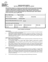
JACC Vol. 54, No. 18, 2009October 27, 2009:1722–9Zahn et al.Early Results From <strong>the</strong> Transca<strong>the</strong>ter <strong>Pulmonary</strong> <strong>Valve</strong> Trial1725Clinical Characteristics <strong>of</strong> Study Patients (n 34)Table 4 Clinical Characteristics <strong>of</strong> Study Patients (n 34)Variable/CategoryWeight, kg 61.1 15.3SexMale 26 (77)Female 8 (23)Age, yrs 19.4 7.7Original diagnosisTetralogy <strong>of</strong> Fallot 18 (52)Truncus arteriosus 4 (12)Aortic valve disease (Ross procedure) 4 (12)Transposition <strong>of</strong> <strong>the</strong> great arteries 3 (9)O<strong>the</strong>r 5 (15)NYHA functional classificationI 6 (18)II 23 (67)III 5 (15)IV 0 (0)RVOT conduit typeHomograft 27 (79)Biologic valved conduit 3 (9)O<strong>the</strong>r* 4 (12)RVOT conduit size 20.0 2.1Primary indication for implantationStenosis 6 (18)Regurgitation 23 (67)Mixed stenosis and regurgitation 5 (15)Pre-existing stents in RVOT conduitNo pre-existing stents in RVOT conduit 19 (56)Single stent in RVOT conduit 12 (35)Multiple stents in RVOT conduit 3 (9)Number <strong>of</strong> previous open surgeries 2.2 1.0PR by echocardiographyNone 0 (0)Trace 0 (0)Mild 1 (3)Moderate 12 (35)Severe 20 (59)Unable to assess 1 (3)Average conduit Doppler mean gradient (mm Hg) 28.4 11.4Data are presented as mean SD for continuous data or n (%) <strong>of</strong> subjects for categorical data.*O<strong>the</strong>r conduit types were nonvalved “tube grafts,” nonvalved homograft, and biopros<strong>the</strong>sis.Abbreviations as in Table 1.discharged within 7 days. Criteria for procedural successwere met in 93% (28 <strong>of</strong> 30) <strong>of</strong> attempted implantations. Inaddition to <strong>the</strong> patient with conduit rupture, <strong>the</strong> o<strong>the</strong>rpatient categorized as a procedural failure had a postimplantationRV-PA peak gradient <strong>of</strong> 37 mm Hg. Thegradient across <strong>the</strong> <strong>Melody</strong> valve was only 5 mm Hg, but<strong>the</strong>re was subvalvar muscular obstruction, which resulted in<strong>the</strong> elevated RV-PA gradient. In subsequent follow-up, thispatient’s conduit Doppler mean gradient was consistently25 mm Hg, and no fur<strong>the</strong>r intervention was performed.Short-term effectiveness. In terms <strong>of</strong> <strong>the</strong> short-term effectivenesscriteria defined in Table 3, 29 patients retained<strong>the</strong> <strong>Melody</strong> valve at 24 h, and 24 (83%) had acceptablehemodynamics at <strong>the</strong> 6-month time point. Follow-up dataare presented for <strong>the</strong> 29 patients who retained a <strong>Melody</strong>valve at 24 h.ACUTE HEMODYNAMIC RESULTS. After valve implantation,RV systolic pressure, RV-PA gradient, and <strong>the</strong> RV/aorticpressure ratio were significantly lower, and <strong>the</strong> PA diastolicpressure was significantly higher than before implantation(Table 5). Pre-discharge echocardiography demonstrated noPR or trivial PR in 86% <strong>of</strong> patients and mild PR in 14%.CLINICAL ASSESSMENT. NYHA functional class improvedafter <strong>Melody</strong> valve implantation (Fig. 1). Of 24 patientswho were NYHA functional class II or III before implantation,19 (79%) had an improvement <strong>of</strong> at least 1 class; nopatient experienced a decline in NYHA functional class.ECHOCARDIOGRAPHY. <strong>Valve</strong> competence was maintainedduring follow-up, with 93% <strong>of</strong> patients having no/trivialPR and no patient having more than mild PR 6 monthsafter implantation. Improvement <strong>of</strong> conduit obstructionwas maintained for <strong>the</strong> group as a whole, with averageDoppler mean gradient improving from 28.8 10 mmHg before implantation to 22.4 8.1 mm Hg at 6months (p 0.001). Five patients had Doppler meangradients 30 mm Hg at 6-month follow-up. One <strong>of</strong><strong>the</strong>se patients had a major stent fracture and underwent asuccessful second <strong>Melody</strong> valve implantation, 1 hadasymmetric expansion <strong>of</strong> <strong>the</strong> <strong>Melody</strong> valve and wastreated successfully using high-pressure balloon angioplasty,1 was discovered at subsequent ca<strong>the</strong>terization tohave obstruction below <strong>the</strong> conduit, 1 had stent fracturebut had not undergone repeat ca<strong>the</strong>terization, and in 1patient <strong>the</strong>re was no obvious explanation. All 5 were inNYHA functional class I at most recent follow-up.cMRI. Twenty-five patients (83%) had paired cMRI databefore and 6 months after implantation (Table 6). Significantreductions in PR fraction and RV end-diastolic volumewere seen. The median PR fraction was 3.1% at 6 monthsAcute Hemodynamic Ca<strong>the</strong>terization Data (n 29)Table 5 Acute Hemodynamic Ca<strong>the</strong>terization Data (n 29)Variable Pre-<strong>Implantation</strong> Post-<strong>Implantation</strong> p ValueRV systolic pressure, mm Hg 67.7 16.1 (68; 32–108) 48.9 13.7 (46; 26–82) 0.001RV/aortic systolic pressure ratio 0.7 0.2 (0.7; 0.3–1.0) 0.4 0.1 (0.4; 0.3–0.75) 0.001RV-PA gradient, mm Hg 37.2 16.3 (39; 9–69) 17.3 7.3 (18; 5–37) 0.001PA diastolic pressure, mm Hg 11.0 5.2 (10; 1–28) 14.7 5.1 (15; 4–28) 0.001Data are presented as mean SD (median; minimum–maximum).Abbreviations as in Table 3.
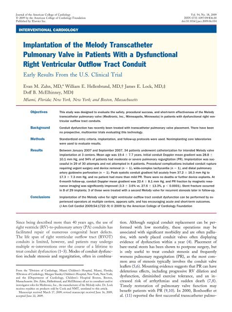
1726 Zahn et al. JACC Vol. 54, No. 18, 2009Early Results From <strong>the</strong> Transca<strong>the</strong>ter <strong>Pulmonary</strong> <strong>Valve</strong> Trial October 27, 2009:1722–9Figure 1New York Heart Association Functional Classification as a Function<strong>of</strong> Time for Patients Receiving a <strong>Melody</strong> <strong>Valve</strong> (Medtronic, Inc.) (n 29)and was no higher than 11.6% in any patient. No significantchanges were noted in RV or left ventricular ejectionfraction or left ventricular end-diastolic volume.CPET. Twenty-nine patients had paired CPET data frombefore and 6 months after implantation. There were nodifferences between pre-implantation and 6-month follow-uppeak VO 2 (21.6 6.8 ml/kg/min vs. 22.7 7.1 ml/kg/min,p 1.0) or anaerobic threshold (15.0 4.7 ml/kg/min vs.15.5 5.6 ml/kg/min, p 1.0).CT PULMONARY ANGIOGRAPHY. Twenty-nine patients hadpaired CT pulmonary angiography before and 6 monthsafter implantation, and no new findings <strong>of</strong> pulmonaryemboli were discovered. One patient had findings <strong>of</strong> chronicpulmonary emboli in <strong>the</strong> right upper lobe noted on <strong>the</strong>pre-implantation CT, which remained unchanged at6-month follow-up.RADIOGRAPHY/CINEFLUOROSCOPY. Stent fractures wereobserved in 8 <strong>of</strong> 29 patients (28%) during follow-up, 1 at 3months and <strong>the</strong> o<strong>the</strong>rs at 6 months. When diagnosed, all 8patients were NYHA functional class I, and all but 1 hadless than trace PR and a conduit Doppler mean gradient30 mm Hg. Although most <strong>of</strong> <strong>the</strong>se fractures were minor(i.e., a single wire break with no loss <strong>of</strong> stent integrity), 3patients had a second <strong>Melody</strong> valve implanted 11, 12, and14 months after <strong>the</strong> initial implantation to treat progressiveconduit obstruction associated with multiple fracture points,loss <strong>of</strong> stent integrity, and progression to NYHA functionalclass II. <strong>Implantation</strong> <strong>of</strong> a second <strong>Melody</strong> valve was uncomplicatedand preceded by placement <strong>of</strong> bare-metal stainlesssteel stents in all 3 cases. Hemodynamic results after repeat<strong>Melody</strong> valve implantation were similar to initial implantation.The o<strong>the</strong>r 5 patients with minor stent fracturesremained stable in NYHA functional class I without evidence<strong>of</strong> a loss <strong>of</strong> stent integrity or progressive RVOTobstruction. Freedom from re-intervention for stent fractureassociated with recurrent RVOT obstruction was 96.6% at 1year (Fig. 2).Safety. Aside from <strong>the</strong> procedural adverse events, stentfractures, and re-interventions summarized above, no o<strong>the</strong>rCardiac 6 Months Magnetic After Cardiac <strong>Melody</strong> Resonance Magnetic <strong>Valve</strong> (Medtronic, Imaging Resonance Results Inc.) Imaging Before <strong>Implantation</strong> Results and Before (n 25) andTable 66 Months After <strong>Melody</strong> <strong>Valve</strong> (Medtronic, Inc.) <strong>Implantation</strong> (n 25)Variable Pre-<strong>Implantation</strong> Post-<strong>Implantation</strong> p ValuePR fraction, % 27.6 13.3 (29.8; 4.3–61.0) 3.3 3.6 (3.1; 0.0 - 11.6) 0.001RV EDV index, ml/m 2 144.7 54.8 (133.5; 72.5–290.8) 117.3 48.3 (109.5; 54.3–256.1) 0.001RV ejection fraction, % 40.8 14.6 (39.8; 10.5–72.7) 42.0 12.1 (39.8; 23.8–63.8) 0.86LV EDV index, ml/m 2 84.5 18.9 (85.7; 43.9–121.4) 90.7 20.4 (90.8; 58.5–132.9) 0.30LV ejection fraction, % 52.6 10.5 (54.3; 31.3–68.8) 52.4 8.6 (52.1; 33.3–68.5) 1.0Data are presented as mean SD (median; minimum–maximum).EDV end-diastolic volume; LV left ventricle; PR pulmonary regurgitation; RV right ventricle.
JACC Vol. 54, No. 18, 2009October 27, 2009:1722–9Zahn et al.Early Results From <strong>the</strong> Transca<strong>the</strong>ter <strong>Pulmonary</strong> <strong>Valve</strong> Trial1727Figure 2adverse events were reported. With respect to <strong>the</strong> safetyoutcome criteria, 100% <strong>of</strong> patients were free fromprocedure- or device-related death, and 91% were free fromserious procedure- or device-related events.DiscussionKaplan-Meier Curve DepictingFreedom From Re-Intervention forStent Fracture and Associated RVOT ObstructionError bars shown are at <strong>the</strong> 95%confidence intervals. RVOT right ventricular outflow tract.To date, <strong>the</strong> only published series <strong>of</strong> <strong>Melody</strong> valveimplantation have been from <strong>the</strong> Bonhoeffer group anddemonstrate excellent short- and intermediate-term results(11,12). This study confirms that equally good acute andshort-term results can be achieved by trained, experiencedoperators at multiple o<strong>the</strong>r centers. Fur<strong>the</strong>rmore, it is <strong>the</strong>first prospective multicenter clinical trial <strong>of</strong> <strong>the</strong> <strong>Melody</strong>valve that uses defined entry criteria, standardized implantationand follow-up protocols, independent core laboratoriesfor assessment <strong>of</strong> outcomes, and cinefluoroscopy andCT pulmonary angiography to evaluate for stent fractureand pulmonary embolism.Procedural success and short-term effectiveness. Theprocedural success rate in this series was high (29 <strong>of</strong> 30attempts) and compared favorably with results published byexperienced operators (12,15). The current system designmakes <strong>Melody</strong> valve implantation simple and comparabletechnically to bare-metal stent implantation, a commonintervention performed in most congenital ca<strong>the</strong>terizationlaboratories. There were no device malpositions or embolizationsin this series. The current delivery system, designedspecifically for this application, protects <strong>the</strong> stent andpreserves <strong>the</strong> balloon–stent relationship during advancementto <strong>the</strong> RVOT and may <strong>of</strong>fer some advantages overconventional methods <strong>of</strong> stent implantation. The averagehospital stay <strong>of</strong> just over 1 day in this study comparesfavorably with both surgical conduit replacement and baremetalstenting.In <strong>the</strong> current study, a minimum sizing balloon waist<strong>of</strong> 14 mm was required to undergo valve implantation.Using this approach, only 1 patient had a postimplantationRV-PA gradient that was considered inadequateby pre-determined criteria. Pre-dilation <strong>of</strong> <strong>the</strong>conduit before sizing was a required component <strong>of</strong> <strong>the</strong>protocol for conduits with an angiographic diameter 18mm, but pre-stenting was not allowed in this initialcohort (<strong>the</strong> protocol has since been modified to allowconcomitant procedures, including pre-stenting <strong>of</strong> <strong>the</strong> conduit).The rationale for pre-dilation was to optimize conduitdiameter before balloon sizing and <strong>Melody</strong> valve implantationand to assess compliance <strong>of</strong> <strong>the</strong> conduit and identify multipleor highly resistant stenoses. As Sugiyama et al. (16) confirmedin <strong>the</strong>ir series <strong>of</strong> bare-metal stenting for conduit obstruction,<strong>the</strong> smaller <strong>the</strong> achieved diameter <strong>of</strong> <strong>the</strong> conduit relative to <strong>the</strong>normal pulmonary valve size for <strong>the</strong> patient, <strong>the</strong> higher <strong>the</strong>post-intervention RVOT gradient. This may be more importantwith an implantable valve, in which <strong>the</strong> valve andvein segment add bulk to <strong>the</strong> implant site and may contributeto luminal narrowing and obstruction if <strong>the</strong> stent isunder-expanded.In <strong>the</strong> patient with a post-implantation peak RVOTgradient <strong>of</strong> 37 mm Hg, <strong>the</strong> obstruction was below <strong>the</strong>conduit and unrelated to <strong>the</strong> <strong>Melody</strong> valve itself. Four o<strong>the</strong>rpatients developed Doppler mean gradients 30 mm Hg by6-month follow-up. These outcomes highlight <strong>the</strong> importance<strong>of</strong> appropriate patient selection for <strong>the</strong> <strong>Melody</strong> valve.As more data become available, it will be important toevaluate patient-related and procedural data associated wi<strong>the</strong>arly recurrence <strong>of</strong> conduit obstruction to improve patientselection and execution <strong>of</strong> <strong>the</strong> procedure. It will also beimportant to determine <strong>the</strong> role <strong>of</strong> pre-dilation and/orpre-stenting in preventing recurrent RVOT obstruction.Consistent with prior reports, function <strong>of</strong> <strong>the</strong> <strong>Melody</strong>valve was excellent in short-term follow-up, with no ortrivial PR in almost all patients, and none with more thanmild regurgitation (11,12,15). RVOT gradients were significantlylower at 6 months than before implantation, andMRI demonstrated favorable RV volume remodeling. Amajority <strong>of</strong> patients improved in NYHA functional class,and none declined, consistent with prior reports (12).Although symptomatic improvement was typically noted 1month after implantation, some patients continued to improveas late as 6 months after implantation, raising <strong>the</strong>possibility that cardiac remodeling and conditioning maycontinue beyond <strong>the</strong> acute restoration <strong>of</strong> pulmonary valvecompetence (21). In <strong>the</strong> current series, we did not observesignificant improvements in maximal VO 2 and anaerobicthreshold, although <strong>the</strong> preponderance <strong>of</strong> patients with PRas <strong>the</strong> primary indication for valve placement may be afactor in this finding. Coats et al. (22) reported that patientswith PR have minimal improvement in maximal VO 2 andanaerobic threshold after <strong>Melody</strong> valve implantation, in
1728 Zahn et al. JACC Vol. 54, No. 18, 2009Early Results From <strong>the</strong> Transca<strong>the</strong>ter <strong>Pulmonary</strong> <strong>Valve</strong> Trial October 27, 2009:1722–9contrast to patients who receive a <strong>Melody</strong> valve primarily forstenosis (22). Fur<strong>the</strong>r examination <strong>of</strong> <strong>the</strong> physiologic mechanismsunderlying changes in exercise performance andsymptomatic improvement is warranted.Stent fracture. Stent fracture, well described after bothbare-metal stenting <strong>of</strong> RVOT conduits and <strong>Melody</strong> valveimplantation, was an important finding in this study(19,23). Peng et al. (19) described 221 patients treated forconduit obstruction with bare-metal stenting and found a43% incidence <strong>of</strong> stent fracture, with external compressionand a substernal conduit location identified as importantrisk factors. In a series <strong>of</strong> 123 patients who underwent<strong>Melody</strong> valve placement, Nordmeyer et al. (23) reported a75% freedom from <strong>Melody</strong> stent fracture at 2 years, withimplantation in <strong>the</strong> native RVOT, no calcification along <strong>the</strong>RVOT, and greater recoil after balloon deflation associatedwith a higher risk <strong>of</strong> fracture. It is logical to postulate from<strong>the</strong>se 2 studies that when excessive external loading forcesare applied to stents in <strong>the</strong> RVOT, ei<strong>the</strong>r from dynamiccompression or o<strong>the</strong>r cyclic stresses, <strong>the</strong>re is an importantrisk <strong>of</strong> stent fracture. The current series includes too fewpatients to provide robust insight into this issue, but <strong>the</strong>frequency <strong>of</strong> stent fracture appears consistent with <strong>the</strong>seearlier reports. Nordmeyer et al. (24) recently reported aseries <strong>of</strong> patients who underwent successful treatment <strong>of</strong>re-stenosis after <strong>Melody</strong> stent fracture, with bare-metalstenting followed by implantation <strong>of</strong> a second <strong>Melody</strong> valve.The same approach was used in 3 patients in this series,with good acute results. Fur<strong>the</strong>r attention is needed tobetter understand <strong>the</strong> risk factors for, prevention <strong>of</strong>, andtreatment <strong>of</strong> stent fracture in patients undergoing transca<strong>the</strong>terpulmonary valve placement.Safety. The procedural adverse event rate <strong>of</strong> 9% in thiscohort is acceptable for a new procedure <strong>of</strong> this magnitudeand compares favorably with previously published reportsinvolving ca<strong>the</strong>ter or surgical intervention for conduit dysfunction(1–3,16–20). The large pr<strong>of</strong>ile <strong>of</strong> <strong>the</strong> deliverysystem did not result in any obvious vascular damage orprohibit delivery <strong>of</strong> <strong>the</strong> device in any patient, includingchildren as young as 10 years. Importantly, none <strong>of</strong> <strong>the</strong>adverse events in this study resulted in death or long-termsequelae. Aside from stent fractures and re-interventions, asdiscussed above, <strong>the</strong>re were no device- or procedure-relatedadverse events reported during follow-up.Protocol. There are challenges to developing a standardized,rigorous protocol for percutaneous pulmonary valveplacement in individuals with dysfunctional RVOT conduitsand a spectrum <strong>of</strong> hemodynamic disease that includesvariable degrees <strong>of</strong> PR and obstruction. There are limiteddata from which to define rigorous, outcome-based indicationsfor valve implantation. Moreover, <strong>the</strong> indications forimplantation and metrics <strong>of</strong> success will necessarily differ forpatients at different points on this disease spectrum, and<strong>the</strong>re is no simple means <strong>of</strong> capturing <strong>the</strong> complexities <strong>of</strong>mixed disease. The inclusion criteria for this protocol weredesigned to reflect general practice on <strong>the</strong> basis <strong>of</strong> <strong>the</strong>published literature and <strong>the</strong> experience <strong>of</strong> <strong>the</strong> participatingcenters and were graduated according to symptomatic status.To address <strong>the</strong> question <strong>of</strong> clinical equipoise, 2 independentphysicians, 1 congenital cardiac surgeon and 1pediatric cardiologist, reviewed prospective patients andprovided written support for <strong>the</strong> decision to implant a<strong>Melody</strong> valve. Never<strong>the</strong>less, individuals varied within <strong>the</strong>cohort, and our population may have differed in compositionfrom that <strong>of</strong> previously reported series (12,21). Echocardiographicassessment <strong>of</strong> hemodynamic entry criteriamay be considered a limitation <strong>of</strong> <strong>the</strong> study protocol and waschosen for logistical reasons. The ca<strong>the</strong>terization protocolused in this study was successful in identifying 2 patients atrisk for coronary compression, a potentially fatal complication.Pre-implantation balloon inflation with simultaneouscoronary angiography should be used in any case <strong>of</strong> conduitstenting or transca<strong>the</strong>ter valve implantation when coronaryartery compression is suspected. Fur<strong>the</strong>r, <strong>the</strong> protocol specifiedcinefluoroscopy at <strong>the</strong> 6-month follow-up visit toassess for stent fracture. The decision to use cinefluoroscopywas based on its anticipated high sensitivity for detectingeven minor stent fractures.ConclusionsThis is <strong>the</strong> first multicenter prospective trial <strong>of</strong> an implantabletransca<strong>the</strong>ter pulmonary valve in a population <strong>of</strong>patients with congenital heart disease and RVOT conduitdysfunction. The implantation success rate was high, <strong>the</strong>frequency <strong>of</strong> adverse events was acceptable, and short-termoutcomes were encouraging, supporting <strong>the</strong> conclusion thatthis technology can be translated effectively to experienced,properly trained interventional cardiologists and is nothighly operator dependent. Longer follow-up and a largerpatient experience are needed to determine <strong>the</strong> ultimate role<strong>of</strong> this <strong>the</strong>rapy in <strong>the</strong> treatment <strong>of</strong> conduit dysfunction.Reprint requests and correspondence: Dr. D<strong>of</strong>f B. McElhinney,Department <strong>of</strong> Cardiology, Children’s Hospital, 300 LongwoodAvenue, Boston, Massachusetts 02115. E-mail: d<strong>of</strong>f.mcelhinney@cardio.chboston.org.REFERENCES1. Stark J, Bull C, Stajevic M, Jothi M, Elliott M, de Leval M. Fate <strong>of</strong>subpulmonary homograft conduits: determinants <strong>of</strong> late homograftfailure. J Thorac Cardiovasc Surg 1998;115:506–16.2. Bielefeld M, Bishop D, Campbell D, Mitchell M, Grover F, ClarkeD. Reoperative homograft right ventricular outflow tract reconstruction.Ann Thorac Surg 2001;71:482–7.3. Gerestein C, Takkenberg J, Oei F, et al. Right ventricular outflow tractreconstruction with an allograft conduit. Ann Thorac Surg 2001;71:911–7.4. Wells WJ, Arroyo H Jr., Bremner RM, Wood J, Starnes VA.Homograft conduit failure in infants is not due to somatic outgrowth.J Thorac Cardiovasc Surg 2002;124:88–96.5. Powell A, Lock J, Keane J, Perry S. Prolongation <strong>of</strong> RV-PA conduitlife span by percutaneous stent implantation: intermediate long-termresults. Circulation 1995;92:3282–8.6. Ovaert C, Caldarone CA, McCrindle BW, et al. Endovascular stentimplantation for <strong>the</strong> management <strong>of</strong> postoperative right ventricular
JACC Vol. 54, No. 18, 2009October 27, 2009:1722–9Zahn et al.Early Results From <strong>the</strong> Transca<strong>the</strong>ter <strong>Pulmonary</strong> <strong>Valve</strong> Trial1729outflow tract obstruction: clinical efficacy. J Thorac Cardiovasc Surg1999;118:886–93.7. Carvalho JS, Shinebourne EA, Busst C, Rigby ML, Redington AN.Exercise capacity after complete repair <strong>of</strong> tetralogy <strong>of</strong> Fallot: deleteriouseffects <strong>of</strong> residual pulmonary regurgitation. Br Heart J 1992;67:470–3.8. Gatzoulis MA, Balaji S, Webber SA, et al. Risk factors for arrhythmiaand sudden cardiac death late after repair <strong>of</strong> tetralogy <strong>of</strong> Fallot: amulticentre study. Lancet 2000;356:975–81.9. Therrien J, Siu S, Harris L, et al. Impact <strong>of</strong> pulmonary valvereplacement on arrhythmia propensity late after repair <strong>of</strong> tetralogy <strong>of</strong>Fallot. Circulation 2001;103:2489–94.10. Eyskens B, Reybrouck T, Bogaert J, et al. Homograft insertion forpulmonary regurgitation after repair <strong>of</strong> tetralogy <strong>of</strong> Fallot improvescardiorespiratory exercise performance. Am J Cardiol 2000;85:221–5.11. Bonhoeffer P, Boudjemline Y, Saliba Z, et al. Percutaneous replacement<strong>of</strong> pulmonary valve in a right-ventricle to pulmonary-arterypros<strong>the</strong>tic conduit with valve dysfunction. Lancet 2000;356:1403–5.12. Khambadkone S, Coats L, Taylor AM, et al. Transca<strong>the</strong>ter pulmonaryvalve implantation in humans: initial results in 59 consecutive patients.Circulation 2005;112:1189–97.13. Hochberg Y. A sharper Bonferroni procedure for multiple tests <strong>of</strong>significance. Biometrics 1988;75:800–2.14. Wright P. Adjusted p-values for simultaneous inference. Biometrics1992;48:1005–13.15. Almeida CL, Chaturvedi R, Lee KJ, et al. Percutaneous pulmonaryvalve implantation: <strong>the</strong> first North American trial. Circulation 2006;114 Suppl S:389.16. Sugiyama H, Williams W, Benson LN. <strong>Implantation</strong> <strong>of</strong> endovascularstents for <strong>the</strong> obstructive right ventricular outflow tract. Heart 2005;91:1058–63.17. Almagor Y, Prevosti LG, Bartorelli AL, et al. Balloon expandablestent implantation in stenotic right heart valved conduits. J Am CollCardiol 1990;16:1310–4.18. Hosking MC, Benson LN, Nakanishi T, et al. Intravascular stentpros<strong>the</strong>sis for right ventricular outflow obstruction. J Am Coll Cardiol1992;20:373–80.19. Peng L, McElhinney D, Nugent A, et al. Endovascular stenting <strong>of</strong>obstructed right ventricle-to-pulmonary artery conduits: a fifteen-yearexperience. Circulation 2006;113:2598–605.20. Kostolny M, Tsang V, Nordmeyer J, et al. Rescue surgery followingpercutaneous pulmonary valve implantation. Eur J Cardiovasc Surg2007;134:258–9.21. Odendaal D, Khambadkone S, Coats L, et al. Maintained improvementin maximal exercise and continued improvement in sub-maximalexercise indices are seen during follow-up after percutaneous pulmonaryvalve insertion. Circulation 2006;114 Suppl S:558.22. Coats L, Khambadkone S, Derrick G, et al. Physiological consequences<strong>of</strong> percutaneous pulmonary valve implantation: <strong>the</strong> differentbehaviour <strong>of</strong> volume- and pressure-overloaded ventricles. Eur Heart J2007;28:1886–93.23. Nordmeyer J, Khambadkone S, Coats L, et al. Risk stratification,systematic classification, and anticipatory management strategies forstent fracture after percutaneous pulmonary valve implantation. Circulation2007;115;1392–7.24. Nordmeyer J, Coats L, Lurz P, et al. Percutaneous pulmonaryvalve-in-valve implantation: a successful treatment concept for earlydevice failure. Eur Heart J 2008;20:810–5.25. Schievano S, Petrini L, Migliavacca F, et al. Finite element analysis <strong>of</strong>stent deployment: understanding stent fracture in percutaneous pulmonaryvalve implantation. J Interv Cardiol 2007;20:546–54.Key Words: intervention y pulmonary valve y tetralogy <strong>of</strong> Fallot.