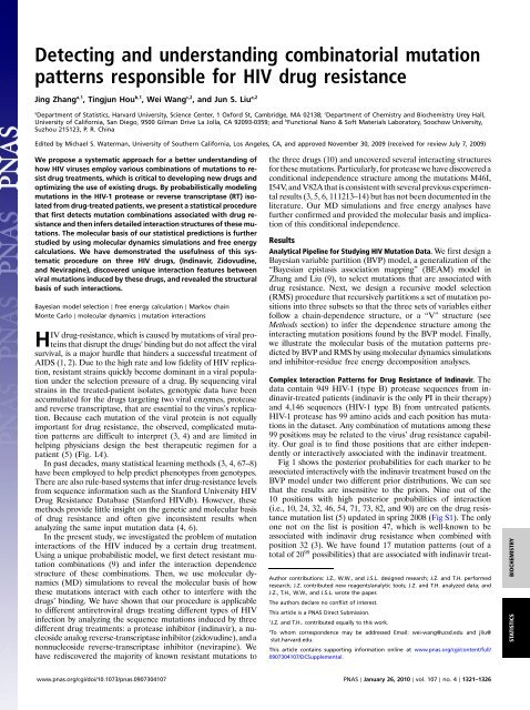
Detecting and understanding combinatorial mutation patterns ...
Detecting and understanding combinatorial mutation patterns ...
Detecting and understanding combinatorial mutation patterns ...

Create successful ePaper yourself
Turn your PDF publications into a flip-book with our unique Google optimized e-Paper software.
<strong>Detecting</strong> <strong>and</strong> underst<strong>and</strong>ing <strong>combinatorial</strong> <strong>mutation</strong><strong>patterns</strong> responsible for HIV drug resistanceJing Zhang a,1 , Tingjun Hou b,1 , Wei Wang c,2 , <strong>and</strong> Jun S. Liu a,2aDepartment of Statistics, Harvard University, Science Center, 1 Oxford St, Cambridge, MA 02138; c Department of Chemistry <strong>and</strong> Biochemistry Urey Hall,University of California, San Diego, 9500 Gilman Drive La Jolla, CA 92093-0359; <strong>and</strong> b Functional Nano & Soft Materials Laboratory, Soochow University,Suzhou 215123, P. R. ChinaEdited by Michael S. Waterman, University of Southern California, Los Angeles, CA, <strong>and</strong> approved November 30, 2009 (received for review July 7, 2009)We propose a systematic approach for a better underst<strong>and</strong>ing ofhow HIV viruses employ various combinations of <strong>mutation</strong>s to resistdrug treatments, which is critical to developing new drugs <strong>and</strong>optimizing the use of existing drugs. By probabilistically modeling<strong>mutation</strong>s in the HIV-1 protease or reverse transcriptase (RT) isolatedfrom drug-treated patients, we present a statistical procedurethat first detects <strong>mutation</strong> combinations associated with drug resistance<strong>and</strong> then infers detailed interaction structures of these <strong>mutation</strong>s.The molecular basis of our statistical predictions is furtherstudied by using molecular dynamics simulations <strong>and</strong> free energycalculations. We have demonstrated the usefulness of this systematicprocedure on three HIV drugs, (Indinavir, Zidovudine,<strong>and</strong> Nevirapine), discovered unique interaction features betweenviral <strong>mutation</strong>s induced by these drugs, <strong>and</strong> revealed the structuralbasis of such interactions.Bayesian model selection ∣ free energy calculation ∣ Markov chainMonte Carlo ∣ molecular dynamics ∣ <strong>mutation</strong> interactionsHIV drug-resistance, which is caused by <strong>mutation</strong>s of viral proteinsthat disrupt the drugs’ binding but do not affect the viralsurvival, is a major hurdle that hinders a successful treatment ofAIDS (1, 2). Due to the high rate <strong>and</strong> low fidelity of HIV replication,resistant strains quickly become dominant in a viral populationunder the selection pressure of a drug. By sequencing viralstrains in the treated-patient isolates, genotypic data have beenaccumulated for the drugs targeting two viral enzymes, protease<strong>and</strong> reverse transcriptase, that are essential to the virus’s replication.Because each <strong>mutation</strong> of the viral protein is not equallyimportant for drug resistance, the observed, complicated <strong>mutation</strong><strong>patterns</strong> are difficult to interpret (3, 4) <strong>and</strong> are limited inhelping physicians design the best therapeutic regimen for apatient (5) (Fig. 1A).In past decades, many statistical learning methods (3, 4, 67–8)have been employed to help predict phenotypes from genotypes.There are also rule-based systems that infer drug-resistance levelsfrom sequence information such as the Stanford University HIVDrug Resistance Database (Stanford HIVdb). However, thesemethods provide little insight on the genetic <strong>and</strong> molecular basisof drug resistance <strong>and</strong> often give inconsistent results whenanalyzing the same input <strong>mutation</strong> data (4, 6).In the present study, we investigated the problem of <strong>mutation</strong>interactions of the HIV induced by a certain drug treatment.Using a unique probabilistic model, we first detect resistant <strong>mutation</strong>combinations (9) <strong>and</strong> infer the interaction dependencestructure of these combinations. Then, we use molecular dynamics(MD) simulations to reveal the molecular basis of howthese <strong>mutation</strong>s interact with each other to interfere with thedrugs’ binding. We have shown that our procedure is applicableto different antiretroviral drugs treating different types of HIVinfection by analyzing the sequence <strong>mutation</strong>s induced by threedifferent drug treatments: a protease inhibitor (indinavir), a nucleosideanalog reverse-transcriptase inhibitor (zidovudine), <strong>and</strong> anonnucleoside reverse-transcriptase inhibitor (nevirapine). Wehave rediscovered the majority of known resistant <strong>mutation</strong>s tothe three drugs (10) <strong>and</strong> uncovered several interacting structuresfor these <strong>mutation</strong>s. Particularly, for protease we have discovered aconditional independence structure among the <strong>mutation</strong>s M46I,I54V, <strong>and</strong> V82A that is consistent with several previous experimentalresults (3, 5, 6, 111213–14) but has not been documented in theliterature. Our MD simulations <strong>and</strong> free energy analyses havefurther confirmed <strong>and</strong> provided the molecular basis <strong>and</strong> implicationof this conditional independence.ResultsAnalytical Pipeline for Studying HIV Mutation Data. We first design aBayesian variable partition (BVP) model, a generalization of the“Bayesian epistasis association mapping” (BEAM) model inZhang <strong>and</strong> Liu (9), to select <strong>mutation</strong>s that are associated withdrug resistance. Next, we design a recursive model selection(RMS) procedure that recursively partitions a set of <strong>mutation</strong> positionsinto three subsets so that the three sets of variables eitherfollow a chain-dependence structure, or a “V” structure (seeMethods section) to infer the dependence structure among theinteracting <strong>mutation</strong> positions found by the BVP model. Finally,we illustrate the molecular basis of the <strong>mutation</strong> <strong>patterns</strong> predictedby BVP <strong>and</strong> RMS by using molecular dynamics simulations<strong>and</strong> inhibitor-residue free energy decomposition analyses.Complex Interaction Patterns for Drug Resistance of Indinavir. Thedata contain 949 HIV-1 (type B) protease sequences from indinavir-treatedpatients (indinavir is the only PI in their therapy)<strong>and</strong> 4,146 sequences (HIV-1 type B) from untreated patients.HIV-1 protease has 99 amino acids <strong>and</strong> each position has <strong>mutation</strong>sin the dataset. Any combination of <strong>mutation</strong>s among these99 positions may be related to the virus’ drug resistance capability.Our goal is to find those positions that are either independentlyor interactively associated with the indinavir treatment.Fig 1 shows the posterior probabilities for each marker to beassociated interactively with the indinavir treatment based on theBVP model under two different prior distributions. We can seethat the results are insensitive to the priors. Nine out of the10 positions with high posterior probabilities of interaction(i.e., 10, 24, 32, 46, 54, 71, 73, 82, <strong>and</strong> 90) are on the drug resistance<strong>mutation</strong> list (5) updated in spring 2008 (Fig S1). The onlyone not on the list is position 47, which is well-known to beassociated with indinavir drug resistance when combined withposition 32 (3). We have found 17 <strong>mutation</strong> <strong>patterns</strong> (out of atotal of 20 99 possibilities) that are associated with indinavir treat-Author contributions: J.Z., W.W., <strong>and</strong> J.S.L. designed research; J.Z. <strong>and</strong> T.H. performedresearch; J.Z. contributed new reagents/analytic tools; J.Z. <strong>and</strong> T.H. analyzed data; <strong>and</strong>J.Z., T.H., W.W., <strong>and</strong> J.S.L. wrote the paper.The authors declare no conflict of interest.This article is a PNAS Direct Submission.1J.Z. <strong>and</strong> T.H.. contributed equally to this work.2To whom correspondence may be addressed Email: wei-wang@ucsd.edu <strong>and</strong> jliu@stat.harvard.edu.This article contains supporting information online at www.pnas.org/cgi/content/full/0907304107/DCSupplemental.STATISTICS BIOCHEMISTRYwww.pnas.org/cgi/doi/10.1073/pnas.0907304107 PNAS ∣ January 26, 2010 ∣ vol. 107 ∣ no. 4 ∣ 1321–1326
Fig. 1. The posterior probabilities for each <strong>mutation</strong> to be associated interactivelywith indinavir treatment. The Upper shows the posterior probabilitiesusing prior one, which assumes that it is equally likely (1∕3) for a<strong>mutation</strong> to be unassociated, individually associated, <strong>and</strong> interactively associatedwith the drug treatment. The Lower shows the posterior probabilitiesusing a more stringent prior (prior two) assuming that only two makers areexpected to be associated with the drug, either individually or interactively.ment with a posterior probability >0.0001 (this cutoff is muchhigher than the equally likely probability 1∕20 99 ). Table S1 inSI Text tabulates these <strong>patterns</strong> <strong>and</strong> their respective posteriorprobabilities. Phenotypic data from Stanford HIVdb providesconfirming evidence for the configurations of the top interactionpattern f24; 32; 46; 54; 82g (SI Text). Many of these <strong>mutation</strong>s arewell-known for their drug resistance effects. For example, it isknown that the <strong>mutation</strong>s of V82A\F\T or L90M are necessarybut not sufficient for measurable resistance to indinavir (11).Dependence Structure of Interaction for Drug Resistance of Indinavir.We applied the RMS procedure to infer the detailed dependencestructure among the interacting positions 10, 24, 32, 46, 47, 54, 71,73, 82, <strong>and</strong> 90 (Fig. 2). Two marginally independent interactiongroups were found with high confidence: one is composed mainlyof 46, 54, <strong>and</strong> 82; <strong>and</strong> the other of 73 <strong>and</strong> 90 (more details aregiven in SI Text). Interestingly, we found a strong conditional independencestructure in group f46; 52; 82g. Given the amino acidat position 82, <strong>mutation</strong>s at 46 <strong>and</strong> 54 are mutually independent.The data did not provide strong enough information regardingthe structures for other variables (<strong>mutation</strong>s) in this group, forexample, 24, 32, <strong>and</strong> 47. For the second group, 73 <strong>and</strong> 90 stronglyinteract with each other.Fig. 2. Detection of a detailed <strong>mutation</strong> interaction structure for resistingindinavir. Positions 46 <strong>and</strong> 54 are conditionally independent given position82, denoted as 42⊥54j82. The ? indicates where we are not able to confidentlyinfer the dependence structure (SI Text).In the study of Zhang et al. (13), a rebound in virus levels inplasma following the initial sharp decline at the beginning ofindinavir therapy was found to be associated with a sequentialacquisition of <strong>mutation</strong>s at the protease positions of46 → 82 → 54. We further searched the Stanford HIVdb, <strong>and</strong>found that 112 patients from the treated group (HIV-1, maingroup, <strong>and</strong> subtype B) had indinavir as their only PI in theirtherapy <strong>and</strong> also have detailed <strong>mutation</strong> records (more thanone complete protease sequences) during the course of therapy.Among them 53.6% (60 patients) have at least one of the <strong>mutation</strong>sat positions 46, 54, <strong>and</strong> 82. We observed no patient with thesingle <strong>mutation</strong> V82I, with the single <strong>mutation</strong> at 54, or with thedouble <strong>mutation</strong>s at 46 <strong>and</strong> 54. Among the 21 patients who haveall three <strong>mutation</strong>s, only six of them have detailed <strong>mutation</strong> recordssuch that we can tell the exact order of sequential acquisitionof these three <strong>mutation</strong>s. Four out of the six have the order46 → 82 → 54, one has the order 82 → 46 → 54, <strong>and</strong> another hasthe order 82 → 54 → 46. Whereas all these observed orders areconsistent with our inferred conditional independence structure,the non-observed orders, 46 → 54 → 82 <strong>and</strong> 54 → 46 → 82, arenot. This suggests that the conditional independence is a directconsequence of sequential acquisition of the three <strong>mutation</strong>s.Molecular Basis of Interacting Mutations Revealed by MD Simulations<strong>and</strong> Free Energy Calculations. To further investigate the molecularimplication of the <strong>mutation</strong> interactions within the f46; 54; 82ggroup, <strong>and</strong> within the f73; 90g group, we conducted MD simulationsto analyze the binding free energies of the protease/indinavir complexes (SI Text). The free energy decompositionanalyses for the wild-type <strong>and</strong> ten mutant proteases (Table 1) showthat the drug resistant <strong>mutation</strong>s primarily affect the van der Waalsinteractions between indinavir <strong>and</strong> the protease. Most of the<strong>mutation</strong>s in the f46; 54; 82g group show positive relative bindingfree energies, that is, decrease of indinavir’s binding affinity.Among the three single <strong>mutation</strong>s, two of them (M46I <strong>and</strong>V82A) substantially increase the indinavir binding free energies(−77.30 0.45 <strong>and</strong> −75.67 1.50), whereas I54V does not impairthe binding. This result is consistent with our observationin the Stanford HIVdb. Among the 112 patients who have morethan one <strong>mutation</strong> record during their indinavir therapy, 10 havethe single <strong>mutation</strong> at 46, 10 have the single <strong>mutation</strong> at 82, <strong>and</strong>zero have the single <strong>mutation</strong> at 54.Among the double <strong>mutation</strong>s, M46I/V82A <strong>and</strong> I54V/V82Aseverely impair the binding of indinavir whereas M46I/I54V doesnot significantly weaken the binding of indinavir. Incidentally,among the 112 patients in the Stanford HIVdb, 11 have double<strong>mutation</strong>s at positions 46 <strong>and</strong> 82, eight at positions 54 <strong>and</strong> 82, <strong>and</strong>zero at positions 46 <strong>and</strong> 54. It appears that 46 <strong>and</strong> 54 cannot interactto resist to indinavir without the <strong>mutation</strong> at 82. The observationsthat double <strong>mutation</strong>s M46I/V82A <strong>and</strong> I54V/V82Aare the two strongest resistant mutants may have important implicationsfor improving the potency of indinavir to combat resistance.If we can decrease the interaction between V82 <strong>and</strong> aderivative of indinavir without affecting the total binding affinityof the inhibitor, the resistant effects of 46 <strong>and</strong> 54 will be reduced,as well. This example highlights the usefulness of our approach foruncovering the interaction structure between <strong>mutation</strong>s in developingpotent drugs.The triple <strong>mutation</strong> M46I/I54V/V82A impairs the binding ofindinavir. As mentioned before, these three <strong>mutation</strong>s occur sequentiallyin specific orders. Because single <strong>mutation</strong> at 54 isnot able to resist indinavir, the first <strong>mutation</strong> has to be at either46 or 82 so that the mutant virus can have a better chance to survivethe attack of indinavir. If the first <strong>mutation</strong> occurs at 46, thesecond <strong>mutation</strong> has to be at 82 because the double <strong>mutation</strong>s at46 <strong>and</strong> 54 cannot resist to indinavir, as well. If the first <strong>mutation</strong> isat 82, the subsequent <strong>mutation</strong> can be at either 46 or 54. We observedexactly these (<strong>and</strong> only these) three possible orders1322 ∣ www.pnas.org/cgi/doi/10.1073/pnas.0907304107 Zhang et al.
Table 1. The binding free energies <strong>and</strong> the energy components calculated by MM/GBSA (kcal∕mol)No. ΔE vdw ΔE ele ΔG GB ΔG SA ΔE ele þ ΔG GB ΔE vdw þ ΔG SA ΔG cal ΔΔG cal *WT −80.00 ± 0.16 † −25.59 ± 0.35 33.97 ± 0.35 −9.87 ± 0.06 8.37 ± 0.00 −89.87 ± 0.22 −81.50 ± 0.46 0.00Group 1M46I −77.43 ± 1.85 −24.01 ± 0.39 34.46 ± 0.37 −10.32 ± 0.64 10.44 ± 0.76 −87.74 ± 1.21 −77.30 ± 0.45 4.20I54V −82.99 ± 0.26 −25.38 ± 0.21 36.13 ± 0.16 −9.25 ± 0.57 10.74 ± 0.37 −92.24 ± 0.83 −81.50 ± 0.46 0.00V82A −75.98 ± 1.27 −24.67 ± 0.10 34.54 ± 0.13 −9.56 ± 0.00 9.87 ± 0.23 −85.54 ± 1.27 −75.67 ± 1.50 5.83M46I/I54V −83.26 ± 1.05 −22.01 ± 1.23 34.19 ± 1.44 −9.98 ± 0.06 12.18 ± 0.21 −93.24 ± 1.11 −81.06 ± 0.90 0.44M46I/V82A −70.84 ± 2.86 −24.92 ± 1.87 33.96 ± 1.40 −9.67 ± 1.43 9.04 ± 0.47 −80.52 ± 4.29 −71.48 ± 4.76 10.02I54V/82A −71.84 ± 0.64 −19.62 ± 0.92 31.08 ± 1.06 −9.82 ± 0.01 11.46 ± 0.13 −81.66 ± 0.63 −70.20 ± 0.77 11.30M46I/I54V/ −79.58 ± 0.91 −19.09 ± 0.17 29.89 ± 0.41 −8.92 ± 0.59 10.79 ± 0.24 −88.50 ± 0.33 −77.71 ± 0.08 3.79V82AGroup 2G73S −78.79 ± 0.50 −24.76 ± 0.51 33.26 ± 0.14 −10.36 ± 0.67 8.50 ± 0.37 −89.15 ± 0.17 −80.65 ± 0.54 0.85L90M −80.56 ± 0.32 −27.13 ± 0.33 35.59 ± 0.06 −9.78 ± 0.06 8.46 ± 0.39 −90.33 ± 0.38 −81.87 ± 0.00 −0.37G73S/L90M −81.31 ± 0.37 −22.89 ± 0.19 33.01 ± 0.05 −9.80 ± 0.00 10.12 ± 0.24 −91.11 ± 0.38 −80.99 ± 0.14 0.51*ΔΔG cal is the difference between the binding free energy of the mutated complex <strong>and</strong> that of the wild-type.†St<strong>and</strong>ard deviations were estimated from two block average values.(46 → 82 → 54, 82 → 46 → 54 <strong>and</strong> 82 → 54 → 46) in the StanfordHIVdb database. Our energy calculation <strong>and</strong> probabilistic modelingare all consistent with this sequential acquisition observation(Fig. 3).Compared to the protease with a single <strong>mutation</strong> M46I, theadditional <strong>mutation</strong> I54V makes more residues contributing favorablyto the indinavir binding (nine vs. five). From Fig. 3B1 <strong>and</strong>C1 we can see that these nine favorable residues spread aroundthe binding pocket, <strong>and</strong> thus enhance the binding of indinavirright in the pocket <strong>and</strong> block the function of protease. However,with V82A, the additional <strong>mutation</strong> I54V does not make indinavirinteract more or less favorably with residues (seven vs. seven),which may superficially suggest that I54V would not affect theresistance caused by V82A. However, we can see from Fig. 3C2that the seven favorable residues cluster tightly at one side of thebinding pocket <strong>and</strong> the seven unfavorable ones at the other side.We speculate that such an uneven or asymmetric distribution offavorable/unfavorable residues may have pushed indinavir asidefrom blocking the binding pocket <strong>and</strong> thus reduced the potency ofthe drug.The resistance caused by 70 <strong>and</strong> 90 cannot be explained by thebinding free energy analysis that is consistent with observationsmade in the previous experiments (15, 16), suggesting that thegroup f73; 90g may follow a different resistant mechanism ratherthan impairing the binding affinity (SI Text).Two Drugs Attacking Reverse Transcriptase. HIV-1 RT is a heterodimerconsisting of p66 <strong>and</strong> p51 subunits. The p66 subunit is composedof all 560 amino acids of RT whereas p51 subunit iscomposed of the first 440 amino acids. RT is critical for RNAdependentDNA polymerization <strong>and</strong> DNA-dependent DNApolymerization. We analyzed drug resistant <strong>mutation</strong> data oftwo drugs targeting RT: Zidovudine, a nucleoside analog reversetranscriptase inhibitor (NRTI), <strong>and</strong> Nevirapine, a non-nucleosidereverse transcriptase inhibitor (NNRTI).Zidovudine is not designed to bind with RT<strong>and</strong> block the functionof RT (unlike indinavir <strong>and</strong> nevirapine in the following) butrather to compete with natural dNTPs for incorporation into thenewly synthesized DNA chains where it causes chain termination.Therefore, we cannot investigate its structural basis of resistant<strong>mutation</strong>s by using MD simulations <strong>and</strong> free energy decompositions.To date, three biochemical mechanisms of NRTI drugresistance have been uncovered or proposed (3, 17). Thesedifferent resistance mechanisms seem to correlate with differentsets of <strong>mutation</strong>s in RT (17), but further biochemical investigationsare needed to confirm which mechanism corresponds towhich independent <strong>mutation</strong> set (SI Text). Unlike NRTIs,NNRTIs bind to a hydrophobic pocket in RT close to the activesite <strong>and</strong> their binding can block the catalytic activity of RT. TheRT <strong>mutation</strong>s resistant to NNRTIs often occur in the hydrophobicbinding pocket to deteriorate the inhibitors’ binding.We have analyzed two RT-related datasets in the StanfordHIVdb by using our statistical procedure: for zidovudine, 339HIV-1 type B RT sequences from zidovudine-treated patients<strong>and</strong> 2187 sequences (HIV-1 type B) from untreated patientscontain <strong>mutation</strong>s at each position of the 190aa-long polypeptidesequences (from position 31 to 220 of RT); for nevirapine, 380 RTsequences from nevirapine-treated patients <strong>and</strong> 1622 RT sequencesfrom untreated patients (both HIV-1 type C) correspondto the same 190aa-long region as in the zidovudien data. Anycombination of <strong>mutation</strong>s among these 190 positions may berelated to the virus’ drug resistance capability. Our goal is to findthose positions that are either independently or interactivelyassociated with each of the treatments.Interaction Patterns for Drug Resistance of Zidovudine. Fig. S3Ashows the interactively associated <strong>mutation</strong>s the BVP modelfound, all of which are on the drug resistance <strong>mutation</strong> list.TableS2 shows all the <strong>mutation</strong> interaction <strong>patterns</strong> we found.The top three have a posterior probability >0.25. We have alsochecked the detailed configurations of the top interaction <strong>patterns</strong>with the phenotypic data [fold resistance from the StanfordHIVdb (Table S4), which provide confirming evidence for the significantones (after Bonferroni corrections).As shown in Fig. S3B, the RMS procedure decomposed the setof interacting <strong>mutation</strong>s f41; 67; 70; 210; 215; 219g into three independentgroups: f41; 210; 215g for group one, f67; 219g for grouptwo, <strong>and</strong> 70 for group three. For group one, it has been observedthat <strong>mutation</strong>s between M41L, L210W, <strong>and</strong> T215Y/F tend to occurtogether (3, 1819–20). We also inferred that L210Wappears afterT215Y/F, which is consistent with crystallographic studies. Thearomatic side chain of Trp 210 can stabilize the interaction ofPhe/Tyr215 with the dNTP-binding pocket (19). For group two,it has been observed earlier that these two <strong>mutation</strong>s usually occurtogether (3) The finding that position 70 is independent of theothers suggests that the R → K reversion of residue 70 may representa compensatory mechanism allowing a functional rearrangementof the dNTP-binding pocket in the mutated RT (19).No Interactions Among Nevirapine-Resistant Mutations. Our analysesof the nevirapine data suggested that the interactions among nevirapine-resistant<strong>mutation</strong>s are very weak. As shown in Fig. S4A,the posterior probabilities for <strong>mutation</strong>s 103, 181, 188, <strong>and</strong> 190 tointeract are reasonably high under one prior distribution, whereasthese probabilities diminished to near zero when another prior isused. Fig. S4B shows the total posterior probability for a <strong>mutation</strong>to resist to the drug, indicating that the results from using the twopriors are consistent. Six <strong>mutation</strong>s, 103, 106, 135, 181, 188, <strong>and</strong>STATISTICS BIOCHEMISTRYZhang et al. PNAS ∣ January 26, 2010 ∣ vol. 107 ∣ no. 4 ∣ 1323
Fig. 3. Energetic <strong>and</strong> structural insight of the resistance mechanism. A1: The difference between each residue’s contribution to the interaction with indinavirin (A1) the M46I/I54V <strong>and</strong> the M46I; (A2) the I54V/V82A <strong>and</strong> the V82A. ΔΔG was calculated by subtracting each residue’s interaction energy in the single mutant(e.g., M46I) from the double mutant (M46I/I54V). Residues with absolute value greater than 0.75 kcal∕mol are labeled. Structural distributions of importantresidues in Fig 3 A1 <strong>and</strong> A2 are shown in B1 <strong>and</strong> B2, resp. The protease is shown in Blue Str<strong>and</strong> <strong>and</strong> indinavir in Green Stick. Residues with negative <strong>and</strong> positiveΔΔG’s, which represent residues contributing more <strong>and</strong> less favorably to binding with indinavir in the double mutant (e.g., M46I/I54V) than in the singlemutant (e.g., M46I) resp., are shown in Red <strong>and</strong> Green CPK models, resp. The favorable residues to the binding of indinavir to the M46I/54V mutant areshown as the Red CPK model <strong>and</strong> those of the unfavorable residues as the Green CPK model. Alignment of the average structure of the double <strong>and</strong> singlemutant complexes (C1) between M46I/54V <strong>and</strong> M46I mutated; (C2) between I54V/V82A <strong>and</strong> V82A. The average structure was obtained by averaging the 125snapshots taken from 0.5–3.0 ns MD simulations. The double (e.g., M46I/I54V) <strong>and</strong> single (e.g., M46I) protease mutants are shown in Blue <strong>and</strong> Green str<strong>and</strong>s,resp. Indinavirs bound to the double (e.g., M46I/I54V) <strong>and</strong> single (e.g., M46I) are shown in Red <strong>and</strong> Green Sticks, resp. The Pink Arrow shows the configurationalchange of indinavir in the two complexes. The cooperation between, for example, V54 <strong>and</strong> A82 significantly changes the active site’s conformation that furtherenhances resistance caused by the <strong>mutation</strong> at position 82 alone. The conformational change is manifested in the alignment of the average structures of thedouble (e.g., I54V/V82A) <strong>and</strong> single (e.g., V82A) mutant complexes.190, have posterior probabilities >0.99 under both prior distributions.All but <strong>mutation</strong> 135 are on the drug resistant <strong>mutation</strong>list (5, 20). Some other positions with slightly lower posteriorprobabilities are also of interest. For example, it was known thatK101E causes low-level resistance to each of the NNRTIs (3).The independent effects of the <strong>mutation</strong>s 103, 106, 181, 188,<strong>and</strong> 190 are further confirmed by using the RMS procedure.We have conducted molecular dynamics simulations <strong>and</strong> freeenergy calculations for the single <strong>mutation</strong>s we found for nevirapine.The predicted binding free energies <strong>and</strong> the correspondingenergy components for the wild-type <strong>and</strong> five mutated RT/nevirapinecomplexes are shown in Table 2. The nevirapine/RTresidue interactions in each of the mutated complexes <strong>and</strong> thewild-type complex were decomposed <strong>and</strong> compared systematicallyin Fig. S2. Interestingly, we found that K103N <strong>mutation</strong> doesnot significantly change the binding mode of nevirapine in theactive site of RT, which is consistent with previous studies(21). For the other <strong>mutation</strong>s, the loss of the binding of the mutatedresidue is an important contributor to the loss of the bindingfree energies of nevirapine (SI Text).1324 ∣ www.pnas.org/cgi/doi/10.1073/pnas.0907304107 Zhang et al.
Table 2. The binding free energies <strong>and</strong> the energy components calculated by MM/GBSA (kcal∕mol)ΔE vdw ΔE ele ΔG GB ΔG SA ΔE ele þ ΔG GB ΔE vdw þ ΔG SA ΔG cal ΔΔG cal *Wild −41.90 ± 0.04 † −3.75 ± 0.50 17.62 ± 0.23 −4.92 ± 0.03 13.88 ± 0.27 −46.81 ± 0.01 −32.94 ± 0.26 0.00G190A −39.67 ± 0.27 −1.82 ± 0.48 16.47 ± 0.42 −4.90 ± 0.02 14.64 ± 0.06 −44.57 ± 0.25 −29.92 ± 0.18 3.02K103N −42.57 ± 0.13 −3.59 ± 0.13 18.83 ± 0.02 −4.87 ± 0.01 15.24 ± 0.15 −47.46 ± 0.14 −32.22 ± 0.29 0.72V106A −41.31 ± 0.27 −5.31 ± 0.95 18.25 ± 0.49 −4.87 ± 0.02 12.94 ± 0.46 −46.18 ± 0.26 −33.24 ± 0.72 −0.30Y181C −41.53 ± 0.56 −6.47 ± 0.46 19.73 ± 0.22 −4.92 ± 0.02 13.62 ± 0.24 −46.45 ± 0.58 −33.19 ± 0.34 −0.25Y188C −39.42 ± 0.27 −4.28 ± 0.92 19.21 ± 0.73 −5.11 ± 0.00 14.92 ± 0.18 −44.53 ± 0.27 −29.61 ± 0.09 3.33*ΔΔG cal is the difference between the binding free energy of the mutated complex <strong>and</strong> that of the wild-type.†St<strong>and</strong>ard deviations were estimated from two block average values.DiscussionWe have proposed a unique procedure that combines Bayesianstatistical modeling with molecular dynamic simulations to investigatecomplex interactions of drug resistance <strong>mutation</strong>s of theHIV-1 protease <strong>and</strong> reverse transcriptase. The interacting <strong>mutation</strong>swe have inferred, solely based on the data of treated <strong>and</strong>untreated HIV-1 sequences isolated from AIDS patients, agreevery well with the drug resistance <strong>mutation</strong> list (updated spring2008) (5). More importantly, our method can also delineate thecomplicated interactions among these <strong>mutation</strong>s, revealing independentgroups (related with different resistant mechanisms) <strong>and</strong>conditional independence relationship (indicating sequentialoccurrence of <strong>mutation</strong>s). The follow-up MD simulations <strong>and</strong>free energy analyses reveal that <strong>mutation</strong>s at positions 46, 54,<strong>and</strong> 82 of the protease directly affect the binding of indinavir,whereas <strong>mutation</strong>s at 73 <strong>and</strong> 90 do not, <strong>and</strong> the additional <strong>mutation</strong>I54V neutralizes the resistance caused by M46I while amplifyingthe one caused by V82A.Most published works (7, 8, 23) attempted to predict phenotype(e.g., fold change) from genotype by using genotype-phenotypedata from Stanford HIVdb. The phenotype data, unfortunately,were measured in vitro. Due to complex disease progression<strong>and</strong> other pharmacokinetic factors, the fold change measured invitro does not necessary imply virologic failure in vivo (3). In contrast,our Bayesian method is not designed to predict phenotypes,but constructed to detect <strong>mutation</strong> <strong>patterns</strong> associated with drugtreatment by using only the genotype-treatment data.Among all the published methods (7, 8, 23, 24) that are relatedto our study, the one by Haq et al.(24) is most closely related.They attempted to achieve a similar goal by using similar datasets.Technically, their method tests individually all two-way <strong>and</strong> threeway<strong>mutation</strong> interactions for associations with drug treatment,<strong>and</strong> then selects significant terms to fit a full log-linear modelwith up to three-way interactions. They found many significanttwo- <strong>and</strong> three-way interactions, <strong>and</strong> a log-linear model with15 positions. Although many of their findings are consistent withours, Haq et al. (23) did not aim to <strong>and</strong> could not pin down thedetailed interaction structures as we reported here that can leadto testable biological hypotheses (12, 13) <strong>and</strong> are to be verifiedbiophysically. The lack of such an interaction structure, generally,makes it difficult to interpret the results. In addition, their exhaustivesearch <strong>and</strong> model building strategies may both be expensiveto scale up <strong>and</strong> tend to miss high-order dependencestructures (9) that are critical in revealing the molecular basisof drug resistance (e.g., the order of <strong>mutation</strong>s of positionsf46; 54; 82g to cause resistance).There are still many complications that have not been consideredin our current model. For example, our HIV data are fromall over the world (downloaded from the Stanford HIVdb). It ispossible that there are multiple subpopulations in both treated<strong>and</strong> untreated populations. Thus, population structure <strong>and</strong> possiblyother factors may bias our statistical analysis, which is why itis important to conduct the follow-up molecular dynamic computations.Another issue is the quasi-species nature of HIV-1. TheHIV-1 population within an individual consists of innumerablevariants <strong>and</strong> minor variants that often go undetected (3). It ispossible that our data underrepresented those minor variants.Furthermore, HIV-1 drug resistance can be not only acquired(developing in a person receiving antiretroviral treatment) butalso transmitted (occurring because a virus with drug-resistance<strong>mutation</strong>s was transmitted to a drug-naive person) (14). In recentyears, the transmitted resistance occurrence has been increasingdue to scaled-up antiretroviral treatments. In Europe, NorthAmerica, <strong>and</strong> Brazil, it has been reported that the prevalenceof drug resistance ranges from 5–15% in newly diagnosedindividuals (14). Because our untreated sequence data were collectedfrom 1982 to 2005, it is possible that there are severaltransmitted drug resistant sequences in the untreated group thatmay affect both the sensitivity of our BVP algorithm <strong>and</strong> thepower of our Bayesian model structure inference method.Because there are many antiretroviral drugs, cross-resistance isa severe <strong>and</strong> practical problem (3).Nevertheless, this proof-of-concept study has demonstratedthat the insights obtained from MD simulations guided by theBayesian inference can shed light on how to improve the potencyof drugs to combat resistance. We believe that this procedurecan be generalized <strong>and</strong> applied to study drug resistance in otherinfectious diseases, antibiotics, or cancer cells.Methods <strong>and</strong> MaterialsBayesian Variable Partition Model. Suppose there are N t sequences in thedrug-treated sample <strong>and</strong> N u sequences in the untreated sample. Each sequenceis of p-residues long, <strong>and</strong> residue type X j at position j can be oneof L j -possible amino acids. The dataset consists of observations on the status(or response) variable Y of each sequence, that is , zero if it is from an untreatedperson <strong>and</strong> one if from a treated person, <strong>and</strong> its p, “explanatory”variables, X 1 ; …;X p (i.e., the sequence). The N t sequences are assumed tobe independent <strong>and</strong> identically distributed (IID) observations of the variablesX 1 ; …;X p from the treated population <strong>and</strong> the N u sequences are IID observationsof these variables from the untreated population.The Bayesian variable partition (BVP) model seeks to partition the p variablesinto three groups: G 0 for variables unlinked to the response variableY,G 1 for variables associated independently with Y, <strong>and</strong> G 2 for variablesjointly associated with Y. Let the vector I ¼ðI 1 ; …;I p Þ indicate membershipsso that I j ¼ k if X j is in group k. The BVP model postulates that, for individuali, Y YPðX i1 ;…;X ip jY i ;IÞ¼ PðX ij Þ PðX ij jY i Þ PðX i;G2 jY i Þ;I j ¼0I j ¼1where we define X i;G2 ¼fX ij ∶I j ¼ 2g. LetðX; YÞ be the observed data withN ¼ N t þ N u including both treated <strong>and</strong> untreated. We have the joint posteriordistribution:PðX;IjYÞ¼PðXjI;YÞπðIÞ¼πðIÞ YNi¼1 YI j ¼0P 0 ðX ij Þ Y× P 1 ðX ij jY i Þ P 2 ðX i;G2 jY i Þ ;I j ¼1assuming that the partition indicator I <strong>and</strong> Y are mutually independent apriori. Note that I is the same for all individuals <strong>and</strong> πðIÞ is its prior distribu-STATISTICS BIOCHEMISTRYZhang et al. PNAS ∣ January 26, 2010 ∣ vol. 107 ∣ no. 4 ∣ 1325
tion. We model P 0 ðX ij Þ by a multinomial distribution (it is independent of Y ibecause the variable is in group zero), denoted as multinom (θ j ), with θ j followinga Dirichlet distribution a priori. Similarly, we model P 1 ðX ij jY i Þ by multinom(θ j;Yi ) with θ j;Yi following a Dirichlet prior <strong>and</strong> model P 2 ðX i;G2 jY i Þ bymultinom (Θ G2 ;Y i). For Y i ¼ 1 (treated), the dimension of Θ G2 ;1 is equal tothe cardinality of the support of X i;G2 <strong>and</strong> Θ G2 ;1 follows a Dirichlet prior.Ideally, all <strong>mutation</strong> positions among the untreated sequences (Y i ¼ 0)should be mutually independent. Complications may arise, however. We thusintroduce a model indicator variable J un (same for all untreated individuals)so that the independence prior model Θ G2;0 ¼ Q j∈G 2θ j;0 holds only whenJ un ¼ 0, with θ j;0 following a Dirichlet distribution; Θ G2;0 is fully saturatedas Θ G2;1 when J un ¼ 1, following a full Dirichlet distribution. We observedthat J un ¼ 0 in most cases, that is, the <strong>mutation</strong>s in G 2 are mutually independentfor untreated individuals. Conditional on I <strong>and</strong> J un , we can integrate outall the multinomial parameters so as to have the posterior distribution ofðI;J un Þ. A Markov chain Monte Carlo (MCMC) algorithm (9) can be designedto sample from this posterior distribution so as to infer which variables areassociated with the treatment status. More details on BVP can be found inSI Text.Recursive Model Selection. In the above BVP model, variables in G 2 are notgiven any simplifying dependence structure, which in statistical term meansthat a “fully saturated” model was used. However, in practice, often a muchmore desirable <strong>and</strong> simpler model that takes advantage of conditional independencerelationships among the variables can fit the data well. A possibleapproach is to infer a complete Bayesian network for all the variables in G 2 .But this is computationally expensive <strong>and</strong> tends to over fit the limitedamount of data. Our strategy is to first infer among two classes of crudermodels, that is, the chain-dependence model <strong>and</strong> the V-dependence model,<strong>and</strong> then recursively apply this strategy until the data do not support moredetailed models.We say that a group of variables X G follow a chain-dependence model ifthe index set G can be partitioned into three subgroups A, B, <strong>and</strong> C such thatX A <strong>and</strong> X C are independent given X B ,suchasX A → X B → X C . Only set C isallowed to be empty, in which case this model degenerates to the saturatedmodel. Under the chain-dependence model, we can decompose the joint distributionof X G as: PðX G Þ¼PðX A ÞPðX B jX A ÞPðX C jX B Þ (Fig. S5A). We say thatX G follow a V-dependence model if X A <strong>and</strong> X C are mutually independent,that is, PðX G Þ¼PðX A ÞPðX C ÞPðX B jX A ;X C Þ. In this case, X B can be viewed as“children” of X A <strong>and</strong> X C (Fig. S5B). Although these models are not fully identifiable,RMS attempts to l<strong>and</strong> in the best equivalent class of models.We define a model indicator I CV , which is equal to one for the chaindependencemodel <strong>and</strong> zero for the V-dependence model. We let Π denotethe set partition, that is, indicating which indices in G belong to which subset.In SI Text, we detailed the model likelihoods for the two competing modelsconditional on the partition Π, that is, PðDjΠ;I CV ¼ 1Þ <strong>and</strong> PðDjΠ;I CV ¼ 0Þ,where D denotes all the data. Assuming an equal prior probability for I CV ,we have that:PðΠ;I CV jDataÞ∝PðDatajΠ;I CV ÞPðΠÞPðI CV Þ: [1]Here PðDjΠ;I CV ¼ 1Þ <strong>and</strong> PðDjΠ;I CV ¼ 0Þ can be computed, respectively, byusing formulas (S5) <strong>and</strong> (S9) ofSI Text. An MCMC algorithm is designed tosimulate from (1) <strong>and</strong> to find the optimal model type <strong>and</strong> variable partition.The procedure is applied recursively until only single-variable nodes are available.We applied RMS to both treated data <strong>and</strong> untreated data separately.Fig. 2 illustrates the structure we found in the treated data (Fig. S7 shows thedetails of recursion). In contrast, we could not find an unambiguous structurein the untreated data.ACKNOWLEDGMENTS. This work was supported in part by the National Institutesof Health Grant R01-HG02518-02 <strong>and</strong> R01GM085188, the NationalScience Foundation (NSF) Grant DMS-0706989, <strong>and</strong> NSF Physics Frontiers Center-sponsoredCenter for Theoretical Biological Physics (CTBP) Grants PHY-0216576 <strong>and</strong> PHY-0225630. MD simulations were performed on the Linuxcluster in the CTBP at University of California, San Diego. T. H. was supportedby a CTBP postdoctoral scholarship.1. Lengauer T, S<strong>and</strong>er O, Sierra S, Thielen A, Kaiser R (2007) Bioinformatics prediction ofHIV coreceptor usage. Nat Biotechnol, 25:1407–1410.2. Lengauer T, Sing L (2006) Bioinformatics-assisted anti-HIV therapy. Nat Rev Microbiol,4:790–797.3. Shafer RW (2002) Genotypic testing for Human Immunodeficiency Virus type 1 drugresistance. Clin Microbiol Rev, 15:247–277.4. Liu TF, Shafer RW (2006) Web resources for HIV type 1 genotypic-resistance testinterpretation. Clin Infect Diseases, 42:1608–1618.5. Johnson VA, et al. (2008) Update of the drug resistance <strong>mutation</strong>s in HIV-1: Spring2008. Top HIV Med, 16:62–68.6. Ravela J, et al. (2003) HIV-1 protease <strong>and</strong> reverse transcriptase <strong>mutation</strong> <strong>patterns</strong>responsible for discordances between genotypic drug resistance interpretationalgorithms. J Acq Immun Def Synd, 33:8–14.7. Beerenwinkel N, et al. (2002) Diversity <strong>and</strong> complexity of HIV-1 drug resistance:A bioinformatics approach to predicting phenotype from genotype. P Natl AcadSci USA, 99:8271–8276.8. Rhee SY, et al. (2006) Genotypic predictors of human immunodeficiency virus type 1drug resistance. P Natl Acad Sci USA, 103:17355–17360.9. Zhang Y, Liu JS (2007) Bayesian inference of epistatic interactions in case-controlstudies. Nat Genet, 39:1167–1173.10. Rhee SY, et al. (2003) Human immunodeficiency virus reverse transcriptase <strong>and</strong>protease sequence database. Nucleic Acids Res, 31:298–303.11. Condra JH, et al. (1995) In vivo emergence of HIV-1 variants resistant to multipleprotease inhibitors. Nature, 374:569–571.12. Condra JH, et al. (1996) Genetic correlatets of in vivo viral resistance to indinavir, aHuman Immunodeficiency Virus type 1 protease inhibitor. J Virol, 70:8270–8276.13. Zhang YM, et al. (1997) Drug resistance during indinavir therapy is caused by<strong>mutation</strong>s in the protease gene <strong>and</strong> in its Gag substrate cleavage sites. J Virol,71:6662–6670.14. Shafer RW, et al. (2007) HIV-1 protease <strong>and</strong> reverse transcriptase <strong>mutation</strong>s for drugresistance surveillance. AIDS, 21:215–223.15. Liu FL, , Boross PI, Wang YF, Tozser J, Louis JMet al. (2005) Kinetic, stability, <strong>and</strong>structural changes in high-resolution crystal structures of HIV-1 protease with drugresistant<strong>mutation</strong>s L241, 150V, <strong>and</strong> G73S. J Mol Bio, 354:789–800.16. Mahalingam B, , Wang YF, Boross PI, Tozser J, Louis JMet al. (2004) Crystal structures ofHIV protease V82A <strong>and</strong> L90M mutants reveal changes in the indinavir-binding site.Eur J of Biochem, 271:1516–1524.17. Sluis-Cremer N, Arion D, Parniak MA (2000) Molecular mechanisms of HIV-1 resistanceto nucleoside reverse transcriptase inhibitors (NRTIs). CMLS, Cell Mol Life S,57:1408–1422.18. Harrigan PR, et al. (1996) Significance of amino acid variation at human immunodeficiencyvirus type 1 reverse transcriptase residue 210 for zidovudine susceptibility.J Virol, 70:5930–5934.19. Hooker DJ, et al. (1996) An in vivo <strong>mutation</strong> from Leucine to tryptophan at position210 in human immunodeficiency virus type 1 reverse transcriptase contributes tohigh-level resistance to 3 0 -azido-3 0 -deoxythymidine. J Virol, 70:8010–8018.20. Yahi N, Tamalet C, Tourres C, Tivoli N, Fantini J (2000) Mutation L210W of HIV-1 reversetranscriptase in patients receiving combination therapy: Incidence, association withother <strong>mutation</strong>s, <strong>and</strong> effects on the structure of mutated reverse transcriptase.J Biomed Sci, 7:507–513.21. Deeks SG (2001) Nonnucleoside reverse transcriptase inhibitor resistance. J Acq ImmunDef Synd, 26:S25–33.22. Hsiou Y, et al. (2001) The Lys103Asn <strong>mutation</strong> of HIV-1 RT: a novel mechanism of drugresistance. J Mol Biol, 309:437–445.23. Saigo H, Uno T, Tsuda K (2007) Mining complex genotypic features for predicting HIV-1drug resistance. Bioinformatics, 23:2455–2462.24. Haq O, Levy RM, Morozov AV, Andrec M (2009) Pairwise <strong>and</strong> higher-order correlationsamong drug-resistance <strong>mutation</strong>s in HIV-1 subtype B protease. BMC Bioinformatics,10(Suppl 8):S10.1326 ∣ www.pnas.org/cgi/doi/10.1073/pnas.0907304107 Zhang et al.