Queensland News - Australian Veterinary Association
Queensland News - Australian Veterinary Association
Queensland News - Australian Veterinary Association
- No tags were found...
You also want an ePaper? Increase the reach of your titles
YUMPU automatically turns print PDFs into web optimized ePapers that Google loves.
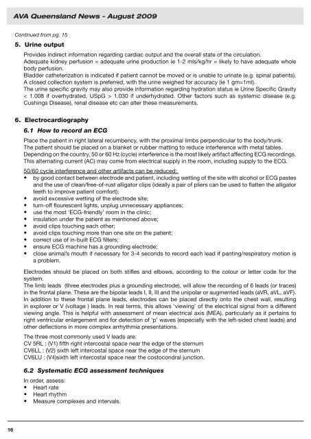
AVA <strong>Queensland</strong> <strong>News</strong> - August 2009Continued from pg. 155. Urine outputProvides indirect information regarding cardiac output and the overall state of the circulation.Adequate kidney perfusion = adequate urine production ie 1-2 mls/kg/hr = likely to have adequate wholebody perfusion.Bladder catheterization is indicated if patient cannot be moved or is unable to urinate (e.g. spinal patients).A closed collection system is preferred, with the urine weighed for accuracy (ie 1 gm=1ml).The urine specific gravity may also provide information regarding hydration status ie Urine Specific Gravity< 1.008 if overhydrated, USpG > 1.030 if underhydrated. Other factors such as systemic disease (e.g.Cushings Disease), renal disease etc can alter these measurements.6. Electrocardiography6.1 How to record an ECGPlace the patient in right lateral recumbency, with the proximal limbs perpendicular to the body/trunk.The patient should be placed on a blanket or rubber matting to reduce interference with metal tables.Depending on the country, 50 or 60 Hz (cycle) interference is the most likely artifact affecting ECG recordings.This alternating current (AC) may come from electrical supply in the room, including supply to the ECG.50/60 cycle interference and other artifacts can be reduced:• by good contact between electrode and patient, including wetting of the site with alcohol or ECG pastesand the use of clean/free-of-rust alligator clips (ideally a pair of pliers can be used to flatten the alligatorteeth to improve patient comfort);• avoid excessive wetting of the electrode site;• turn-off flourescent lights, unplug unnecessary appliances;• use the most ‘ECG-friendly’ room in the clinic;• insulation under the patient as mentioned above;• avoid clips touching each other;• avoid clips touching more than one site on the patient;• correct use of in-built ECG filters;• ensure ECG machine has a grounding electrode;• close animal’s mouth if necessary for 3-4 seconds to record each lead if panting/respiratory motion isa problem.Electrodes should be placed on both stifles and elbows, according to the colour or letter code for thesystem.The limb leads (three electrodes plus a grounding electrode), will allow the recording of 6 leads (or traces)in the frontal plane. These are the bipolar leads I, II, III and the unipolar or augmented leads (aVR, aVL, aVF).In addition to these frontal plane leads, electrodes can be placed directly onto the chest wall, resultingin explorer or V (voltage ) leads. In real terms, this allows ‘viewing’ of the electrical signal from a differentviewing angle. This is helpful with assessment of mean electrical axis (MEA), particularly as it pertains toright ventricular enlargement and for detection of ‘p’ waves (especially with the left-sided chest leads) andother deflections in more complex arrhythmia presentations.The three most commonly used V leads are:CV 5RL : (V1) fifth right intercostal space near the edge of the sternumCV6LL : (V2) sixth left intercostal space near the edge of the sternumCV6LU : (V4)sixth left intercostal space near the costocondral junction.6.2 Systematic ECG assessment techniquesIn order, assess:• Heart rate• Heart rhythm• Measure complexes and intervals.16


