Magnetic Resonance Imaging (MRI), Low-Field
Magnetic Resonance Imaging (MRI), Low-Field
Magnetic Resonance Imaging (MRI), Low-Field
You also want an ePaper? Increase the reach of your titles
YUMPU automatically turns print PDFs into web optimized ePapers that Google loves.
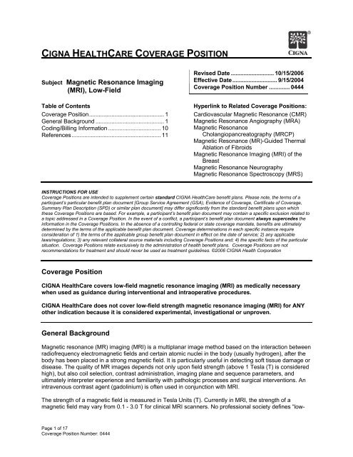
CIGNA HEALTHCARE COVERAGE POSITIONSubject <strong>Magnetic</strong> <strong>Resonance</strong> <strong>Imaging</strong>(<strong>MRI</strong>), <strong>Low</strong>-<strong>Field</strong>Table of ContentsCoverage Position............................................... 1General Background ........................................... 1Coding/Billing Information ................................. 10References........................................................ 11Revised Date ........................... 10/15/2006Effective Date............................ 9/15/2004Coverage Position Number ............. 0444Hyperlink to Related Coverage Positions:Cardiovascular <strong>Magnetic</strong> <strong>Resonance</strong> (CMR)<strong>Magnetic</strong> <strong>Resonance</strong> Angiography (MRA)<strong>Magnetic</strong> <strong>Resonance</strong>Cholangiopancreatography (MRCP)<strong>Magnetic</strong> <strong>Resonance</strong> (MR)-Guided ThermalAblation of Fibroids<strong>Magnetic</strong> <strong>Resonance</strong> <strong>Imaging</strong> (<strong>MRI</strong>) of theBreast<strong>Magnetic</strong> <strong>Resonance</strong> Neurography<strong>Magnetic</strong> <strong>Resonance</strong> Spectroscopy (MRS)INSTRUCTIONS FOR USECoverage Positions are intended to supplement certain standard CIGNA HealthCare benefit plans. Please note, the terms of aparticipant’s particular benefit plan document [Group Service Agreement (GSA), Evidence of Coverage, Certificate of Coverage,Summary Plan Description (SPD) or similar plan document] may differ significantly from the standard benefit plans upon whichthese Coverage Positions are based. For example, a participant’s benefit plan document may contain a specific exclusion related toa topic addressed in a Coverage Position. In the event of a conflict, a participant’s benefit plan document always supercedes theinformation in the Coverage Positions. In the absence of a controlling federal or state coverage mandate, benefits are ultimatelydetermined by the terms of the applicable benefit plan document. Coverage determinations in each specific instance requireconsideration of 1) the terms of the applicable group benefit plan document in effect on the date of service; 2) any applicablelaws/regulations; 3) any relevant collateral source materials including Coverage Positions and; 4) the specific facts of the particularsituation. Coverage Positions relate exclusively to the administration of health benefit plans. Coverage Positions are notrecommendations for treatment and should never be used as treatment guidelines. ©2006 CIGNA Health CorporationCoverage PositionCIGNA HealthCare covers low-field magnetic resonance imaging (<strong>MRI</strong>) as medically necessarywhen used as guidance during interventional and intraoperative procedures.CIGNA HealthCare does not cover low-field strength magnetic resonance imaging (<strong>MRI</strong>) for ANYother indication because it is considered experimental, investigational or unproven.General Background<strong>Magnetic</strong> resonance (MR) imaging (<strong>MRI</strong>) is a multiplanar image method based on the interaction betweenradiofrequency electromagnetic fields and certain atomic nuclei in the body (usually hydrogen), after thebody has been placed in a strong magnetic field. It is particularly useful in detecting soft tissue damage ordisease. The quality of MR images depends not only upon field strength (above 1 Tesla (T) is consideredhigh), but also coil selection, contrast administration, imaging plane and sequence parameters, andultimately interpreter experience and familiarity with pathologic processes and surgical interventions. Anintravenous contrast agent (gadolinium) is often used in conjunction with <strong>MRI</strong>.The strength of a magnetic field is measured in Tesla Units (T). Currently in <strong>MRI</strong>, the strength of amagnetic field may vary from 0.1 - 3.0 T for clinical <strong>MRI</strong> scanners. No professional society defines “low-Page 1 of 17Coverage Position Number: 0444
Tavernier and Cotton et al. (2005) state the primary limitation of low-field <strong>MRI</strong> is lower SNR, which has tobe compensated for by increasing the section thickness, reducing the in-plane resolution, increasing thenumber of acquisitions (and consecutively the acquisition time), and decreasing the bandwidth. Tavernierand Cotton et al. (2005) conclude that implementation of low-field <strong>MRI</strong> systems may be useful, especiallyin orthopedic centers, or if installation of an additional high-field scanner is not possible because ofeconomic considerations. Blanco et al. (2005) noted there is trade-off in image quality towards lessresolution due to open structure of these systems. The image quality of low-field scanners is, however,sufficient for interventional use.In a review article, Hayashi et al. (2004) noted that no reliable efficacy studies exist comparing thediagnostic capabilities of low- versus high-field scanners. In order to compensate for lower SNR, scannerswith low-field strength tend to have longer acquisition times, often resulting in greater image degradationdue to patient movement. However, it is widely accepted that today’s low-field scanners provide sufficientdiagnostic information when spin-echo and gradient-echo techniques are used. Hayashi et al. concluderecent technological developments in the realm of low-field MR scanners will lead to higher image quality,shorter scan times, and refined imaging protocols. Interventional and intraoperative use also supports theinstallation of low-field MR scanners. Utilization of low-field systems has the potential to enhance overallcost reductions with little or no loss of diagnostic performance.Potential Indications for <strong>Low</strong>-field <strong>MRI</strong>Most of the <strong>MRI</strong> studies reported in the literature are based on intermediate- or high-field <strong>MRI</strong>. Evidencein the published, peer-reviewed scientific literature identifies a small and varied amount of studiesinvestigating low-field <strong>MRI</strong>. Indications under investigation include, but are not limited to: MR-imageguided interventional and intraoperative procedures; musculoskeletal (knee, shoulder, and spine) andrheumatology.Interventional and intraoperative <strong>MRI</strong> (i<strong>MRI</strong>): Clinical <strong>MRI</strong> is mainly used as a diagnostic imagingmodality but it is increasingly being applied to guide various forms of intervention. Numerous studies haveshown that MR can be used to safely and effectively guide interventional and intraoperative procedures(i<strong>MRI</strong>). Major advantages of MR-guided intervention include the lack of exposure to ionizing radiation toboth staff and patients and the ability of MR to provide various intervention-specific image contrasts. Thephysical nature of MR has led to a range of compatible tools being developed, whose properties arespecific to the MR environment. i<strong>MRI</strong> facilitates better direct access to the patient in the imaging field(open magnet designs), faster fluoroscopic imaging techniques having good spatial resolution, the abilityto interactively control the image scan-plane and the development of novel contrast mechanisms(Grainger, 2001).Interventional MR imaging suites share three fundamental requirements: the availability of an open-fieldscanner configuration to facilitate patient access, the ability to implement rapid pulse sequences toensure safe device placement, and the ability to operate the scanner and to review updated images at thepatient’s bedside without having to remove the operator’s hand at any time from the interventional device.The designs of this equipment do, however, vary according to the manufacturer, nature of proceduresperformed, and user preferences, for example. Most of the open scanners use low- or middle-fieldmagnets. A few institutions have recently installed newer generations of open-bore high-field scanners.The higher the MR imaging field strength, the larger is the apparent width of the needles and otherinterventional devices used. MR imaging guidance is not intended to be a substitute for other lessexpensive and relatively simpler modes of biopsy and/or aspiration guidance whenever their use isappropriate. Rather, interventional MR imaging procedures assume a role when the patient wouldotherwise be subjected to surgical exploration or open biopsy performed solely for the purpose of tissuediagnosis or when procedure safety under the more conventional methods of guidance is considered lessthan optimal (Nour and Lewin, 2005).<strong>MRI</strong> is used for guiding interventional procedures because of its superior soft tissue contrast and lack ofradiation exposure. The introduction of open configuration, low-field <strong>MRI</strong> devices enabled direct patientaccess and the study of <strong>MRI</strong> as an interventional and intraoperative device. Some current interventionsinclude MR-guided biopsy, periradicular nerve root infiltration, guiding modalities for drainage andintravascular therapies, or ablative therapy monitoring and guidance. <strong>MRI</strong> is being utilized for imagePage 3 of 17Coverage Position Number: 0444
guidance in minimally invasive neurosurgery. Intraoperative <strong>MRI</strong> is an imaging tool that can be usedeffectively for navigation with an attached instrument tracking device because a low-field scanner can beturned off during surgery. <strong>MRI</strong> is increasingly used for guiding, monitoring and controlling percutaneousprocedures and surgery. Open configuration <strong>MRI</strong> scanners on which one side is usually open for patientaccess are obviously more suited to interventional procedures than closed systems. Although there istrade-off in image quality towards less resolution due to open structure of these systems, the imagequality of low-field scanners is, however, sufficient for interventional use (Blanco, et. al., 2005).In a review article, Martin et al. (2005) stated that recent development of open-configuration <strong>MRI</strong>scanners—which have allowed improved patient access, near real-time imaging, and more available <strong>MRI</strong>compatibleequipment—has opened up an entirely new area of image-guided surgical and interventionalprocedures. The use of intraoperative <strong>MRI</strong>-guided therapy has continued to grow since initial reportspublished in the 1990s. Intraoperative <strong>MRI</strong> has been reportedly used in hepatic tumor ablations,endometrial treatment, sarcoma resection, perirectal disease, and most commonly in neurosurgicalprocedures.Samdani et al. (2005) used a 0.12 T open-design MR scanner in performing pediatric neurosurgery totreat 20 patients with astrocytoma, craniopharyngioma, cortical dysplasia, and hydrocephalus. Theauthors concluded that in addition to neuronavigation, the MR system provided information on extent ofresection, real-time guided catheter placement, and avoided complications. Nimsky et al. (2002) used a0.2 T open-design MR scanner to perform neurosurgery in 310 patients. There were no adverse effectsdue to intraoperative <strong>MRI</strong>. Image quality was sufficient to evaluate the extent of the tumor resection in themajority of cases. The main indications for intraoperative <strong>MRI</strong> were the evaluation of the extent of aresection in glioma, ventricular tumor, pituitary tumor, and in epilepsy surgery. The authors stated thatintraoperative <strong>MRI</strong> offers the possibility of further tumor removal during the same surgical procedure incase of tumor remnants, increasing the rate of complete tumor removal. Schwartz et al. (1999) used a0.5 T open-design MR scanner to perform 200 intracranial surgical procedures. The authors concludedthat intraoperative <strong>MRI</strong> was successfully implemented for a variety of intracranial procedures andprovided continuous visual feedback, which can be helpful in all stages of neurosurgical interventionwithout affecting the duration of the procedure or the incidence of complications. They noted their systemhas potential advantages over conventional frame-based and frameless stereotactic procedures withrespect to the safety and effectiveness of neurosurgical interventions.<strong>Low</strong>-field MR scanners were used to guide or perform the following: hepatic tumor ablation (Martin, et al.,2005), transgluteal-approach prostate biopsies (Zangos, et al., 2005), microwave thermocoagulationtherapy to treat metastatic liver tumors from breast cancer (Abe, et al., 2005), abdominal biopsy onpatients in whom ultrasound (US)-guided abdominal biopsy was not possible because the lesion was notvisualized in US or an US-guided procedure was not considered safe (Kariniemi, et al., 2005),percutaneous cryosurgery for invasive breast cancer (Morin, et al., 2004), percutaneous transthoracicfine-needle aspiration biopsy of lung masses (Sakarya, et al., 2003), discography (Sequeiros, et al.,(2006), transvaginal cryotherapy for treating intramural, submucosal, and sub-serosal uterine fibroids(Dohi, et al., 2004), and sacro-iliac joint arthrography in patients with low-back pain (Ojala, et al., 2001).Musculoskeletal: <strong>MRI</strong> of the musculoskeletal system is performed most commonly using 1.5 T scanners.In the previous decade much vendor research and development went in to the optimization of low-fieldand open scanners. In the last 5 years, as the focus of <strong>MRI</strong> became less anatomic there has been apronounced trend to high-field strengths; however, higher-field systems of 3 T are becoming more widelyavailable in clinical practice, and developments with 7 T and high-fields are ongoing (Cunningham, et al.,2006).Knee: In a randomized trial of 189 patients with recent knee injury, Oei et al. (2005) utilized a 0.2 Textremity-dedicated MR scanner to assess the predictive value of using the low-field <strong>MRI</strong> in a
in the initial stage after knee trauma is limited. They noted that a short <strong>MRI</strong>, in addition to or instead ofradiography, improved the prediction of the need for additional treatment in patients with traumatic kneeinjury but did not significantly aid in the identification of patients who can be discharged without furtherfollow-up.Oei et al. (2003) conducted a literature review and performed a metaanalysis including 29 articles todetermine the diagnostic performance of all-strength <strong>MRI</strong> of the menisci and cruciate ligaments and toassess the effect of study design characteristics and magnetic field strength on diagnostic performance.Oei et al. stated that higher magnetic field strength improved the diagnostic performance for ACL tears.Cotton et al. (2000) used a 0.2 T scanner to compare the diagnostic efficacy of low- and high-fieldstrengthMR imagers in the diagnosis of anterior cruciate ligament tears and meniscus tears in 219patients with suspected internal derangement of the knee. Selection of patients for surgery wasperformed using only the data from the 1.5-T magnet. <strong>MRI</strong> scans were interpreted by musculoskeletalradiologists in a hospital setting. Therefore in 90 patients, using arthroscopy findings as the standardreference, the authors found no significant difference in diagnostic performance between low and highfield-strengthMR imaging of the knee. Several points should be noted: Gradient-echo vs. spin-echosequences were used with both <strong>MRI</strong>. Results might have been different if spin-echo T2-weightedsequences had been used for both. Also, the low-field scan took 15 minutes longer than 1.5 scan. Inaddition, the two reviewers were musculoskeletal radiologists.Rand et al. (1999) noted that qualitative evaluation of the level of confidence in decision making wassignificantly superior on high-field strength images. Boxheimer et al. (2006) prospectively evaluated ifkinematic <strong>MRI</strong> of the knee can demonstrate displacement of menisci with tears and characterizedisplaceable and non-displaceable meniscal tears in conjunction with the patient’s level of pain. Thisstudy did not address any value of positional low-field <strong>MRI</strong> compared to conventional <strong>MRI</strong> or impact topatient outcomes.Shoulder: Ghazinoor and Crues (2006) state that a handful of studies have been performed comparinghigh-field versus low-field <strong>MRI</strong> of the shoulder. Most have demonstrated no significant difference insensitivity and specificity in detection of rotator cuff and labral lesions. They note their subjectiveexperience is in concordance with many studies demonstrating the high diagnostic value in the use oflow-field scanners in musculoskeletal pathology. In general, the experience and training of the reader islikely to impact the interpretation of these images. <strong>MRI</strong> performed on extremity scanners would beperformed best by musculoskeletal-trained radiologists with experience reading on low-field systems,working closely with referring orthopedic surgeons (Ghazinoor and Crues, 2006).Zlatkin et al. (2004) retrospectively assessed a 0.2 T extremity-dedicated MR scanner in 160 patients withsuspected tears of the rotator cuff and glenoid labrum, compared with surgical findings. Surgical findingsdemonstrated rotator cuff tears in 131 patients and labral tears in 60 patients. For the rotator cuff, thesensitivity, specificity, positive predictive value, and negative predictive value were 90%, 93%, 98%, and68% (two false positives and 13 false negatives), respectively. For the labrum, the sensitivity, specificity,positive predictive value, and negative predictive value were 55%, 100%, 100%, and 82% (24 falsenegatives), respectively. This study was small and retrospective in design and did not include acomparison to conventional <strong>MRI</strong>.In a review article, Kreitner et al. (2003) summarized experiences with low-field MR arthrography of theglenohumeral joint with respect to image quality and diagnostic accuracy in detecting labral and rotatorcuff lesions. “Up to now, there has been only a limited number of studies available dealing with low-fieldMR arthrography of the glenohumeral joint. They reveal that, despite a minor image quality in comparisonwith high-field imaging, low-field MR arthrography of the shoulder allows for sufficient evaluation of intraandextra-articular structures in the detection of major abnormalities such as glenohumeral instability orrotator cuff disease. Furthermore, open-configured MR scanners enable kinematic studies: besides theanalysis of normal motion, pathological findings in patients with instabilities and impingement syndromecan be delineated. They further offer the possibility for performing <strong>MRI</strong>-guided arthrography of theshoulder” (Kreitner, et al., 2003).Page 5 of 17Coverage Position Number: 0444
Magee et al. (2003) used a 0.2 T open-design MR scanner for detection of supraspinatus tendon tearsand labral tears in 40 patients, compared with 1.5 T and arthroscopy. The authors stated that high-fieldstrengthimages altered reviewers' interpretations of low-field-strength scans for nine of 40 patients. Infour patients, full-thickness supraspinatus tendon tears could be diagnosed definitively on the high-fieldstrengthunit but not on the open unit. Three labral tears and two superior labral anteroposterior lesionscould be depicted definitively on the high-field-strength unit but not on the open unit. All tears wereconfirmed at arthroscopy. The authors concluded that high-field-strength <strong>MRI</strong> units provide better spatialand contrast resolution and allow more accurate interpretations than low-field-strength units; thesefindings may affect clinical treatment.Shellock et al. (2001) used a 0.2 T extremity-dedicated MR scanner to detect lesions of the rotator cuffand glenoid labrum in 47 patients, compared to the surgical findings. For the rotator cuff tears, thesensitivity, specificity, positive predictive value, and negative predictive value were 89%, 100%, 100%,and 90% (three false negatives), respectively. For the labral lesions, the sensitivity, specificity, positivepredictive value, and negative predictive value were 89%, 95%, 80%, and 97% ( two false positives, onefalse negative), respectively. The findings indicated that there was good agreement comparing the lowfieldMR results to the surgical findings for determination of lesions of the rotator cuff and glenoid labrum.This study did not evaluate conventional <strong>MRI</strong> compared to low-field or surgical findings.Loew et al. (2000) used a 0.2 T open-design MR scanner to compare the image quality, sensitivity,specificity, and diagnostic accuracy of low-field MR with 1.5 T MR after arthrography of the shoulder.Thirty-eight patients either with suspected chronic instability (n = 12) or rotator cuff abnormalities (n = 26)were examined. Intra-articular injection of diluted Gd-DTPA was followed in randomized order either firstby imaging on an open 0.2 T system or on a 1.5 T system. The image material was evaluatedindependently by two radiologists in a blinded fashion with respect to overall image quality and thedetection of rotator cuff as well as capsular and labral abnormalities. Surgical correlation was available in27 (71%) of 38 patients. For both systems, sensitivity and specificity for rotator cuff tears were 100%each, and for labrum pathologies, these values were 100 and 93%, respectively. The authors concludedthat low-field MR compares favorably to high-field MR in the detection of major abnormalities of theglenohumeral joint, at least when MR arthrography is used. Disadvantages are the duration of theexamination and thus the risk of reduced image quality caused by motion artifacts. It should be noted thatthere was an intentional use of different sequences at each MR unit. Also, the small number of patients(27) evaluated limits the power of the statistical results.Tung et al. (2000) used a 0.2 T open-design MR scanner to diagnose a glenoid superior labralanteroposterior (SLAP) tears compared to 1.5 T MR. Forty-one patients with SLAP tears and 26symptomatic patients with normal superior labra were retrospectively evaluated. Both groups of patientshad either high-field (n = 46) or low-field (n = 21) <strong>MRI</strong> and arthroscopy. For the diagnosis of SLAP tear,the sensitivity of high-field <strong>MRI</strong> was 90%, specificity was 63%, and accuracy was 80%. The sensitivity oflow-field <strong>MRI</strong> was 64%, specificity was 70%, and accuracy was 67%. Authors stated that the performancecharacteristics of high-field <strong>MRI</strong> are superior to those of low-field <strong>MRI</strong> for the diagnosis of a superior labraltear. Again, the small number of patients evaluated limits the power of the statistical results.Merl et al. (1999) prospectively evaluated the diagnostic quality of an open design 0.2 T MR scannercompared with high-field MR scanners measured by the applicable gold standard. Over a period of 3months, 401 patients with suspected renal tumors (n=78), diseases of the capsulolabral complex and therotator cuff of the shoulder joint (n=122), diseases of the spinal canal (n=105) and of the brain with focalneurological deficit (n=96) were prospectively evaluated in four participating centers. They all underwentclinical evaluation, low-field and high-field <strong>MRI</strong> and surgical or follow-up confirmation of diagnosis.Clinical, histopathologic, high-field and low-field <strong>MRI</strong> diagnoses were recorded in standardizedquestionnaires that were centrally evaluated. Statistical evaluation comprised two parts: receiveroperating characteristic (ROC) analysis assessed accuracy of <strong>MRI</strong> and clinical diagnoses; furthermore,rates of concordance of high- and low-field <strong>MRI</strong> diagnosis were calculated. Researchers did not compareimages and record differences between the different systems, but both class of units were measuredagainst the clinical gold standard and their diagnostic accuracy determined. Researchers found nostatistically relevant difference in high-field <strong>MRI</strong> diagnosis compared to low-field <strong>MRI</strong> diagnostic accuracymeasured by clinical or surgical gold standard in three of the four regions examined; in cerebralexaminations there was a small yet significant advantage for the high-field systems. The authorsPage 6 of 17Coverage Position Number: 0444
conclude that the open low-field scanner (using clinical and surgical gold standard as reference) is able toachieve comparable diagnostic accuracy compared to high-field scanners at lower costs and greaterpatient comfort. They stated that limitations due to field strength (signal-to-noise ratio, resolution, scantime) seem to be relevant only in a very small number of cases that warrant high-field examination.Allmann et al. (1998) used a 0.2 T MR scanner to determine the diagnostic accuracy of glenohumeralinstabilities with low-field <strong>MRI</strong> compared to 1.0 T <strong>MRI</strong>. A preselected group of 35 symptomatic patientswith recurrent shoulder luxations underwent imaging and therapeutic arthroscopy, which was used asstandard reference. The sensitivity, specificity, and accuracy of MR images acquired at 1.0 T for labrumpathology were 91%, 67%, 91% and 70%, 80%, 71% for the capsular complex, respectively. Comparedwith the above, the sensitivity, specificity, and accuracy for 0.2 T MR revealed 91%, 67%, 91% for thelabrum pathology and 63%, 80%, 66% for the capsular complex respectively. In the evaluation ofcapsular lesions, a comparison between the 0.2 T and the 1.0 T MR indicated a higher sensitivity andaccuracy for the high-field images. Concerning labral lesions, the sensitivity and accuracy of the 0.2 Tand the 1.0 T MR were comparable. A selection bias could not be avoided because arthroscopy wasused as the gold standard. It should be noted that a highly selected patient population - because onlythose subjects with more than eight luxations and arthroscopy, - impacted results (e.g., lower specificityfor labral lesions).Spine: Jinkins et al. (2005) stated that weight-bearing open-design <strong>MRI</strong> allows for improved sensitivityand specificity; however, no supporting data was provided. Vitaz et al. (2004) used a 0.5 T <strong>MRI</strong> scannerto prospectively evaluate the first 20 patients referred for <strong>MRI</strong> for neck pain. There was no comparator; nocomparison to conventional <strong>MRI</strong> can be drawn.In a review article, Weishaupt et al. (2003) stated that conventional <strong>MRI</strong> of the lumbar spine (i.e., in thesupine position) remains the imaging method of choice for the assessment of degenerative disk disease.Weight-bearing imaging in upright seated or upright standing positions (usually combined with flexion andextension movements) using vertical open-configuration MR scanners may be helpful in patients withequivocal findings on conventional <strong>MRI</strong>, clinically suspected position-dependent nerve root compromise,or in cases of suspected spinal canal or neuroforaminal stenosis with equivocal or borderline findings onconventional <strong>MRI</strong>.Weishaupt et al. (2000) used a 0.5 T open-design MR scanner to evaluate whether positional <strong>MRI</strong> of thelumbar spine demonstrated nerve root compromise not visible on supine 1.0 T MR. A total of 30 patientswith chronic low back pain unresponsive to nonsurgical treatment and with disk abnormalities but withoutcompression of neural structures were included. Results demonstrated nerve root contact withoutdeviation was present in 34 of 152 instances in the supine position, in 62 instances in the seated flexionposition, and in 45 instances in the seated extension position. As compared with the supine position, inthe seated flexion position nerve root deviation decreased from 10 to eight instances; in the seatedextension position, it increased from 10 to 13 instances. Nerve root compression was seen in one patientin the seated extension position. The authors concluded that positional <strong>MRI</strong> more frequentlydemonstrates minor neural compromise than does supine <strong>MRI</strong>. The relevance or value of this finding onlow back pain treatment or patient outcomes is unknown.Vitzthum et al. (2000) used a 0.5 T open-design MR scanner to study healthy volunteers and patientssuffering from degenerative disorders of the lumbar spine to determine the relationship of differentstructures of the lower lumbar spine during interventional movement examination. In 50 patients withdegenerative disorders of the lumbar spine (41 with disc herniation, five with osteogenic spinal stenosis,and four with degenerative spondylolisthesis), the range of rotation was increased in the relevant spinalsegments. Signs of neural compression were increased under motion. The authors concluded thatdynamic examination in which vertical, open 0.5 T <strong>MRI</strong> was used demonstrated that the extent of neuralcompression and the increasing range of rotation are important signs of segmental instability. Therelevance or value of this finding on low back pain treatment or patient outcomes is unknown.Rheumatology: The Outcome MEasures in Rheumatoid Arthritis Clinical Trials (OMERACT) andEuropean League Against Rheumatism (EULAR) initiative is an informal international network of workinggroups and gatherings interested in outcome measurement across the spectrum of rheumatologyintervention studies. The proposed OMERACT <strong>MRI</strong> scoring system is based upon conventional strengthPage 7 of 17Coverage Position Number: 0444
<strong>MRI</strong>, and provides a framework for scoring inflammation and damage in RA and is useful for inclusion asan outcome measure in clinical trials (Ostergaard, et al., 1996; McQueen, et al., 1998; McGonagle, et al.,1999). OMERACT is currently working to standardize image acquisition, terminology, and possiblediagnostic and monitoring indications for <strong>MRI</strong> in rheumatoid arthritis. Lack of standardization contributesto the difficulty in assessing studies regarding the use of conventional strength <strong>MRI</strong> in the diagnosis andtreatment of rheumatoid arthritis.There are numerous, very small, comparative studies that validate low-field <strong>MRI</strong> is superior to x-ray in thedetection of rheumatoid arthritis (RA) findings (e.g., bone erosion, joint-space narrowing, synovitis);Yoshioka et al. (2006), Scheel et al. (2006) Ejbjerg et al. (2005), Crues et al. (2004), Lindegaard et al.(2001). A few small, comparative studies have evaluated the diagnostic capability of low-field MR withconventional MR in the detection of RA findings. Using conventional <strong>MRI</strong> as the standard reference,Ejbjerg et al. (2005) evaluated findings from a 0.2 T extremity-dedicated MR scanner in 37 patients withRA. <strong>Low</strong>-field MR (3D gradient echo sequence) wrist and metacarpophalangeal (MCP) joint imagingdemonstrated sensitivity, specificity, and accuracy for erosions of 94%, 93%, 94%; for synovitis, 90%,96%, and 94%; and for bone marrow edema, 39%, 99%, and 95%. The authors stated that low-fieldextremity-dedicated <strong>MRI</strong> provides similar information on bone erosions and synovitis as high-field <strong>MRI</strong>units. The low sensitivity of the low-field <strong>MRI</strong> unit for the detection of bone marrow edema may limit theusefulness of this type of scanner in RA if bone marrow edema is proved to be a pathological event ofmajor prognostic significance. But if bone marrow edema is only an interim phase between synovitis andbone erosion, this may not have major impact on the usefulness of low-field <strong>MRI</strong> in RA because theprecursor of bone marrow edema is generally accepted as being synovitis. The authors conclude that thelatter statement still needs to be validated in further scientific studies.Taouli et al. (2004) used a 0.2 T extremity-dedicated MR scanner to detect and grade bone erosions,joint-space narrowing, and synovitis in the hands and wrists of 18 patients with rheumatoid arthritis. Theauthors concluded conventional <strong>MRI</strong> and 0.2 T <strong>MRI</strong> showed similar results in terms of cross-sectionalgrading of bone erosions, joint-space narrowing, and synovitis in the hands and wrists of patients withrheumatoid arthritis. Different T2-weighted sequences for conventional and low-field were used. Also, thesmall number of patients evaluated limits the power of the statistical results.Savnik et al. (2001) used a 0.2 T extremity-dedicated MR scanner to compare diagnostic capability of lowto high-field <strong>MRI</strong> of arthritic wrist and finger joints. A total of 103 patients (group 1 = 28 patients with RA
<strong>MRI</strong> of the Knee Practice Guideline states “diagnostic quality knee <strong>MRI</strong> can be performed using a varietyof magnet designs (closed bore, whole body, open whole body, dedicated extremity) and field strengths.The coil’s placement should allow imaging of the major structures in and around the knee, or the coiland/or extremity should be repositioned during the examination to include any pertinent anatomy wherean abnormality is suspected. For example, when a quadriceps tendon abnormality is clinically suspectedor suggested by ancillary imaging findings in the knee, an additional set of images may be necessaryabove the knee after repositioning when using a dedicated extremity magnet. Certain MR systems (e.g.,low-field magnets) have inherently lower signal-to-noise ratios than others. When using such a system toperform knee <strong>MRI</strong>, other imaging parameters – such as the receiver bandwidth and number ofacquisitions – will require modification to ensure adequate spatial and contrast resolution for confidentdiagnosis, often at the expense of longer examination times. It may also be more difficult to achieveuniform chemical fat suppression on low-field systems. For some indications, imaging on a low-fieldsystem may be disadvantageous compared to a high-field system. For example, high-resolution imagesof articular cartilage are more difficult to achieve with low-field systems. Detection of other conditions, likemeniscal and anterior cruciate ligament tears, is probably less influenced by magnet strength and design”(October, 2005).<strong>MRI</strong> of the Shoulder Practice Guideline states “even when the imaging protocol is optimized for shoulderimaging on a low-field open system, subjective image quality will likely be inferior to that obtained with ahigh-field system. Various investigators using different equipment and scanning parameters have reachedcontradictory conclusions regarding the diagnostic performance of low-field-strength MR scanners forshoulder disorders. Some studies have found that the accuracy for complete and partial rotator cuff tearsand for labral abnormalities is not significantly different for open, low-field and closed, high-field systems,with careful attention to technique. MR arthrography can further enhance the diagnostic yield for shoulder<strong>MRI</strong> performed on low-field strength systems. Other investigators have found lower accuracy for theevaluation of disorders like SLAP tears, capsular abnormalities, and small rotator cuff tears with specificlow-field systems compared to high-field ones” (October, 2005).Appropriateness Criteria regarding Acute Hand and Wrist Trauma states “when high-field or low-fieldMR imaging is performed in addition to radiographs, radiographically occult fractures of the distal radiusas well as unsuspected fractures of the carpal bones are frequently demonstrated. MR imagingevaluation for radiographically occult scaphoid fractures can be performed with high-field or low-fieldequipment, using a whole-body imaging system and appropriate local coil, or using a dedicated extremityMR scanner. Unlike the case for the wrist, low-field MR imaging is less sensitive than radiographs forhand and finger fractures.The ACR Practice Guideline for <strong>MRI</strong> of the Adult Spine (2002) does not address weight-bearing,standing, upright, axial-loading or vertical positioning. It does not address field strength. Regardingdegenerative disc disease, ACR states <strong>MRI</strong> has proven to be the technique of choice for imaging ofintervertebral disc degeneration. Because of its greater contrast resolution and the ability to image in thesagittal as well as the axial planes, <strong>MRI</strong> is regarded as the diagnostic modality of choice for evaluation ofpossible disc herniation with a high sensitivity for demonstrating the presence of nerve root compression.As compared with CT (with or without myelography), the greater contrast resolution and multiplanarimaging capabilities of <strong>MRI</strong> enable more accurate demonstration of nerve root impingement beyond theroot sleeve and neural foramen. Regarding spinal stenosis, ACR states on both axial and sagittal MRimages, the sizes of both the spinal canal and the thecal sac are well demonstrated. The contents of thethecal sac (spinal cord, nerve roots) can also be assessed. These capabilities enable the use of <strong>MRI</strong> forthe evaluation of possible spinal stenosis with high sensitivity and specificity.American College of Rheumatology: A 2006 report of the American College of Rheumatology onextremity <strong>MRI</strong> in rheumatology notes that most of the literature “assessing the utility of peripheral joint<strong>MRI</strong> has used high-field, not low-field extremity <strong>MRI</strong>; therefore, actual sensitivity, specificity, andpredictive value of the low-field scanners available for the practicing rheumatologists are not known. Thebenefits of low-field strength extremity <strong>MRI</strong> for the diagnosis and management of rheumatoid arthritis arestill being elucidated”. In the American College of Rheumatology guidelines for the management ofrheumatoid arthritis (2002), radiography of selected involved joints is addressed, but no other type ofimaging is discussed.Page 9 of 17Coverage Position Number: 0444
National Agency for Accreditation and Evaluation in Health (ANAES): ANAES states that “as therehas not yet been any explicit clinical proof of the diagnostic performance of low-field <strong>MRI</strong>, ANAESrecommends that a rigorous clinical evaluation of low-field <strong>MRI</strong> should be carried out in a valid, controlledprospective study, comparing low-field and high-field <strong>MRI</strong> in the most relevant clinical indications to date.In this regard, as the diagnostic efficacy of <strong>MRI</strong> investigation of traumatic knees appears to besatisfactory, this is the indication that should be used in the clinical studies to be undertaken as a matterof priority” (ANAES, 1999).Washington State Department of Labor and Industries: Washington State published a HealthTechnology Assessment on Standing, Weight-Bearing, Positional, or Upright <strong>MRI</strong> (2006). Someconclusions included:• There is limited scientific data available on the accuracy and diagnostic utility of standing, upright,weight-bearing or positional <strong>MRI</strong>.• There is no evidence from well-designed clinical trials demonstrating the accuracy or effectiveness ofweight-bearing <strong>MRI</strong> for specific conditions or patient populations.• Due to the lack of evidence addressing diagnostic accuracy or diagnostic utility, standing, weightbearing,positional <strong>MRI</strong> is considered investigational and experimental.SummaryInterventional and Intraoperative <strong>Low</strong>-field <strong>MRI</strong>: <strong>MRI</strong> as a guidance modality quickly progressed fromresearch sites to clinical practice; therefore, there is limited evidence in the published peer-reviewedscientific literature providing direct comparisons with conventionally-approached interventions. However,small observational trials and textbooks indicate the use of <strong>MRI</strong> in guiding interventional andintraoperative procedures (i<strong>MRI</strong>) has become widely accepted as standard of care in equipped facilities.Knee and Shoulder: Evidence in the published peer-reviewed scientific literature does not clearly andconsistently demonstrate that low-field <strong>MRI</strong> diagnostic capability is equivalent to the scope ofconventional <strong>MRI</strong> diagnostic capability. There is a lack of data clarifying the impact of treatment decisionsbased upon low-field interpretation on patient outcomes. There is a lack of data regarding the accuracyand impact of interpretation of low-field MR images outside the hospital setting (i.e., non-radiologistinterpretation). There is a lack of data clarifying what role low-field imaging should hold in the diagnosticalgorithm of knee or shoulder injuries.Spine: There is a lack of evidence in the published peer-reviewed scientific literature validating theaccuracy, relevance or value of positional <strong>MRI</strong> in the diagnosis and treatment of patients with neck orback pain. The few small studies on positional <strong>MRI</strong> do not address the relevance, value or impact ofpositional <strong>MRI</strong> in the diagnosis, treatment or outcomes of patients with neck or back pain.Rheumatology: The group currently working to standardize use of <strong>MRI</strong> in rheumatoid arthritis isevaluating conventional strength <strong>MRI</strong>. Lack of standardization contributes to the difficulty in assessingstudies regarding the use of conventional strength or low-field strength <strong>MRI</strong> in the diagnosis andtreatment of rheumatoid arthritis.Other: Most <strong>MRI</strong> studies reported in the literature are based on intermediate- or high-field <strong>MRI</strong>. There isinsufficient evidence in the published peer-reviewed literature to support the use of low-field <strong>MRI</strong> for anyindication other than i<strong>MRI</strong>, including but not limited to the following: breast (Paakko, et al., 2005); cardiac(Rupprecht, et al., 2002); pulmonary (Abolmaali, et al., 2004; Wagner, et al., 2001), renal (Kajander, etal., 2000), multiple sclerosis (Ertl-Wagner, et al., 2001; Lee, et al., 1995), ankle and wrist trauma (Nikken,et al., 2005; Brydie, et al., 2003; Remplik, et al., 2004) and retrocochlear disorders (Dubrulle, et al., 2002).There is a lack of evidence that the use of low-field <strong>MRI</strong> in place of conventional <strong>MRI</strong> could not negativelyimpact accuracy of diagnosis, proposed treatment, and thus long-term patient outcomes.Coding/Billing InformationPage 10 of 17Coverage Position Number: 0444
9. American College of Rheumatology Subcommittee on Rheumatoid Arthritis Guidelines.Guidelines for the management of rheumatoid arthritis: 2002 update. Arthritis Rheum 2002;46:328–310. Besier TF, Draper CE, Gold GE, Beaupre GS, Delp SL. Patellofemoral joint contact areaincreases with knee flexion and weight-bearing. J Orthop Res. 2005 Mar;23(2):345-50.11. Blanco RT, Ojala R, Kariniemi J, Perala J, Niinimaki J, Tervonen O. Interventional andintraoperative <strong>MRI</strong> at low field scanner--a review. Eur J Radiol. 2005 Nov;56(2):130-42.12. Boxheimer L, Lutz AM, Zanetti M, Treiber K, Labler L, Marincek B, et al. Characteristics ofdisplaceable and nondisplaceable meniscal tears at kinematic <strong>MRI</strong> of the knee. Radiology. 2006Jan;238(1):221-31.13. Brydie A, Raby N. Early <strong>MRI</strong> in the management of clinical scaphoid fracture. Br J Radiol. 2003May;76(905):296-30014. Centers for Medicare & Medicaid Services (CMS). Medicare coverage database NationalCoverage Determination (NCD). NCD for magnetic resonance imaging (<strong>MRI</strong>) (220.2). UpdatedSeptember 10, 2004a. Accessed August 2006. Available at:http://www.cms.hhs.gov/mcd/viewncd.asp?ncd_id=220.2&ncd_version=1&basket=ncd%3A220%2E2%3A1%3A<strong>Magnetic</strong>+<strong>Resonance</strong>+<strong>Imaging</strong>+%28<strong>MRI</strong>%29.15. Cimmino MA, Parodi M, Innocenti S, Succio G, Banderali S, Silvestri E, Garlaschi G. Dynamicmagnetic resonance of the wrist in psoriatic arthritis reveals imaging patterns similar to those ofrheumatoid arthritis. Arthritis Res Ther. 2005;7(4):R725-31. Epub 2005 Apr 1.16. Cotten A, Delfaut E, Demondion X, Lapegue F, Boukhelifa M, Boutry N, et al. MR imaging of theknee at 0.2 and 1.5 T: correlation with surgery. AJR Am J Roentgenol. 2000 Apr;174(4):1093-7.17. Crues JV, Shellock FG, Dardashti S, James TW, Troum OM. Identification of wrist andmetacarpophalangeal joint erosions using a portable magnetic resonance imaging systemcompared to conventional radiographs. J Rheumatol. 2004 Apr;31(4):676-85.18. Cunningham PM, Law M, Schweitzer ME. High-field <strong>MRI</strong>. Orthop Clin North Am. 2006Jul;37(3):321-9.19. Dohi M, Harada J, Mogami T, Fukuda K, Kobayashi S, Yasuda M. MR-guided transvaginalcryotherapy of uterine fibroids with a horizontal open <strong>MRI</strong> system: initial experience. Radiat Med.2004 Nov-Dec;22(6):391-7.20. Dubrulle F, Delomez J, Kiaei A, Berger P, Vincent C, Vaneecloo FM, et al. Mass screening forretrocochlear disorders: low-field-strength (0.2-T) versus high-field-strength (1.5-T) <strong>MRI</strong>. AJNRAm J Neuroradiol. 2002 Jun-Jul;23(6):918-23.21. Ejbjerg BJ, Narvestad E, Jacobsen S, Thomsen HS, Ostergaard M. Optimised, low cost, low-fielddedicated extremity <strong>MRI</strong> is highly specific and sensitive for synovitis and bone erosions inrheumatoid arthritis wrist and finger joints: comparison with conventional high-field <strong>MRI</strong> andradiography. Ann Rheum Dis. 2005 Sep;64(9):1280-7. Epub 2005 Jan 1322. Ertl-Wagner BB, Reith W, Sartor K. <strong>Low</strong> field-low cost: can low-field magnetic resonance systemsreplace high-field magnetic resonance systems in the diagnostic assessment of multiple sclerosispatients? Eur Radiol. 2001;11(8):1490-4.23. Eshed I, Althoff CE, Schink T, Scheel AK, Schirmer C, Backhaus M, et al. <strong>Low</strong>-field <strong>MRI</strong> forassessing synovitis in patients with rheumatoid arthritis. Impact of Gd-DTPA dose on synovitisscoring. Scand J Rheumatol. 2006 Jul-Aug;35(4):277-82.Page 12 of 17Coverage Position Number: 0444
24. FONAR. Index of Upright <strong>MRI</strong> Research Publications. Accessed August 2006. Available atURL address: http://www.fonar.com/research_index.htm25. GE Healthcare. Applause. Accessed August 2006. Available at URL address:http://www.gehealthcare.com/usen/bone_densitometry/products/applause.htmlhttp://www.gehealthcare.com/company/pressroom/releases/pr_release_9223.html26. GE Healthcare. GE 0.35T Signa ® Ovation with Excite <strong>Magnetic</strong> <strong>Resonance</strong> System. AccessedAugust 2006. Available at URL address: http://www.gehealthcare.com/usen/mr/ovation/index.html27. Ghazinoor S, Crues JV 3rd. <strong>Low</strong> field <strong>MRI</strong>: a review of the literature and our experience in upperextremity imaging. Clin Sports Med. 2006 Jul;25(3):591-606, viii.28. Grainger RG, Allison D, Adam A, Dixon AK. <strong>Magnetic</strong> resonance imaging: Basic principles. In:Grainger & Allison's diagnostic radiology: a textbook of medical imaging. 4th ed. London, UK:Churchill Livingstone; 2001.29. Hayashi N, Watanabe Y, Masumoto T, Mori H, Aoki S, Ohtomo K, Okitsu O, et al. Utilization oflow-field MR scanners. Magn Reson Med Sci. 2004 Apr 1;3(1):27-38.30. Hayes Search and Summary. Upright (Standing and Seated) <strong>Magnetic</strong> <strong>Resonance</strong> <strong>Imaging</strong>(<strong>MRI</strong>). April 25, 2006. Accessed August 2006.31. Jinkins JR, Dworkin JS, Damadian RV. Upright, weight-bearing, dynamic-kinetic <strong>MRI</strong> of the spine:initial results. Eur Radiol. 2005 Sep;15(9):1815-25. Epub 2005 May 20.32. Jinkins JR, Dworkin J. Proceedings of the State-of-the-Art Symposium on Diagnostic andInterventional Radiology of the Spine, Antwerp, September 7, 2002 (Part two). Upright, weightbearing,dynamic-kinetic <strong>MRI</strong> of the spine: p<strong>MRI</strong>/k<strong>MRI</strong>. JBR-BTR. 2003 Sep-Oct;86(5):286-93.(abstract only)33. Kajander S, Kallio T, Alanen A, Komu M, Forsstrom J. <strong>Imaging</strong> end-stage kidney disease inadults. <strong>Low</strong>-field <strong>MRI</strong> with magnetization transfer vs. ultrasonography.Acta Radiol. 2000Jul;41(4):357-60.34. Kariniemi J, Blanco Sequeiros R, Ojala R, Tervonen O. <strong>MRI</strong>-guided abdominal biopsy in a 0.23-Topen-configuration <strong>MRI</strong> system. Eur Radiol. 2005 Jun;15(6):1256-62. Epub 2004 Dec 31.35. Keen HI, Brown AK, Wakefield RJ, Conaghan PG. <strong>MRI</strong> and musculoskeletal ultrasonography asdiagnostic tools in early arthritis. Rheum Dis Clin North Am. 2005 Nov;31(4):699-714.36. Kettenbach J, Kacher DF, Kanan AR, Rostenberg B, Fairhurst J, Stadler A, et al. Intraoperativeand interventional <strong>MRI</strong>: recommendations for a safe environment. Minim Invasive Ther AlliedTechnol. 2006;15(2):53-64.37. Kinnunen J, Bondestam S, Kivioja A, Ahovuo J, Toivakka SK, et al. Diagnostic performance oflow field <strong>MRI</strong> in acute knee injuries. Magn Reson <strong>Imaging</strong>. 1994;12(8):1155-60. (abstract only)38. Kreitner KF, Loew R, Runkel M, Zollner J, Thelen M. <strong>Low</strong>-field MR arthrography of the shoulderjoint: technique, indications, and clinical results. Eur Radiol. 2003 Feb;13(2):320-9. Epub 2002Aug 2839. Lee DH, Vellet AD, Eliasziw M, Vidito L, Ebers GC, Rice GP, et al. <strong>MRI</strong> field strength: prospectiveevaluation of the diagnostic accuracy of MR for diagnosis of multiple sclerosis at 0.5 and 1.5 T.Radiology. 1995 Jan;194(1):257-62.(abstract only)Page 13 of 17Coverage Position Number: 0444
40. Lindegaard H, Vallo J, Horslev-Petersen K, Junker P, Ostergaard M. <strong>Low</strong>-field dedicatedmagnetic resonance imaging in untreated rheumatoid arthritis of recent onset. Ann Rheum Dis.2001 Aug;60(8):770-6.41. Loew R, Kreitner KF, Runkel M, Zoellner J, Thelen M. MR arthrography of the shoulder:comparison of low-field (0.2 T) vs high-field (1.5 T) imaging. Eur Radiol. 2000;10(6):989-96.42. Magee T, Shapiro M, Williams D. Comparison of high-field-strength versus low-field-strength <strong>MRI</strong>of the shoulder. AJR Am J Roentgenol. 2003 Nov;181(5):1211-5.43. <strong>Magnetic</strong> <strong>Resonance</strong> Technology Information Portal (MR TIP). Accessed August 2006. Availableat URL address: http://www.mr-tip.com/serv1.php?type=db1&dbs=Open%20<strong>MRI</strong> http://www.mrtip.com/serv1.php?type=db1&dbs=<strong>Low</strong>%20<strong>Field</strong>%20<strong>MRI</strong>44. MagneVu Inc. Accessed February 2006. Available at URL address: http://www.mri4ra.com/45. Martin RC 2nd. Intraoperative magnetic resonance imaging ablation of hepatic tumors. Am JSurg. 2005 Apr;189(4):388-94.46. McGonagle D, Conaghan PG, O'Connor P, Gibbon W, Green M, Wakefield R, et al. Therelationship between synovitis and bone changes in early untreated rheumatoid arthritis: acontrolled magnetic resonance imaging study. Arthritis Rheum. 1999 Aug;42(8):1706-11.(abstract only)47. McQueen FM, Stewart N, Crabbe J, Robinson E, Yeoman S, Tan PL, McLean L. <strong>Magnetic</strong>resonance imaging of the wrist in early rheumatoid arthritis reveals a high prevalence of erosionsat four months after symptom onset. Ann Rheum Dis. 1998 Jun;57(6):350-6.48. Merl T, Scholz M, Gerhardt P, Langer M, Laubenberger J, Weiss HD, et al. Results of aprospective multicenter study for evaluation of the diagnostic quality of an open whole-body lowfield<strong>MRI</strong> unit. A comparison with high-field <strong>MRI</strong> measured by the applicable gold standard. Eur JRadiol. 1999 Apr;30(1):43-53.49. Morin J, Traore A, Dionne G, Dumont M, Fouquette B, Dufour M, et a. <strong>Magnetic</strong> resonanceguidedpercutaneous cryosurgery of breast carcinoma: technique and early clinical results. Can JSurg. 2004 Oct;47(5):347-51.50. National Agency for Accreditation and Evaluation in Health (ANAES). Clinical and economicevaluation of low-field magnetic resonance imaging systems (< 0.5 tesla). November 1999.Accessed August 2006.Available at URL address:http://www.anaes.fr/anaes/Publications.nsf/nPDFFile/SU_LILF4ZDCD6/$File/IRM.pdf?OpenElement51. Nikken JJ, Oei EH, Ginai AZ, Krestin GP, Verhaar JA, van Vugt AB, et al. Acute ankle trauma:value of a short dedicated extremity MR imaging examination in prediction of need for treatment.Radiology. 2005 Jan;234(1):134-42.52. Nikken JJ, Oei EH, Ginai AZ, Krestin GP, Verhaar JA, van Vugt AB, et al. Acute wrist trauma:value of a short dedicated extremity MR imaging examination in prediction of need for treatment.Radiology. 2005 Jan;234(1):116-24.53. Nimsky C, Ganslandt O, Fahlbusch R. Comparing 0.2 tesla with 1.5 tesla intraoperative magneticresonance imaging analysis of setup, workflow, and efficiency. Acad Radiol. 2005Sep;12(9):1065-79.54. Nimsky C, Ganslandt O, Tomandl B, Buchfelder M, Fahlbusch R. <strong>Low</strong>-field magnetic resonanceimaging for intraoperative use in neurosurgery: a 5-year experience. Eur Radiol. 2002Nov;12(11):2690-703. Epub 2002 May 1.Page 14 of 17Coverage Position Number: 0444
55. Nour SG, Lewin JS. Percutaneous biopsy from blinded to MR guided: an update on currenttechniques and applications. Magn Reson <strong>Imaging</strong> Clin N Am. 2005 Aug;13(3):441-64.56. Oei EH, Nikken JJ, Ginai AZ, Krestin GP, Verhaar JA, van Vugt AB, et al. Acute knee trauma:value of a short dedicated extremity <strong>MRI</strong> examination for prediction of subsequent treatment.Radiology. 2005 Jan;234(1):125-33.57. Oei EH, Nikken JJ, Verstijnen AC, Ginai AZ, Myriam Hunink MG. <strong>MRI</strong> of the menisci and cruciateligaments: a systematic review.Radiology. 2003 Mar;226(3):837-48. Epub 2003 Jan 15.58. Ojala R, Klemola R, Karppinen J, Sequeiros RB, Tervonen O. Sacro-iliac joint arthrography in lowback pain: feasibility of <strong>MRI</strong> guidance. Eur J Radiol. 2001 Dec;40(3):236-959. OMERACT. Accessed August 2006. Available at URL address: http://www.omeract.org/60. Open Stand Up <strong>MRI</strong>. Accessed August 2006. Available at URL address:http://www.openstandupmri.com/medical_lit.php61. Ostergaard M, Hansen M, Stoltenberg M, Lorenzen I. Quantitative assessment of the synovialmembrane in the rheumatoid wrist: an easily obtained <strong>MRI</strong> score reflects the synovial volume. BrJ Rheumatol. 1996 Oct;35(10):965-71.62. Paakko E, Reinikainen H, Lindholm EL, Rissanen T. <strong>Low</strong>-field versus high-field <strong>MRI</strong> in diagnosingbreast disorders. Eur Radiol. 2005 Jul;15(7):1361-8. Epub 2005 Feb 12.63. Rand T, Imhof H, Turetschek K, Schneider B, Vogele T, Gabler C, et al. Comparison of low field(0.2T) and high field (1.5T) <strong>MRI</strong> in the differentiation of torned from intact menisci. Eur J Radiol.1999 Apr;30(1):22-7.64. Remplik P, Stabler A, Merl T, Roemer F, Bohndorf K. Diagnosis of acute fractures of theextremities: comparison of low-field <strong>MRI</strong> and conventional radiography. Eur Radiol. 2004Apr;14(4):625-30. Epub 2003 Nov 5.65. Rupprecht T, Nitz W, Wagner M, Kreissler P, Rascher W, et al. Determination of the pressuregradient in children with coarctation of the aorta by low-field magnetic resonance imaging. PediatrCardiol. 2002 Mar-Apr;23(2):127-31. Epub 2002 Feb 19.66. Sakarya ME, Unal O, Ozbay B, Uzun K, Kati I, Ozen S, Etlik O. MR fluoroscopy-guidedtransthoracic fine-needle aspiration biopsy: feasibility. Radiology. 2003 Aug;228(2):589-92. Epub2003 Jun 20.67. Samdani AF, Schulder M, Catrambone JE, Carmel PW. Use of a compact intraoperative low-fieldmagnetic imager in pediatric neurosurgery. Childs Nerv Syst. 2005 Feb;21(2):108-13; discussion114. Epub 2004 Nov 25.68. Savnik A, Malmskov H, Thomsen HS, Bretlau T, Graff LB, Nielsen H, Danneskiold-Samsoe B,Boesen J, Bliddal H. <strong>MRI</strong> of the arthritic small joints: comparison of extremity <strong>MRI</strong> (0.2 T) vshigh-field <strong>MRI</strong> (1.5 T). Eur Radiol. 2001;11(6):1030-8.69. Scheel AK, Hermann KG, Ohrndorf S, Werner C, Schirmer C, Detert J, et al. Prospective 7 yearfollow up imaging study comparing radiography, ultrasonography, and magnetic resonanceimaging in rheumatoid arthritis finger joints. Ann Rheum Dis. 2006 May;65(5):595-600. Epub2005 Sep 28.70. Schwartz RB, Hsu L, Wong TZ, Kacher DF, Zamani AA, Black PM, et al. Intraoperative <strong>MRI</strong>guidance for intracranial neurosurgery: experience with the first 200 cases. Radiology. 1999May;211(2):477-88Page 15 of 17Coverage Position Number: 0444
71. Sequeiros RB, Niinimaki J, Ojala R, Haapea M, Vaara T, Klemola R et al. <strong>Magnetic</strong> resonanceimaging-guided diskography and diagnostic lumbar 0.23T <strong>MRI</strong>: an assessment study. ActaRadiol. 2006 Apr;47(3):272-80.72. Shellock FG, Bert JM, Fritts HM, Gundry CR, Easton R, Crues JV 3rd. Evaluation of the rotatorcuff and glenoid labrum using a 0.2-Tesla extremity magnetic resonance (MR) system: MRresults compared to surgical findings. J Magn Reson <strong>Imaging</strong>. 2001 Dec;14(6):763-70.73. Taouli B, Zaim S, Peterfy CG, Lynch JA, Stork A, Guermazi A, Fan B, Fye KH, Genant HK.Rheumatoid arthritis of the hand and wrist: comparison of three imaging techniques. AJR Am JRoentgenol. 2004 Apr;182(4):937-43.74. Tavernier T, Cotten A. High- versus low-field MR imaging. Radiol Clin North Am. 2005Jul;43(4):673-81, viii.75. The Tesla Society. Accessed August 2006. Available at URL address:http://www.teslasociety.com/mri.htm76. Tung GA, Entzian D, Green A, Brody JM. High-field and low-field <strong>MRI</strong> of superior glenoid labraltears and associated tendon injuries. AJR Am J Roentgenol. 2000 Apr;174(4):1107-14.77. U.S. Food and Drug Administration. Accessed August 2006. Available at URL address:http://www.fda.gov/cdrh/pdf/k994287.pdf Fonar http://www.fda.gov/cdrh/pdf6/K060956.pdf (E-Scan opera) http://www.fda.gov/cdrh/pdf3/k033504.pdf SIGNA ovationhttp://www.fda.gov/cdrh/pdf3/k032541.pdf Odin http://www.fda.gov/cdrh/pdf5/K050602.pdfALTAIRE http://www.fda.gov/cdrh/pdf6/K061132.pdf Superopen Neusoft78. Vitaz TW, Shields CB, Raque GH, Hushek SG, Moser R, Hoerter N, et al. Dynamic weightbearingcervical magnetic resonance imaging: technical review and preliminary results. SouthMed J. 2004 May;97(5):456-6179. Vitzthum HE, Konig A, Seifert V. Dynamic examination of the lumbar spine by using vertical, openmagnetic resonance imaging. J Neurosurg. 2000 Jul;93(1 Suppl):58-64. (abstract only)80. Wagner M, Bowing B, Kuth R, Deimling M, Rascher W, Rupprecht T. <strong>Low</strong> field thoracic <strong>MRI</strong>--afast and radiation free routine imaging modality in children. Magn Reson <strong>Imaging</strong>. 2001Sep;19(7):975-83.81. Washington State Department of Labor and Industries. Health Technology Assessment Standing,Weight-Bearing, Positional, or Upright <strong>Magnetic</strong> <strong>Resonance</strong> <strong>Imaging</strong>. May 31, 2006. AccessedAugust 2006. Available at URL address:http://www.lni.wa.gov/ClaimsIns/Files/OMD/StandMriTAMay2006.pdf82. Weishaupt D, Boxheimer L. <strong>Magnetic</strong> resonance imaging of the weight-bearing spine. SeminMusculoskelet Radiol. 2003 Dec;7(4):277-86.83. Weishaupt D, Schmid MR, Zanetti M, Boos N, Romanowski B, Kissling RO, et al. Positional <strong>MRI</strong>of the lumbar spine: does it demonstrate nerve root compromise not visible at conventional <strong>MRI</strong>?Radiology. 2000 Apr;215(1):247-53.84. Yoshioka H, Ito S, Handa S, Tomiha S, Kose K, Haishi T, Tsutsumi A, Sumida T. <strong>Low</strong>-fieldcompact magnetic resonance imaging system for the hand and wrist in rheumatoid arthritis. JMagn Reson <strong>Imaging</strong>. 2006 Feb 2;23(3):370-376 [Epub ahead of print]85. Zangos S, Eichler K, Engelmann K, Ahmed M, Dettmer S, Herzog C, et al. MR-guidedtransgluteal biopsies with an open low-field system in patients with clinically suspected prostatecancer: technique and preliminary results. Eur Radiol. 2005 Jan;15(1):174-82. Epub 2004 Sep 4.Page 16 of 17Coverage Position Number: 0444
86. Zlatkin MB, Hoffman C, Shellock FG. Assessment of the rotator cuff and glenoid labrum using anextremity MR system: MR results compared to surgical findings from a multi-center study. J MagnReson <strong>Imaging</strong>. 2004 May;19(5):623-31.Page 17 of 17Coverage Position Number: 0444


