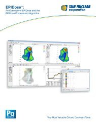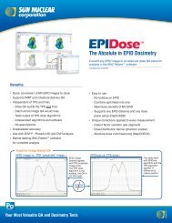
EPID Dosimetry for IMRT QA - Sun Nuclear
EPID Dosimetry for IMRT QA - Sun Nuclear
EPID Dosimetry for IMRT QA - Sun Nuclear

Create successful ePaper yourself
Turn your PDF publications into a flip-book with our unique Google optimized e-Paper software.
<strong>EPID</strong> <strong>Dosimetry</strong> <strong>for</strong> <strong>IMRT</strong> <strong>QA</strong><br />
Karl Rasmussen, Ph.D.<br />
Long Island Radiation Oncology<br />
Garden City, NY
Overview<br />
�� What is <strong>EPID</strong> dosimetry?<br />
�� Methods of <strong>EPID</strong> dosimetry<br />
�� <strong>EPID</strong>ose Algorithm<br />
�� <strong>EPID</strong>ose Physics Modeling<br />
�� Our study<br />
�� Examples
About the speaker<br />
�� Ph.D. in Medical Physics at UW<br />
�� >2 years of clinical <strong>IMRT</strong> <strong>QA</strong> experience<br />
using MapCheck at multiple centers<br />
�� Researched accuracy of <strong>EPID</strong>ose<br />
�� Currently at a center per<strong>for</strong>ming<br />
RapidArc<strong>QA</strong> using <strong>Dosimetry</strong>Check<br />
�� Disclosure<br />
– Not being paid to give this talk (Flight/hotel)<br />
– No financial interest in <strong>EPID</strong>ose or<br />
<strong>Sun</strong><strong>Nuclear</strong>
<strong>EPID</strong> Background<br />
�� <strong>EPID</strong> = Electronic Portal Imaging Device<br />
�� Used <strong>for</strong> 2D digital imaging <strong>for</strong> patient<br />
localization<br />
– kV <strong>EPID</strong><br />
�� On Board Imaging (OBI)<br />
�� Some soft tissue contrast<br />
�� Additional kV generator required<br />
– MV <strong>EPID</strong><br />
�� Portal Imaging<br />
�� Bony anatomy<br />
�� Uses treatment beam <strong>for</strong> imaging<br />
�� Most commercial <strong>EPID</strong> dosimetry solutions<br />
use MV <strong>EPID</strong> only
Current MV <strong>EPID</strong> models<br />
�� Varian – aS500/aS1000<br />
�� Siemens - Optivue<br />
�� Elekta – iViewGT<br />
Party<br />
�� 3 rd Party<br />
– TheraView<br />
– Kodak 2000RT CR Plus<br />
�� Most are standard on new linacs, many<br />
can be retrofitted on older linacs
<strong>EPID</strong> Clinical Application<br />
�� <strong>EPID</strong> Cost > $500,000<br />
�� Used to allow <strong>for</strong> digital imaging of patient localization<br />
– Faster 1 st day setup due to no film development<br />
– Digital record keeping easier than maintaining storage rooms<br />
devoted to patient films<br />
– Helps in transition to paperless charts
Not to scale<br />
How an MV <strong>EPID</strong> works<br />
�� Electrons generated in Cu<br />
plate<br />
�� Cause visible light in<br />
phosphor screen<br />
�� Photodiode measures light<br />
�� Complicated<br />
scatter/backscatter due to<br />
additional layers
How an MV <strong>EPID</strong> works<br />
�� Individual photodiode<br />
signals are sent to X and<br />
Y coordinates<br />
�� Measures charge<br />
collected in multiple 2D<br />
discrete locations<br />
�� Resolution is<br />
approximately 0.7mm
Benefits/limitations of <strong>IMRT</strong> <strong>QA</strong> techniques<br />
Method Efficiency<br />
Film<br />
IC Array<br />
Diode Array<br />
Detector<br />
Resolution<br />
Detector<br />
Density<br />
Measures<br />
Dose<br />
MV <strong>EPID</strong> ? ? ? ? ?<br />
4D
�� Easy Setup<br />
Benefits of <strong>EPID</strong> Based <strong>Dosimetry</strong><br />
– Automatic SSD positioning<br />
– Fixed relative to treatment beam<br />
�� Measurable at any gantry angle<br />
�� Image instantly available<br />
�� High data density and resolution
Problems of <strong>EPID</strong> Based <strong>Dosimetry</strong><br />
�� Fixed location relative to beam prohibits use <strong>for</strong> 4D<br />
<strong>IMRT</strong> <strong>QA</strong><br />
�� Designed <strong>for</strong> imaging, not <strong>QA</strong><br />
– Complicated scatter characteristics<br />
– Not a water phantom<br />
�� Field size response differs between <strong>EPID</strong> and water (S c,p )<br />
�� MLC transmission response differs between <strong>EPID</strong> and water<br />
�� Scatter Kernel <strong>for</strong> generated electrons differs between <strong>EPID</strong><br />
and water<br />
�� Pixel responses vary across the detector<br />
– Charge collected in an <strong>EPID</strong> is not dose
Methods of <strong>EPID</strong> <strong>Dosimetry</strong><br />
�� Varian<br />
�� RIT<br />
�� <strong>Sun</strong> <strong>Nuclear</strong><br />
�� Math Resolutions LLC<br />
All methods are arguably valid
Varian Method – Portal <strong>Dosimetry</strong><br />
�� Acquire MV <strong>EPID</strong><br />
image <strong>for</strong> each<br />
beam<br />
�� TPS calculates a<br />
simulated <strong>EPID</strong><br />
image based on the<br />
expected response<br />
of the <strong>EPID</strong><br />
Image http://www.varian.com
�� TPS calculates a<br />
simulated <strong>EPID</strong><br />
image based on<br />
the expected<br />
response of the<br />
<strong>EPID</strong><br />
Varian Method – Portal <strong>Dosimetry</strong><br />
Right image http://www.wienkav.at/kav/kfj/91033454/physik/images/eval_ws.png
�� Positives<br />
Varian Method – Portal <strong>Dosimetry</strong><br />
– Integrated with Varis/Aria systems<br />
– Fully integrated with rapidarc<br />
– Fast<br />
�� Negatives<br />
– Does not <strong>QA</strong> water-based water based TPS plan directly<br />
�� Not measuring or comparing dose<br />
�� Image to image comparison<br />
�� Creates planar image map based on <strong>EPID</strong> algorithm,<br />
not the water based TPS<br />
�� Possibility of systematic errors between water model and aSi <strong>EPID</strong> <strong>EPID</strong><br />
model<br />
– One vendor solution <strong>for</strong> TPS, <strong>QA</strong>, and delivery may not be best <strong>for</strong> <strong>for</strong><br />
independent second checks
Varian Method – Portal <strong>Dosimetry</strong><br />
�� Does not <strong>QA</strong> water-based water based TPS plan directly<br />
– Not measuring or comparing dose<br />
– Image to image comparison<br />
– Creates planar image map based on <strong>EPID</strong> algorithm, not the<br />
water based TPS<br />
�� Possibility of systematic errors between water model<br />
and aSi <strong>EPID</strong> model<br />
�� One vendor solution <strong>for</strong> TPS, <strong>QA</strong>, and delivery may not<br />
be best <strong>for</strong> independent second checks
RIT Method – RIT113<br />
�� Image is taken with 2cm of solid water<br />
buildup placed directly on the <strong>EPID</strong><br />
�� Image is calibrated using a wedged<br />
field and ION chamber measurements<br />
�� Assumes uni<strong>for</strong>m response across<br />
<strong>EPID</strong><br />
�� This is compared directly to water-<br />
based fluence from original TPS<br />
image http://www.radimage.com/epid/index.php
<strong>Sun</strong><strong>Nuclear</strong> - <strong>EPID</strong>ose<br />
�� <strong>EPID</strong> Image acquired without buildup<br />
�� Algorithm applied to raw <strong>EPID</strong><br />
– Output/FS Correction<br />
– Dose Redistribution kernel<br />
– 2D wide field calibration map<br />
�� Compared to water-based water based TPS-<br />
generated Planar Fluence<br />
�� Fast conversion/analysis process
<strong>EPID</strong>ose Physics Modeling<br />
�� Need to acquire with MapCHECK/<strong>EPID</strong>:<br />
– MLC defined field sizes at 100MU:<br />
�� 1x1<br />
�� 2x2<br />
�� 5x5<br />
�� 10x10<br />
�� 15X15<br />
�� 20X20<br />
�� 25x25<br />
�� 30x30<br />
– 20x20 MLC defined FS at 25, 50, 100, and 200 MU
<strong>EPID</strong>ose Physics Modeling<br />
�� All <strong>EPID</strong> measurements need to be at the same<br />
distance to be used <strong>for</strong> <strong>IMRT</strong> <strong>QA</strong> (140cm)<br />
�� All MapCHECK measurements need to be acquired<br />
at the TPS dose plane distance (100cm) and<br />
depth (5cm)<br />
�� Additional setups can be defined, but must have<br />
separate models (Example: a large H&N field can<br />
be measured in full at a smaller SDD)
Basic<br />
in<strong>for</strong>mation to<br />
allow <strong>for</strong> MLC<br />
type and dose<br />
plane setup<br />
conditions<br />
Used to<br />
geometrically<br />
backproject<br />
acquired image<br />
to desired dose<br />
plane<br />
<strong>EPID</strong>ose Physics Modeling
MLC-based MLC based<br />
correction<br />
<strong>for</strong> field size<br />
response<br />
differences<br />
between<br />
<strong>EPID</strong> and<br />
water<br />
phantom<br />
<strong>EPID</strong>ose - Output/FS Correction
Redistribution<br />
kernel to<br />
simulate wider<br />
electron spread<br />
in water<br />
<strong>EPID</strong><br />
(Not to scale)<br />
Water<br />
<strong>EPID</strong>ose Dose Redistribution kernel
Individual<br />
pixel dose<br />
response<br />
correction<br />
across the<br />
<strong>EPID</strong> field<br />
correlated to<br />
MapCHECK<br />
dose points<br />
<strong>EPID</strong>ose 2D wide field calibration map<br />
>1%<br />
<strong>EPID</strong> response is NOT always uni<strong>for</strong>m across the field
How do you acquire an <strong>EPID</strong> image? (Varian)<br />
�� Move <strong>EPID</strong> to standard SSD (Typically 140<br />
or 145 SSD)<br />
�� Deliver treatment beams (with couch<br />
retracted)<br />
– Aria<br />
– Portal Vision<br />
– AM Maintenance<br />
�� No additional licenses should * be required<br />
from Varian <strong>for</strong> these methods<br />
* I am not employed by either Varian or <strong>Sun</strong> <strong>Nuclear</strong>, so this is not a guarantee.
�� Aria<br />
<strong>EPID</strong>ose Acquisition<br />
– Create verification plan<br />
– Schedule plan in RT chart<br />
– Schedule Integrated Images <strong>for</strong> the fields<br />
– Deliver <strong>EPID</strong><br />
– Export portal images from the ‘review review’ tab<br />
to a DICOM file
�� Portal Vision<br />
<strong>EPID</strong>ose Acquisition<br />
– Prepare Portal Vision:<br />
�� Set up sequence template<br />
�� Set up acquisition template<br />
�� Set up DICOM export<br />
– Collect <strong>EPID</strong> images<br />
�� Create verification plan<br />
�� Treat the plan fields<br />
�� Export the portal images individually
�� AM Maintenance<br />
<strong>EPID</strong>ose Acquisition<br />
– Export MLC Files to MLC workstation<br />
– Manually enter field info in service mode<br />
– Deliver <strong>EPID</strong> image<br />
– Export acquired image
�� Load data (<strong>EPID</strong>s,<br />
MLCs, planar doses)<br />
into MapCHECK<br />
�� Batch calculate<br />
<strong>EPID</strong>ose conversion<br />
(~45 seconds to<br />
convert 7 beams)<br />
�� Compare to TPS planar<br />
dose like MapCHECK<br />
measurements<br />
<strong>EPID</strong>ose Analysis
Prostate - MapCHECK/Pinnacle TPS
Prostate – <strong>EPID</strong>ose/Pinnacle TPS
�� HN diff split<br />
H & N - MapCHECK/Eclipse TPS<br />
124
�� HN diff split<br />
H & N – <strong>EPID</strong>ose/Eclipse TPS<br />
124
<strong>EPID</strong>ose Study<br />
�� 28 <strong>IMRT</strong> patients from 6 centers were used (16 UW patients)<br />
�� Treatment sites included:<br />
– Prostate<br />
– Brain<br />
– Lung<br />
– Head & Neck<br />
�� High (10 & 15MV) and low (6MV) energy beams were used in<br />
treatments<br />
�� Comparison was made with MapCHECK diode array<br />
measurements and <strong>EPID</strong>ose converted dose planes<br />
– Allowed <strong>for</strong> measured to measured dose comparison<br />
– Fields had a uni<strong>for</strong>m shift applied to all measurements from the<br />
same setup
Results<br />
�� Results based on 26,207 points and 188<br />
fields of MapCheck/<strong>EPID</strong>ose comparison:<br />
– 1%/1mmDTA<br />
�� 84.6% ±1.3 1.3 agreement<br />
– 2%/2mmDTA<br />
�� 97.8% ±0.4 0.4 agreement<br />
– 3%/3mm DTA<br />
�� 99.7% ±0.1 0.1 agreement
�� 6MV<br />
Energy (In)Dependence<br />
– 1%/1mmDTA = 84.7% agreement<br />
– 2%/2mmDTA = 97.8% agreement<br />
– 3%/3mm DTA = 99.7% agreement<br />
�� 10MV/15MV<br />
– 1%/1mmDTA = 84.5% agreement<br />
– 2%/2mmDTA = 98.0% agreement<br />
– 3%/3mm DTA = 99.9% agreement
Prostate - MapCHECK/CMS Xio TPS
Prostate - EpiDOSE/CMS Xio TPS<br />
?
Prostate - MapCHECK/EpiDOSE<br />
Might miss small<br />
field errors
Math Resolutions LLC – <strong>Dosimetry</strong> Check<br />
�� <strong>EPID</strong> Image acquired<br />
�� Deconvolves image to primary<br />
fluence<br />
�� Backprojects secondary dose<br />
calculation <strong>for</strong> 3D dose calc (Pencil<br />
beam)<br />
�� Allows <strong>for</strong> single beam or 3D<br />
dose/DVH comparisons using patient<br />
CT<br />
�� Deconvolution/Analysis takes from 1<br />
to 9 hours to complete per patient
�� Example case:<br />
– 45Gy to Rectum<br />
– RapidArc Delivery<br />
– 2 Arcs<br />
– Calc time ~2 hours<br />
<strong>Dosimetry</strong>Check
�� Transverse slice<br />
– Purple is TPS<br />
generated Isodose<br />
– Green is<br />
reconstructed dose<br />
from <strong>EPID</strong> image<br />
– CT is patient- patient<br />
specific<br />
<strong>Dosimetry</strong>Check
�� Coronal slice<br />
– Purple is TPS<br />
generated Isodose<br />
– Green is<br />
reconstructed dose<br />
from <strong>EPID</strong> image<br />
<strong>Dosimetry</strong>Check
Note that a<br />
pencil beam<br />
algorithm<br />
will be less<br />
accurate<br />
than<br />
convolution-<br />
convolution<br />
based<br />
methods<br />
Better at<br />
showing<br />
“ballpark ballpark”<br />
in<strong>for</strong>mation<br />
<strong>Dosimetry</strong>Check
GVH data,<br />
while<br />
interesting,<br />
is new and<br />
common<br />
standards<br />
haven’t haven t been<br />
set<br />
<strong>Dosimetry</strong>Check
Conclusions<br />
�� <strong>EPID</strong>-based <strong>EPID</strong> based <strong>QA</strong> can be a valuable tool<br />
– High Data density<br />
– Easy Setup<br />
�� Many factors need to be considered <strong>for</strong> accurate<br />
comparison to a water based dose plane:<br />
– Water/<strong>EPID</strong> field size responses<br />
– MLC transmission factors<br />
– scatter kernel differences<br />
– individual pixel response variations<br />
�� Multiple methods exist that offer different<br />
benefits and accuracy<br />
– What is best <strong>for</strong> your clinic is ultimately up to you
�� Dr. Wolfgang Tomé Tom<br />
�� Ben Nelms<br />
�� Contributors<br />
– Ben Harris<br />
– Gary Adler<br />
– Shirley Yang<br />
– Bryan Coopey<br />
– David Barbee<br />
– And many others<br />
Acknowledgements