OnSite TB IgG/IgM Combo Rapid Test - Agentúra Harmony vos
OnSite TB IgG/IgM Combo Rapid Test - Agentúra Harmony vos
OnSite TB IgG/IgM Combo Rapid Test - Agentúra Harmony vos
- No tags were found...

Create successful ePaper yourself
Turn your PDF publications into a flip-book with our unique Google optimized e-Paper software.
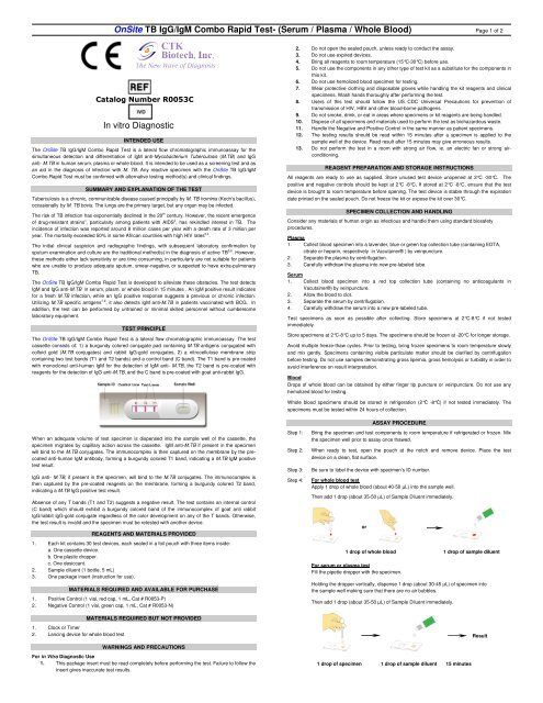
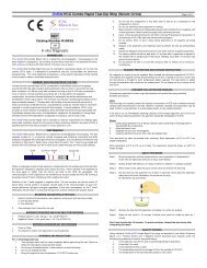
<strong>OnSite</strong> <strong>TB</strong> <strong>IgG</strong>/<strong>IgM</strong> <strong>Combo</strong> <strong>Rapid</strong> <strong>Test</strong>- (Serum / Plasma / Whole Blood) Page 1 of 2Catalog Number R0053CIn vitro DiagnosticINTENDED USEThe <strong>OnSite</strong> <strong>TB</strong> <strong>IgG</strong>/<strong>IgM</strong> <strong>Combo</strong> <strong>Rapid</strong> <strong>Test</strong> is a lateral flow chromatographic immunoassay for thesimultaneous detection and differentiation of <strong>IgM</strong> anti-Mycobacterium Tuberculosis (M.<strong>TB</strong>) and <strong>IgG</strong>anti- M.<strong>TB</strong> in human serum, plasma or whole blood. It is intended to be used as a screening test and asan aid in the diagnosis of infection with M. <strong>TB</strong>. Any reactive specimen with the <strong>OnSite</strong> <strong>TB</strong> <strong>IgG</strong>/<strong>IgM</strong><strong>Combo</strong> <strong>Rapid</strong> <strong>Test</strong> must be confirmed with alternative testing method(s) and clinical findings.SUMMARY AND EXPLANATION OF THE TESTTuberculosis is a chronic, communicable disease caused principally by M. <strong>TB</strong> hominis (Koch’s bacillus),occasionally by M. <strong>TB</strong> bovis. The lungs are the primary target, but any organ may be infected.The risk of <strong>TB</strong> infection has exponentially declined in the 20 th century. However, the recent emergenceof drug-resistant strains 1 , particularly among patients with AIDS 2 , has rekindled interest in <strong>TB</strong>. Theincidence of infection was reported around 8 million cases per year with a death rate of 3 million peryear. The mortality exceeded 50% in some African countries with high HIV rates 3,4 .The initial clinical suspicion and radiographic findings, with subsequent laboratory confirmation bysputum examination and culture are the traditional method(s) in the diagnosis of active <strong>TB</strong> 5,6 . However,these methods either lack sensitivity or are time consuming, in particularly are not suitable for patientswho are unable to produce adequate sputum, smear-negative, or suspected to have extra-pulmonary<strong>TB</strong>.The <strong>OnSite</strong> <strong>TB</strong> <strong>IgG</strong>/<strong>IgM</strong> <strong>Combo</strong> <strong>Rapid</strong> <strong>Test</strong> is developed to alleviate these obstacles. The test detects<strong>IgM</strong> and <strong>IgG</strong> anti-M.<strong>TB</strong> in serum, plasm, or whole blood in 15 minutes . An <strong>IgM</strong> positive result indicatesfor a fresh M.<strong>TB</strong> infection, while an <strong>IgG</strong> positive response suggests a previous or chronic infection.Utilizing M.<strong>TB</strong> specific antigens 7-9 , it also detects <strong>IgM</strong> anti-M.<strong>TB</strong> in patients vaccinated with BCG. Inaddition, the test can be performed by untrained or minimal skilled personnel without cumbersomelaboratory equipment.TEST PRINCIPLEThe <strong>OnSite</strong> <strong>TB</strong> <strong>IgG</strong>/<strong>IgM</strong> <strong>Combo</strong> <strong>Rapid</strong> <strong>Test</strong> is a lateral flow chromatographic immunoassay. The testcassette consists of: 1) a burgundy colored conjugate pad containing M.<strong>TB</strong> antigens conjugated withcolloid gold (M.<strong>TB</strong> conjugates) and rabbit <strong>IgG</strong>-gold conjugates, 2) a nitrocellulose membrane stripcontaining two test bands (T1 and T2 bands) and a control band (C band). The T1 band is pre-coatedwith monoclonal anti-human <strong>IgM</strong> for the detection of <strong>IgM</strong> anti- M.<strong>TB</strong>, the T2 band is pre-coated withreagents for the detection of <strong>IgG</strong> anti-M.<strong>TB</strong>, and the C band is pre-coated with goat anti-rabbit <strong>IgG</strong>.2. Do not open the sealed pouch, unless ready to conduct the assay.3. Do not use expired devices.4. Bring all reagents to room temperature (15°C-30°C) before use.5. Do not use the components in any other type of test kit as a substitute for the components inthis kit.6. Do not use hemolized blood specimen for testing.7. Wear protective clothing and disposable gloves while handling the kit reagents and clinicalspecimens. Wash hands thoroughly after performing the test.8. Users of this test should follow the US CDC Universal Precautions for prevention oftransmission of HIV, HBV and other blood-borne pathogens.9. Do not smoke, drink, or eat in areas where specimens or kit reagents are being handled.10. Dispose of all specimens and materials used to perform the test as biohazardous waste.11. Handle the Negative and Positive Control in the same manner as patient specimens.12. The testing results should be read within 15 minutes after a specimen is applied to thesample well of the device. Read result after 15 minutes may give erroneous results.13. Do not perform the test in a room with strong air flow, ie. an electric fan or strong airconditioning.REAGENT PREPARATION AND STORAGE INSTRUCTIONSAll reagents are ready to use as supplied. Store unused test device unopened at 2°C -30°C. Thepositive and negative controls should be kept at 2°C -8°C. If stored at 2°C -8°C, ensure that the testdevice is brought to room temperature before opening. The test device is stable through the expirationdate printed on the sealed pouch. Do not freeze the kit or expose the kit over 30°C.SPECIMEN COLLECTION AND HANDLINGConsider any materials of human origin as infectious and handle them using standard biosafetyprocedures.Plasma1. Collect blood specimen into a lavender, blue or green top collection tube (containing EDTA,citrate or heparin, respectively in Vacutainer® ) by veinpuncture.2. Separate the plasma by centrifugation.3. Carefully withdraw the plasma into new pre-labeled tube.Serum1. Collect blood specimen into a red top collection tube (containing no anticoagulants inVacutainer®) by veinpuncture.2. Allow the blood to clot.3. Separate the serum by centrifugation.4. Carefully withdraw the serum into a new pre-labeled tube.<strong>Test</strong> specimens as soon as possible after collecting. Store specimens at 2°C-8°C if not testedimmediately.Store specimens at 2°C-8°C up to 5 days. The specimens should be frozen at -20°C for longer storage.Avoid multiple freeze-thaw cycles. Prior to testing, bring frozen specimens to room temperature slowlyand mix gently. Specimens containing visible particulate matter should be clarified by centrifugationbefore testing. Do not use samples demonstrating gross lipemia, gross hemolysis or turbidity in order toavoid interference on result interpretation.BloodDrops of whole blood can be obtained by either finger tip puncture or veinpuncture. Do not use anyhemolized blood for testing.Whole blood specimens should be stored in refrigeration (2°Ċ -8°Ċ) if not tested immediately. Thespecimens must be tested within 24 hours of collection.ASSAY PROCEDUREWhen an adequate volume of test specimen is dispensed into the sample well of the cassette, thespecimen migrates by capillary action across the cassette. <strong>IgM</strong> anti-M.<strong>TB</strong> if present in the specimenwill bind to the M.<strong>TB</strong> conjugates. The immunocomplex is then captured on the membrane by the precoatedanti-human <strong>IgM</strong> antibody, forming a burgundy colored T1 band, indicating a M.<strong>TB</strong> <strong>IgM</strong> positivetest result.<strong>IgG</strong> anti- M.<strong>TB</strong>, if present in the specimen, will bind to the M.<strong>TB</strong> conjugates. The immunocomplex isthen captured by the pre-coated reagents on the membrane, forming a burgundy colored T2 band,indicating a M.<strong>TB</strong> <strong>IgG</strong> positive test result.Absence of any T bands (T1 and T2) suggests a negative result. The test contains an internal control(C band) which should exhibit a burgundy colored band of the immunocomplex of goat anti rabbit<strong>IgG</strong>/rabbit <strong>IgG</strong>-gold conjugate regardless of the color development on any of the T bands. Otherwise,the test result is invalid and the specimen must be retested with another device.REAGENTS AND MATERIALS PROVIDED1. Each kit contains 30 test devices, each sealed in a foil pouch with three items inside:a. One cassette device.b. One plastic dropper.c. One desiccant.2. Sample diluent (1 bottle, 5 mL)3. One package insert (instruction for use).MATERIALS REQUIRED AND AVAILABLE FOR PURCHASE1. Positive Control (1 vial, red cap, 1 mL, Cat # R0053-P)2. Negative Control (1 vial, green cap, 1 mL, Cat # R0053-N)1. Clock or Timer2. Lancing device for whole blood testMATERIALS REQUIRED BUT NOT PROVIDEDWARNINGS AND PRECAUTIONSFor in Vitro Diagnostic Use1. This package insert must be read completely before performing the test. Failure to follow theinsert gives inaccurate test results.Step 1:Step 2:Step 3:Step 4:Bring the specimen and test components to room temperature if refrigerated or frozen. Mixthe specimen well prior to assay once thawed.When ready to test, open the pouch at the notch and remove device. Place the testdevice on a clean, flat surface.Be sure to label the device with specimen’s ID number.For whole blood testApply 1 drop of whole blood (about 40-50 µL) into the sample well.Then add 1 drop (about 35-50 µL) of Sample Diluent immediately.or1 drop of whole blood 1 drop of sample diluentFor serum or plasma testFill the pipette dropper with the specimen.Holding the dropper vertically, dispense 1 drop (about 30-45 µL) of specimen intothe sample well making sure that there are no air bubbles.Then add 1 drop (about 35-50 µL) of Sample Diluent immediately.1 drop of specimen 1 drop of sample diluent 15 minutesResult
<strong>OnSite</strong> <strong>TB</strong> <strong>IgG</strong>/<strong>IgM</strong> <strong>Combo</strong> <strong>Rapid</strong> <strong>Test</strong>- (Serum / Plasma / Whole Blood) Page 2 of 2Step 5:Step 6:Set up timer.Results can be read in 15 minutes. Positive results can be visible in as short as 1 minute.Don’t read result after 15 minutes. To avoid confusion, discard the test device after interpretingthe result.QUALITY CONTROLUsing individual <strong>OnSite</strong> <strong>TB</strong> <strong>IgG</strong>/<strong>IgM</strong> <strong>Rapid</strong> <strong>Test</strong> cassettes as described in the Assay Procedure above,run 1 Positive Control and 1 Negative Control (provided upon request) under the followingcircumstances to monitor test performance:1. A new operator uses the kit, prior to performing testing of specimens.2. A new test kit is used.3. A new shipment of kits is used.4. The temperature used during storage of the kit falls outside of 2°C-30°C.5. The temperature of the test area falls outside of 15°C-30°C.Expected results are as follows:Negative ControlOnly the C band shows color development, the two T bands (T1 and T2) show no colordevelopment.Positive ControlThe C band and two T bands (T1 and T2) show color development.The appearance of any burgundy color in the T bands, regardless of intensity, must beconsidered as presence of the band.INTERPRETATION OF ASSAY RESULT1. NEGATIVE RESULT: If only the C band is present, the absence of any burgundy color in theboth T bands (T1 and T2) indicates that no anti- M.<strong>TB</strong> antibodies are detected. The result isnegative.2. POSITIVE RESULT:2.1 In addition to the presence of C band, if only T1 band is developed, indicates for thepresence of <strong>IgM</strong> anti- M.<strong>TB</strong>. The result is positive.2.2 In addition to the presence of C band, if only T2 band is developed, the test indicates forthe presence of <strong>IgG</strong> anti- M.<strong>TB</strong>. The result is positive.2.3 In addition to the presence of C band, both T1 and T2 bands are developed, indicatesfor the presence of <strong>IgG</strong> and <strong>IgM</strong> anti- M.<strong>TB</strong> . The result is also positive.PERFORMANCE CHARACTERISTICS1. Clinical Performance For <strong>IgM</strong> <strong>Test</strong>A total of 200 specimens from non-<strong>TB</strong> patients and 35 specimens from patients under anti <strong>TB</strong> treatmentwere tested by the <strong>OnSite</strong> <strong>TB</strong> <strong>IgG</strong>/<strong>IgM</strong> <strong>Combo</strong> <strong>Rapid</strong> <strong>Test</strong> and a commercial <strong>TB</strong> <strong>IgM</strong> ELISA kit.Comparison for all subjects is showed in the following table.<strong>OnSite</strong> <strong>TB</strong> <strong>IgG</strong>/<strong>IgM</strong> <strong>Combo</strong> <strong>Rapid</strong> <strong>Test</strong><strong>IgM</strong> ELISA <strong>Test</strong> Positive Negative TotalPositive 30 5 35Negative 7 193 200Total 37 198 235Relative Sensitivity: 85.7%, Relative Specificity: 96.5%, Overall Agreement: 94.9%2. Clinical Performance For <strong>IgG</strong> <strong>Test</strong>A total of 200 specimens from the non-<strong>TB</strong> patients and 35 specimens from the patients under anti <strong>TB</strong>treatment were tested by the <strong>OnSite</strong> <strong>TB</strong> <strong>IgG</strong>/<strong>IgM</strong> <strong>Combo</strong> <strong>Rapid</strong> <strong>Test</strong> and a commercial <strong>TB</strong> <strong>IgG</strong> ELISAkit. Comparison for all subjects is showed in the following table.<strong>OnSite</strong> <strong>TB</strong> <strong>IgG</strong>/<strong>IgM</strong> <strong>Combo</strong> <strong>Rapid</strong> <strong>Test</strong><strong>IgG</strong> ELISA <strong>Test</strong> Positive Negative TotalPositive 31 4 35Negative 7 193 200Total 38 197 235Relative Sensitivity: 88.6%, Relative Specificity: 96.5%, Overall Agreement: 95.3%LIMITATIONS OF TEST1. The Assay Procedure and the Assay Result Interpretation must be followed closely when testingthe presence of antibodies to M.<strong>TB</strong> in serum or plasma from individual subjects. Failure to followthe procedure may give inaccurate results.2. The <strong>OnSite</strong> <strong>TB</strong> <strong>IgG</strong>/<strong>IgM</strong> <strong>Rapid</strong> <strong>Test</strong> is limited to the qualitative detection of <strong>IgG</strong> and <strong>IgM</strong> anti-M.<strong>TB</strong> in human serum or plasma. The intensity of the test band does not have linear correlationwith the antibody titer in the specimen.3. The test also recognizes antibodies to M. bovis and M. africanum.4. An <strong>IgG</strong> positive response may be detected in BCG vaccinated personnel.5. A negative result for an individual subject indicates absence of detectable antibodies to M.<strong>TB</strong>.However, a negative test result does not preclude the possibility of exposure to or infection withM.<strong>TB</strong>.6. A negative result can occur if the quantity of the antibodies to M.<strong>TB</strong> present in the specimen isbelow the detection limits of the assay, or the antibodies that are detected are not presentduring the stage of disease in which a sample is collected.7. Immunocompromised condition such as HIV infection may reduce the test sensitivity. If HIV coinfectionis highly suspected, the <strong>OnSite</strong> <strong>TB</strong> Plus (R0055C) <strong>Rapid</strong> <strong>Test</strong> is highly recommended.8. Some specimens containing unusually high titer of heterophile antibodies or rheumatoid factormay affect expected results.9. The results obtained with this test should only be interpreted in conjunction with other diagnosticprocedures and clinical findings.REFERENCES1. Schaaf, H. S., P. Botha, N. Beyers, R. P. Gie, H. A. et, al 1996. The 5-year outcome ofmultidrug resistant tuberculosis patients in the Cape Province of South Africa. Trop. Med. Int.Health 1:718-7222. Havlir, D. V., and P. F. Barnes. 1999. Tuberculosis in patients with human immunodeficiencyvirus infection. N. Engl. J. Med. 340:367-3733. Dye L., Scheele S., V. Pathania, et al: 1999 Global Burden of Tuberculosis EstimatedIncidence, Prevalence, and Mortality by Country. WHO Global Surveillance and MonitoringProject. JAMA. 282:677-686.4. Kochi, A. 1991 The global tuberculosis situation and the new control strategy of the WorldHealth Organization. Tubercle 72:1-65. Merlin TL, Gibson DW, and DH Connor 1994. Tuberlulosis, p400-404 in Rubein E and Farber JL(ed)-Pathology, 2 nd edition. J.B. Lippincott Company6. Daniel, T. M. 1996. Immunodiagnosis of tuberculosis, p. 223-231. In W. N. Rom, and S. Garay(ed.), Tuberculosis. Letter, Brown & Co., Boston, Mass.7. Wilkens, E. G. L. 1994. The serodiagnosis of tuberculosis, p. 367-379. In P. D. O. Davies (ed.),Clinical tuberculosis. Chapman & Hall, Ltd., London, England.8. Chan ED., Heifets L., and MD Iseman. 2000 Immunological Diagnosis of tuberculosis: a review.Tuber. Lung Dis. 80: 131-140.9. Foulds, J., and R. O'Brien. 1998. New tools for the diagnosis of tuberculosis: the perspective ofdeveloping countries. Int. J. Tubercle. Lung Dis. 2: 778-783Samples with positive results should be confirmed with alternative testing method(s) and clinicalfindings before a positive determination is made.3. INVALID: If no C band is developed, the assay is invalid regardless of any burgundy color in theT bands as indicated below. Repeat the assay with a new device.European Authorized Representative:CEpartner4U , Esdoornlaan 13, 3951DB Maarn.The Netherlands. Tel.: +31 (0)6.516.536.26Manufacturer:CTK Biotech, Inc.6748 Nancy Ridge Drive, San Diego, CA 92121, USATel: 858-457-8698, Fax: 858-535-1739,E-mail: info@ctkbiotech.comPI-R0053C Rev. A Effective date: June 01-2006English versionFor Export Only, Not For Re-sale In the USAIndex of CE SymbolAttention,see instructions for useFor in vitrodiagnostic use onlyREF Catalog #Lot NumberUse by<strong>Test</strong>s per kitStore between 2-30°CDo not reuseManufacturerDate of manufacture
![[APTT-SiL Plus]. - Agentúra Harmony vos](https://img.yumpu.com/50471461/1/184x260/aptt-sil-plus-agentara-harmony-vos.jpg?quality=85)
![[SAS-1 urine analysis]. - Agentúra Harmony vos](https://img.yumpu.com/47529787/1/185x260/sas-1-urine-analysis-agentara-harmony-vos.jpg?quality=85)
![[SAS-MX Acid Hb]. - Agentúra Harmony vos](https://img.yumpu.com/46129828/1/185x260/sas-mx-acid-hb-agentara-harmony-vos.jpg?quality=85)