Trichophaeopsis tetraspora, a New Coprophilous Discomycete from ...
Trichophaeopsis tetraspora, a New Coprophilous Discomycete from ...
Trichophaeopsis tetraspora, a New Coprophilous Discomycete from ...
- No tags were found...

Create successful ePaper yourself
Turn your PDF publications into a flip-book with our unique Google optimized e-Paper software.
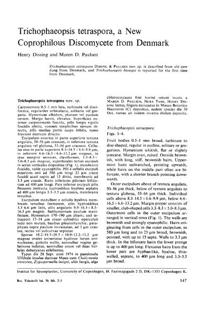
<strong>Trichophaeopsis</strong> <strong>tetraspora</strong>, a <strong>New</strong><strong>Coprophilous</strong> <strong>Discomycete</strong> <strong>from</strong> DenmarkHenry Dissing and Maren D. Paulsen<strong>Trichophaeopsis</strong> <strong>tetraspora</strong> DISSING & PAULSEN novo sp. is described <strong>from</strong> old cowdung <strong>from</strong> Denmark, and <strong>Trichophaeopsis</strong> bicuspis is reported for the first time<strong>from</strong> Denmark.<strong>Trichophaeopsis</strong> <strong>tetraspora</strong> novo sp.Carposomata 0.5-1 mm lata, turbinata vel disciformia,regulariter orbicularia, solitaria vel gregaria.Hymenium albidum, planum vel paulumcavum. Margo laevis, elevatus. Superficies exteriorcarposomatis fuscida, pilis longis rigidisfuscidis obsita, summis simplicibus sursum directis,pilis mediae partis saepe bifidis, ramobreviore deorsum directo.Excipulum exterius in parte superiore texturaangulata, 50-56 ~m crassum, in inferiore texturaangulata vel globosa, 53-66 ~m crassum. Cellulaeeius in parte superiore 8.3-14.5 x 6.6-9.9 ~m,in inferiore 6.6-16.5 x 6.6-13.2 ~m magnae, inipso margine minores, claviformes, 3.3-8.3 x5.0-8.3 ~m magnae, superficiales totius excipuliin series verticales dispositae (Fig. 1), membranisfuscidis, valde cyanophilis. Pili e cellulis excipuliexterioris orti ad 580 ~m longi 23 ~m crassifuscidi acuti septis ad 15 divisi, membranis ad3.3 ~m crassis. Rami inferiores pilorum bifidorumad 400 ~m longi. Pars inferior excipuli pilisflexuosis instructa hyphoidibus hyalinis septatisad 400 ~m longis 3.3-3.5 ~m crassis, membranistenuibus.Excipulum medullare e cellulis hyalinis membranistenuibus formatum, aliis hyphoidibus3.3-6.6 ~m latis, aliis angulatis 9.9-16.5 x 8.316.5 ~m magnis. Subhymenium paulum manifestum.Hymenium 170-190 ~m altum; asci tetraspori13-14 ~m crassi cylindrici operculatiiodo non mutati, basibus pleurorhynchis; paraphysessupra paulum incrassatae, ad 3 ~m crassae,rectae vel subcurvae septatae.Sporae 18.2-19.5-20.5 x 10.9-12.2-13.2 ~mmagnae ovales uniseriatae hyalinae laeves uninucleatae,guttulis nullis, nonnullae vagina gelatinosaindutae, nonnullae unam vel duas bullulasdebaryanas exhibentes.Typus die 28 Sept. anni 1974 in paeninsulaUlfshale insulae danicae M",en cum Cheilymeniastercorea, Zygospermella insigni, aliis fungis, algachlorococcacea fimi bovini vetusti incola aMAREN D. PAULSEN, NORA TAMS, HENRY DISSING lectus, frigore exsiccatus in Museo BotanicoHauniensi (C) depositus, eadem species die 30Oct. rursus ab iisdem inventa ibidem deposita.<strong>Trichophaeopsis</strong> <strong>tetraspora</strong>Figs. 1-4.Fruit bodies 0.5-1 mm broad, turbinate todisc-shaped, regular in outline, solitary or gregarious.Hymenium whitish, flat or slightlyconcave. Margin even, raised. Outside brownish,with long, stiff, brownish hairs. Uppermosthairs unbranched, pointing upwards,while hairs on the middle part often are bifurcate,with a shorter branch pointing downwards.Outer excipulum above of textura angulata,50-56 /lm thick, below of textura angulata totextura globosa, 53-66 /lm thick. Individualcells above 8.3-14.5 x 6.6-9.9 /lm, below 6.616.5 x 6.6-13.2 /lm. Margin proper consists ofsmaller, club-shaped cells 3.3-8.3 x 5.0-8.3 /lm.Outermost cells in the outer excipulum arrangedin vertical rows (Fig. 1). The walls arebrownish and strongly cyanophilic. Hairs originating<strong>from</strong> cells in the outer excipulum, to580 /lm long and to 23 /lm broad, brownish,pointed, with up to 15 septa. Walls to 3.3 /lmthick. In the bifurcate hairs the lower prongeis up to 400 /lm long. Flexuous hairs <strong>from</strong> thelower part are hyphae-like, hyaline, thinwalled,septate, to 400 /lm long and 3.3-3.5/lm broad.Institut for Sporeplanter, University of Copenhagen, 0. Farimagsgade 2 D, DK-1353 Copenhagen K.Bot. Tidsskrift bd. 70, hft. 2-3 147
Medullary excipulum of thin-walled, hyalinecells. Some are hyphoid, 3.3-6.6 J.lmbroad, others angular, 9.9-16.5 x 8.3-16.5J.lm. Subhymenium indistinct. Hymenium 170190 J.lm high; asci 4-spored, 13-14 J.lm broad,cylindrical, operculate, J-, with a pleurorhynchousbase; paraphyses above slightly enlarged,to 3 J.lm broad, straight or slightlycurved, septate.Spores 18.2-19.5-20.5 x 10.9-12.2-13.2 J.lm,ovale, uniseriate, hyaline, smooth, uninucleate,without guttules. Some spores with a gelatinoussheath, and some with one or two deBary bubbles.Material: M0en, Ulfshale, on old cow dung,together with i.a. Cheilymenia stercorea andZygospermella insignis, and a Chlorococcaceousalga, September 28, 1974, leg. MAREND. PAULSEN, NORA TAMs, and HENRY DISSING(Holotype, freeze-dried, in C); - Ibid. October30, idem. (several collections, C).The new species of <strong>Trichophaeopsis</strong> was foundon several samples of old cow dung when the..lc··\,·,.~·.··~.l._. \l.-.~J,l\~..:1, .'. '·"i ...•'cFig. 1. <strong>Trichophaeopsis</strong> <strong>tetraspora</strong>. - vertical rowsof cells in outer excipulum, and hairs. - <strong>from</strong>Ho1otype (C). x 325.148Fig. 2. <strong>Trichophaeopsis</strong> <strong>tetraspora</strong>. - to the left,ascus with spores with a gelatinous sheath; notethe refringent bodies in the spores, X 400. - tothe right, fruit bodies, schematic, x 20. - <strong>from</strong>Holotype (C).Bot. Tidsskrift bd. 70, hft. 2-3
TricIJoplweopsis <strong>tetraspora</strong>"Mykologisk Kongres 1974" visited UlfshaleForest in the northern part of the island ofMeen (see p. 199). More than 50 cows arefeeding in a fenced common outskirt situatedwest of the forest, with scattered groups ofjUlIiperus cOIlIl1/l/llis and PiIlIlS. Fresh cowdung is normally not especially attractive, butthese old samples with numerous fruit bodiesof yellowish-orange Cheilyml'lIia Sfercorea anda reddish colour <strong>from</strong> populations ofa Chlorococcaceousalga were very fascinating. Fur·thermore a closer examination showed a veryrich flora of Pyrenomycetes, i.a. Zygospermellail/sigllis, which is new to Denmark.Fresh material for fixation was obtained ona visit on October 30, 1974. After this visitabout 10 samples were examined in detail.Besides T. ferraspora the following coprophilousfungi were found: Cheilymellia sfercorea,Coprofus grallltli[ormis, Coproflls sexdecimsporus,Lasiobofus ciliarus, Sporormiella megalospora,Sporormiella oCfo1/alis (new to Denmark),Zygospermella insignis (new to Denmark),COlliocltaefe scatigena, Trichodelischiabisporula and COpl'illliS pellucidus. Besides theFig. 4. TricIJoplweopsis tClraspora. - a. EM photoof spore wall, b. detail of the same, arrows indicatethe position. - <strong>from</strong> Holotypc (C). a.X 6000, b. X 26000.(Method: Frceze dried material fixed for 24 h in4% glutaraldehyde in 0.1 M cacodylatc bufferat pH 7. Post fixed in 2% OS04 in the same buffer.Dehydrated in a graded ethanol series and embeddedin Spurr's medium. The sections werestained at 30 C in 2% aqueous uranyl acetalefor 30 mill and subsequently 10 min in Reynold'slead citrate).above mentioned reddish Chlorococcaceousalgaa blue-green alga were seen.Fig. 3. TricIJoplweopsis lelraspora. - a. spore,SEM photo, b. spores with gelatinous sheath,phasc contrast, c. part of sporc with transversewrinkling, in Cotton Blue, Light Microscope,d. ascus with spores, cOnlainillg refringent bodies,in water, Light Microscope. - a. & e. Ulfshale,October 30. 1974 (C), b. & d. <strong>from</strong> Holotype (C).a. x 2000, b. & d. x 650, d. x 1550.Bot. Tidsskrift bd. 70, hfl. 2-3During our studies of the material collectedon October 30, we were surprised to find thaton some spores, mounted in Cotton Blue, thespore walls showed a very delicate transversestriation (Fig. 3 c). A restudy of spores onslides of the Tian-Shan material of T. bicusp;s(see DISSING & RAITVIIR, 1973) and of Danishmaterial collected during the above mentionedcongress revealed the same very delicate striation.Wilh the Light Microscope it was notpossible to get an impression of the true natureof the striation, but the observations re-149
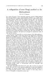
Henry Dissing and Maren D. Paulsencalled the studies by RIFAI (1968) and ERB(1972) on spore material of the genus Rhizoblepharia.This genus was erected (RIFAI, l.c.)because the transverse wrinkling was consideredto be "unique among the Pezizineae".For a number of reasons this genus is consideredto be closely related to the genus<strong>Trichophaeopsis</strong> (KORF & ERB, 1972). Spores<strong>from</strong> fresh material were then studied in theScanning Electron Microscope. The wall wascompletely smooth (Fig. 3 a). This needs not,however, be in discordance with ERB's (l.c.)observations on Rhizoblepharia, since it is herethe inner spore wall which is transversallywrinkled.Spores <strong>from</strong> freeze-dried material were thenstudied with EM. All layers in the wall wereseen to be smooth (Fig. 4).A satisfactory explanation for the curiousstriation in T. bicuspis and T. <strong>tetraspora</strong> wasthus not found. SEM and EM studies providedno support for this as a fact.Presence or not of guttules in the spores ofT. bicuspis has been much disputed (for a reviewof the literature on the subject, see KORF& ERB, 1972 and DISSING & RAITVIIR, 1973).In 1974 we had the opportunity to study, atthe same time, fresh material of T. bicuspisand T. <strong>tetraspora</strong>. No guttules, droplets orgranules were seen in spores mounted in water.For T. <strong>tetraspora</strong> this is no surprise, since, tooUI knowledge, no coprophilous discomyceteshave spores with guttules.A refringent body (Figs. 2 & 3 d) could beseen in the spores of T. <strong>tetraspora</strong>. The positionof this, and its size, were very similar tothat of the nucleus.<strong>Trichophaeopsis</strong> bicuspis has never been described<strong>from</strong> Denmark. It has now been foundon three localities and a description is providedbelow.<strong>Trichophaeopsis</strong> bicuspis (Bouo.) KORF & ERBSyn.: Trichophaea bicuspis (Bouo.) Bouo.Fruit bodies 2-3 mm broad, turbinate to discshaped,regular in outline, solitary or gregarious.Hymenium whitish, flat. Margin even,raised. Outside brownish, with long stiff brown150hairs, some of which are bifurcate, with ashorter branch pointing downwards.Outer excipulum above of textura angulata,39-43 flm thick, below of textura angulatato textura globosa, 50-95 flm thick. Individualcells above 7.3-13.3 x 10.6-15.5 flm, below16-20 x 16-23 flm. Cells in the margin aresmaller, club-shaped, 5.6-9.9 x 5.0-6.6 flm.Outermost cells in outer excipulum are arrangedin vertical rows (as in T. <strong>tetraspora</strong>,see Fig. 1). They are with brownish walls, andstrongly cyanophilic. The hairs originate <strong>from</strong>cells in outer excipulum, they are up to 770 flmlong, 14.9-19.8 flm broad, pointed, with upto 15 septa, the walls up to 5.8 flm thick. Inthe bifurcate hairs the lower branch is up to380 flm long. Flexuous hairs <strong>from</strong> the lowerpart are hyphae-like, hyaline, thin-walled, septate,to 660 flm long and 3-4 flm broad. Medullaryexcipulum of thin-walled cells. Some arehyphae-like, 3-4 flm broad, others angular,6.6-9.9 x 6.6-13.2 flm. Subhymenium indistinct.Hymenium 215-240 flm high; asci 8spored, 13-14 flm broad, cylindrical, operculate,J-, with a pleurorhynchous base. Paraphysesabove slightly enlarged, 2-3 flm broad,septate. Spores 15.2-15.8-16.8 x 9.3-9.5-9.8flm, ovale, uniseriate, hyaline, smooth, withoutguttules. Some spores with one or two deBary bubbles.Material: Island of Sams0, near Tranebjerg,on moist soil and decaying leaves, under adense cover of Ranunculus repens, July 31,1968, leg. L. D0SSING (D0SSING, private herb.);- ibid., August 1968 (C); - Jylland, RingelmoseSkov, under Populus tremula, September21, 1970, leg. H. DISSING (C); - M0en,Ulfshale Skov, on small twigs of Populus sp.,September 28, 1974, leg. L. D0SSING (C).<strong>Trichophaeopsis</strong> bicuspis is new to Denmark.On Sams0 it has been found several times inthe same locality (D0SSING, unpublished). Inmost characters it is much alike T. <strong>tetraspora</strong>.The distinguishing characters may be summarizedas in the following scheme.Bot. Tidsskrift bd. 70, hft. 2-3
habitus. . . . . . . ..T. <strong>tetraspora</strong>cow-dungsize of fruit body 0.5-1 mmthickness of wallsin hairs. . . . . . .. 3.3 Jlmlength ofascus .. 170-190 Jlmnumber of spores 4size of spores ... 18.2-20.5 x10.9-13.2 JlmT. bicuspissoil and decayingleavesor twigs2-3 mm5.8 Jlm215-240 Jlm815.2-16.8 x9.3-9.8 JlmGAMUNDI (1973) described Trichophaeaeguttulispora <strong>from</strong> Argentina. No materialwas seen by us, but the description and drawingsclearly indicate a species congeneric withT. bicuspis and T. <strong>tetraspora</strong>. It is separated<strong>from</strong> both on its ochroleucous to pallid avellaneushymenium.AcknowledgementWe want to express our best thanks to LEIFD0SSING, who generously placed his materialof <strong>Trichophaeopsis</strong> bicuspis at our disposal.ULRIK S0CHTING made the EM studies, andTYGE CHRISTENSEN prepared the Latin diagnosis.J. FUGLSANG NIELSEN, Institut for historiskgeologi og palaeontologi, University ofCopenhagen, operated the Cambridge ScanningElectron Microscope, and LENE CHRISTIANSEN prepared the photographs. We appreciatetheir co-operation.LiteratureDISSING, H. & A. RAITVIIR, 1973: <strong>Discomycete</strong>s ofMiddle Asia 11. Thelebolaceae, Ascobolaceae,Pyronemataceae and Pezizaceae <strong>from</strong> the TienShan Mountains. - Eesti NSV TA Toimet.BioI. 22,2: 124-131.ERB, R. W., 1972: A new species of the genusRhizoblepharia <strong>from</strong> the Neotropics, and aredisposition of the genus in the Pyronemataceae,Pseudombrophileae. - Phytologia 24,1:5-14.GAMUNDI, 1. J., 1973: <strong>Discomycete</strong>s de Tierradel Fuega 11. Especies nuevas de Humariaceae.- BoI. Soc. Argent. Bot. 15,1: 85-92.KORF, R. P. & R. W. ERB, 1972: The genus<strong>Trichophaeopsis</strong>. - Phytologia 24,1: 15-19.RIFAI, M. A., 1968: The Australasian Pezizalesin the Herbarium of the Royal Botanic GardensKew. - Verh. K. Ned. Akad. Wet., Afd.Natuurk. 11, 57,3: 1-295.Bot. Tidsskrift bd. 70, hft. 2-310151