

View - Andreas Prokop Lab
View - Andreas Prokop Lab
View - Andreas Prokop Lab
- No tags were found...

Create successful ePaper yourself
Turn your PDF publications into a flip-book with our unique Google optimized e-Paper software.
Cell Tissue Res (1999) 297:169–186 © Springer-Verlag 1999REVIEW<strong>Andreas</strong> <strong>Prokop</strong>Integrating bits and pieces: synapse structure and formationin Drosophila embryosReceived: 12 August 1998 / Accepted: 17 November 1998Abstract During the development of the nervous system,numerous neurons connect to form complex networks. Inorder to build a functional network each neuron has toestablish contacts with appropriate target cells, and atthese contacts synapses of the right quality and strengthhave to be formed. Gaining insight into the mechanismsunderlying this complex development is an importantstep towards a better understanding of how the nervoussystem is formed and behaviour generated. One modelsystem in which synapse formation can be studied at themorphological, physiological and molecular level is thatof the fruitfly Drosophila, and insights gained from Drosophilaembryos are reviewed here. The first part of thisreview deals with the neuromuscular junction as thebest-known synaptic contact in Drosophila. It describes:(1) its structure, (2) mechanisms underlying the formationof the neuromuscular cell junction and the arborisationof the presynaptic terminal, and (3) our present understandingof signal-dependent and -independent processesduring synapse formation at the neuromuscularjunction. The last part of this review deals with the questionof how particular neurons can adopt specific synapticproperties, stating as an example the development ofthe neural lineage of NB7-3, which gives rise to two serotonergicneurons.Key words Adhesion · Cell lineage · Development ·Neuromuscular junction · SerotoninIntroductionThe nervous system lies at the heart of behaviour, receiving,integrating and processing sensory information,storing information and generating distinct patterns ofThe author is funded by the Deutsche ForschungsgemeinschaftA. <strong>Prokop</strong> (✉)Institut für Genetik-Zellbiologie, Becherweg 32,D-55128 Mainz, Germanye-mail: prokop@mail.uni-mainz.deTel.: +49 6131 394328; Fax: +49 6131 394584motor or gland activity. The cellular basis for this abilitylies in the complex but highly ordered networks consistingof numerous neurons with distinct properties, connectedto one another via synaptic cell junctions. In orderto build these networks, neurons have to carry out atleast three main developmental tasks. Firstly, they have toestablish connections with the right target cells, thus determiningthe paths of information flow. Secondly, theconnecting neurons have to form the correct types of synapsesat the contacts they establish. This can be electricalsynapses, chemical synapses with excitatory, inhibitory ormodulatory characteristics, or they can be of mixed nature.Each type of synapse will have a particular impacton the neuronal circuit, as it transmits and modifies informationin distinct ways. Finally, the strength of transmissionbetween cells has to be regulated, for instance by thenumber of synapses formed between two cells.The nature of synapses seems to depend on propertieswhich the synaptic partner cells have acquired independently,as a function of their developmental history. Inaddition, during the process of cell contact formation,cell signalling is required to align synaptic structures atprecisely apposed sides of the cell junction. Furthermore,cell signalling might influence the specificity of the synapticcomponents. For example, neurons in the sympatheticperipheral ganglia of vertebrates initially acquireadrenergic properties as a function of their developmentalhistory (see for example Hirsch et al. 1998 and referencestherein), but the adrenergic quality can be changedinto a cholinergic fate, depending on the type of targetcells that are innervated (Kuno 1995).In order to understand synapse formation we have to:(1) identify the structural and molecular components requiredat developing and functional synapses, (2) unravelthe signalling and assembly processes involved in theirformation and (3) ask by which gene regulatory eventsthe required molecules are provided. One model systemin which different aspects of synaptogenesis can be studiedand hopefully one day be integrated comprehensivelyis the embryonic nervous system of the fruitfly Drosophilamelanogaster. The first part of this review focuses on
1711 This may be different for some imaginal NMJs; for example inthe moth some NMJs show wrapping by glial processes (Stockerand Nüesch 1975; Rheuben and Reese 1978).2 At the larval NMJ there are two pools of vesicles, a readily releasablepool at the active zone and an actin- and shibire-dependentreserve pool located in the centre of the bouton (Wu and Bellen1997; Kuromi and Kidokoro 1998). In late embryonic boutonsvesicles tend to concentrate mainly at the active zone (Broadie etal. 1995; see Fig. 2).dissect the mechanisms underlying its formation andfunction (Keshishian et al. 1996).In the Drosophila embryo a stereotypical pattern of30 somatic muscles forms per abdominal hemisegment,and each muscle is individually identifiable by its characteristicshape and position. Drosophila muscles aresingle polynucleated fibres and much of their developmentis well studied (Fig. 1; Bate 1993; Baylies et al.1998; Ruiz-Gómez 1998). Each muscle is innervated byat least one identified motoneuron, of which most can beassigned to a particular neural lineage (Landgraf et al.1997; Figs. 1, 5; see later). Motoneurons have a characteristicsoma position in the ventral nerve cord and distinctdendritic and axonal projections. Most (if not all)embryonic neuromuscular terminals are glutamatergic,and they form branches on the muscle surface, with reproducibleshape differences between different muscles(Johansen et al. 1989a; Fig. 2A vs. B). Also the point ofnerve entrance and position of the NMJ on the musclesurface are reproducible, although position does not appearto be essential to neuromuscular transmission, asshown by displaced NMJs in prospero mutant embryos(Broadie and Bate 1993d). The branches of motoneuronalterminals show more or less reproducible numbers ofvaricosities, called boutons (Fig. 2A,B,E; Johansen et al.1989a; Broadie and Bate 1993c). The embryonic boutons,which have a diameter of up to 1 µm, are attachedto the muscle surface on one side and on the other sideface the haemolymph, covered only by basement membrane(Fig. 2F). The attachment between muscle andneuronal surface is a non-specialized junction with acleft of about 15 nm. This junction is interspersed withsynapses, which are stretches of membrane with characteristicstructural specialisations and that are believed tobe the sites of neural transmission (Figs. 2F,R, 3A; Broadieet al. 1995; <strong>Prokop</strong> et al. 1996). Interestingly, and incontrast to vertebrates, the fully functional embryonicDrosophila NMJ is not covered by glial cells, leaving10–20 µm of neuronal surface without insulation fromthe hemolymph 1 (Fig. 2F; Hall and Sanes 1993; Auld etal. 1995). However, after larval hatching, NMJ morphologychanges, and the neuronal terminal submerges belowthe muscle surface, so that the type I boutons appear tobe relatively shielded from the hemolymph by the musclecell (Fig. 2G; Atwood et al. 1993; Jia et al. 1993).Further changes occurring after larval hatching are: (a)immense growth of the NMJ, (b) increase in numbers ofsynapses, (c) more than twofold increase in the numberof vesicles 2 (own observations), and (d) extensive infoldingof the muscle membrane opposite to the neuronal terminal(called subsynaptic reticulum, SSR; see Fig. 2F vs.G; for review, see Budnik 1996). Mechanisms regulatingthese postembryonic changes are under intense investigation,turning the larval NMJ into an important modelsystem to study mechanisms underlying neuronal plasticity(reviewed in Budnik 1996; Davis and Goodman 1998).The structure of neuromuscular synapsesAlthough the architecture of NMJs changes dramaticallyduring postembryonic development, the characteristicsof neuromuscular synapses are similar between embryoand larva (Fig. 3Ai vs. ii, D vs. inset in D). This alsoholds true for NMJs which develop in vitro (Seecof et al.1972). At neuromuscular synapses the membranes appearsmoother and more electron dense compared to theextrasynaptic areas of the neuromuscular junction (Fig.2F,R), suggesting that the molecular composition of synapticmembranes differs from extrasynaptic membranes.The intracellular face of the presynaptic membrane harboursactive zones, which contain T-shaped structures ofelectron-dense material (T-bars) surrounded by clustersof synaptic vesicles (Fig. 3, white arrowheads). T-barscan still be seen in neuronal terminals that are depletedof synaptic vesicles and therefore appear to be independentmolecular structures (Poodry and Edgar 1979). Themolecular composition and function of T-bars remainsunclear, although studies of larval NMJs indicate a correlationbetween the number of T-bars and the strength ofsynaptic transmission (Jia et al. 1993; Stewart et al.1996). In NMJs of the moth Manduca or the lobsterHomarus which are similar to neuromuscular synapsesof Drosophila (by criteria of conventional transmissionelectron microscopy), freeze-fracture analysis reveals T-bars to be associated with clusters of particles within thepresynaptic membrane (arrow in Fig. 3B,C; Rheubenand Reese 1978; Rheuben 1985; Walrond et al. 1993).Similar electron-dense presynaptic particles have beenfound in vertebrates and other invertebrates. They arethought to be calcium channels which, upon stimulation,mediate the presynaptic high calcium peaks that induceexocytosis of synaptic vesicles (Heuser et al. 1979;Pumplin et al. 1981; Robitaille et al. 1990; Llinás et al.1992). Therefore, T-bars may contain intracellular portionsof calcium channels and/or molecules involved intheir clustering, and might thus be a key element of synapticrelease (Rheuben 1985; Walrond et al. 1993). Alternatively,T-bars might be involved in the trapping andclustering of vesicles at the active zone, as vesicles oftenappear to be physically attached to T-bars (Fig. 3A; Osborne1966; Koenig and Ikeda 1996). Similarly, vesiclescluster at presynaptic dense material of vertebrate NMJs,and loss of this dense material (in s-laminin mutantmice) correlates with a failure of vesicle clustering(Noakes et al. 1995). A third potential function of T-barsmight be an involvement in one specific pathway of reformationof synaptic vesicles: In photoreceptor neuronssynaptic vesicles bud off the presynaptic membrane ei-
172
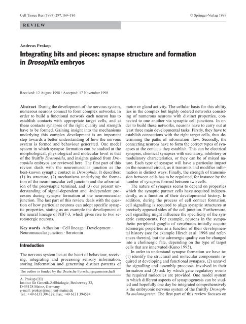
Fig. 2A–U Phenotypes of NMJs, central synapses and other releasesites. All images except E,G,H and N are late-stage 17; A–E lightmicroscopicimages of NMJs stained with synaptic markers (A anti-Kakapo, B–D anti-Synaptotagmin, E anti-cysteine string protein;see Table 1); F–M ultrastructural images of boutons; N–U details ofsynaptic or junctional structures. A,B NMJs (black arrowheads) ondifferent muscles show characteristic shape differences (A dorsal,B ventral muscle field). C,D NMJs in mutant embryos (same area asin B): kakapo mutant NMJs are reduced (C), myoblast city mutantmuscles and NMJs are reduced but ectopic presynaptic terminalsform in the area (D). E NMJ boutons and branches have grown atlate larval stages (same NMJ as in B); unstained line (bent black arrowin close-up) represents most likely vesicle-free axolemma (cf.G). F At late embryonic NMJs, neuronal boutons ( * ) are half coveredby basement membrane (open arrowhead) and half attached tomuscle (mu) via an unspecialised junction (small open arrow),which is interspersed with synapses (black arrow; cf. R). G At latelarval NMJs boutons are completely surrounded by folds of themuscle membrane (SSR) and enriched with vesicles, except in theaxolemma (bent black arrows indicate microtubules in axolemma;cf. E). H At late stage 16, the neuromuscular cell junction (smallopen arrow) consists of only short stretches of apposed neuronaland muscle surfaces and harbours immature synapses (cf. N).J lamininA mutant NMJs lack basement membrane attachment andalso neuromuscular contact is reduced. K At mef2 mutant NMJsboutons are separated from the muscle by basement membrane(open arrowhead) and presynaptic structures are mislocalised (blackarrows). L Classical neurohemal release site (black arrow) of a peptidergicvaricosity with dense-cored vesicles attached to muscle,most likely corresponding to larval-type III boutons (Jia et al. 1993;Martínez-Padrón and Ferrús 1997). M Synapses in the central nervoussystem. N Neuromuscular synapse at late stage 16 exhibiting apresynaptic dense body with clustered vesicles and a stretch of electron-densemembranes. O Neuroneural synapse outside the CNSwith presynaptic T-bar (see also Yoshihara et al. 1997). P Centralsynapse with postsynaptic densities (bent white arrows). Note thatsynapses in N–P lack structured material in the synaptic cleft (incontrast to R). Q Neurohemal active zone in myoblast city mutantembryo covered by basement membrane (open arrowhead; rarecases exist also in wild type; <strong>Prokop</strong> et al. 1996). R,S Neuromuscularsynapse with structured material in the cleft (between white arrowsin R), which appears as a honeycomb-like pattern in obliquesections (white arrow in S). T,U Ribbons of septa (between whitearrows) in synaptic cleft of epidermal pleated septate junctions(pSJ) are similarly spaced as cleft material at NMJs (cf. T with R),and look similar in oblique sections (cf. U with S) [bent black arrowscore of the bouton (axolemma), bent white arrows postsynapticdensity, DO/DA dorsal oblique and acute muscles, black arrowssynapses or release sites, black arrowheads boutons, asterisks presynapticor releasing boutons, ep epidermis, kak kakapo mutant embryo,L3 third larval instar, mbc, myoblast city mutant embryo, mumuscle, open arrowheads basement membrane, pSJ pleated septatejunction, small open arrows extrasynaptic cell junction, SSR subsynapticreticulum, st.16 embryonic stage 16, VL ventral longitudinalmuscles, VO ventral oblique muscles, white arrows demarcate orindicate material in junctional cleft, wt wild type]. Muscle nomenclatureas in Bate 1993. Scale bars 15 µm (A–E), 480 nm (F–M),255 nm (N–U)ther below the T-bar or extrasynaptically, and both populationscan be distinguished (Koenig and Ikeda 1996). Incontrast to extrasynaptic reformation, the vesicles at T-bars form more rapidly, without cisternal intermediatesand can be blocked by high-Mg 2+ /low-Ca 2+ solution.A further feature of neuromuscular synapses in Drosophilaembryos and larvae is the presence of regularlystructured electron-dense material in the synaptic cleftover a distance of several hundred nanometres (betweenbent arrows in Fig. 3). In transverse sections a continuousfine line can be seen in the middle of the cleft, and athick dashed line closely associated with the postsynapticmembrane (Fig. 3A). Oblique sections through thesynaptic cleft show a patch of honeycomb-like pattern(Fig. 3D). Freeze-fracture analyses of comparable neuromuscularsynapses in the moth Manduca and the lobsterHomarus reveal areas of stippled material in the externalleaflet of the muscle membrane opposite the T-bar, suggestingthat the structured material in the cleft is of postsynapticnature and anchors in the muscle membrane(black arrowheads in Fig. 3B,C,E; Rheuben and Reese1978; Walrond et al. 1993). Synaptic proteins likely toform part of these postsynaptic structures are transmembranemolecules like the ionotropic glutamate receptors,voltage-gated Shaker potassium channels and the cell adhesionmolecule (CAM) Fasciclin 2 (Fas2; Fig. 4C; Broadieand Bate 1993c; Saitoe et al. 1997; Tejedor et al.1997; Thomas et al. 1997a; note that Fas2 is expressedalso on the presynaptic side). Interestingly, the honeycomb-likepattern in oblique NMJ sections is very similarto patterns seen in oblique sections through anothertype of cell junction, the so-called pleated septate junction(Fig. 2S vs. U; Baumgartner et al. 1996). In transversesections of pleated septate junctions transverselines can be seen spanning the cleft every 20–25 nm, andthis frequency of repetition is similar to that of the thickdashed line at NMJs (Fig. 2R vs. T). In spite of a similarorganisation, the extracellular material at pleated septatejunctions looks different from that at NMJs and is likelyto include Neurexin IV, an unusual member of theNeurexin family of proteins which has not been found atNMJs (Baumgartner et al. 1996). However, common cytoskeletalelements might arrange the different structuralelements at both junction types into similar honeycomblikepatterns. A likely candidate for such an organisationalcytoskeletal element is the Discs large (Dlg) protein,which is localised both at NMJs and pleated septatejunctions and has been shown to organize transmembraneproteins at junctional sites (Budnik 1996).Formation of the neuromuscular junctionInitiation of neuromuscular contact173In Drosophila, NMJ formation has been studied mostthoroughly on the ventral longitudinal muscles VL3 andVL4 (nomenclature according to Bate 1993). However,all or most embryonic NMJs might develop in a similarway, as they are glutamatergic and share a commonstructural organisation. At stage 15/16 muscles send outfilopodia preferentially from the future site of innervation(Suzuki et al. 1999), and these are contacted by filopodiasent out from the motoneuronal growth cones in arandom fashion (Fig. 4A). Those motoneuronal processeswhich are in contact with inappropriate muscles aresoon withdrawn (Broadie et al. 1993; Yoshihara et al.1997). Selection for the right muscle is thought to be mediatedby repulsion from inappropriate and contact-medi-
174Fig. 3A–E Ultrastructural details of neuromuscular synapses.Images show details of neuromuscular synapses analysed withconventional transmission electron microscopy in Drosophila(A,D) or freeze-fracture technique in Manduca (B,E). C Schematicrepresentation of an attachment between bouton (bo) andmuscle (mu) opened up on two sides towards the front. The muscleexhibits myofilaments (mf), the bouton contains mitochondria(mi), vesicles (v) and T-bars (white arrowhead), and both are coveredby basement membrane (bm). Pre- and postsynaptic membranesare partially unfolded, and external (e) and protoplasmic(p) membrane leaflets shown. Positions of the plates A,B,D and Eare indicated in the scheme; chosen symbols indicate the samedistinct structures throughout the figure. A Transverse sectionthrough a larval (i) or late embryonic (ii) contact between neuronalbouton (bo) and muscle (bottom), forming synapses with similardimensions and features (width of the synapse defined by adashed line of material in the synaptic cleft; between bent arrows).White arrowheads indicate presynaptic T-bars (with a roofand a stem) surrounded by synaptic vesicles ( * indicates fusion orreformation events of vesicles at the presynaptic membrane).Obliquely sectioned T-bars (iii larval example) reveal the prongednature of their horizontal roof. B Freeze-fracture images of comparablesynapses in Manduca show concentrations of membraneparticles (black arrow) on the protoplasmic membrane leaflet (p)believed to be accumulations of calcium channels at the base ofT-bars. D Oblique sections through the dashed material in thesynaptic cleft of the late larva or late embryo (inset) reveal honeycomb-likepatterning of the material (between bent arrows; seealso Fig. 2S). E Freeze-fracture images of the external leaflet ofthe postsynaptic membrane reveal oval particle fields (black arrowhead,cf. B), suggesting that the extracellular material may beanchored in the postsynaptic membrane. (B and E reprinted withpermission from Rheuben and Reese 1978). Scale bar 300 nm
ated attraction to the right target muscle (Fig. 4A; reviewedin Bate and Broadie 1995; Keshishian et al.1996). For example, the RP3 neuron grows past Toll-expressingmuscles VO4 and VO5 to innervate the non-Toll-expressing muscles VL3 and VL4. Taking away expressionof the transmembrane molecule Toll from VO4and VO5 causes some extra branching in that area,whereas misexpression of Toll on VL3 and VL4 will repelthe RP3 motoneuron and retard the neuromuscularcontact (Rose et al. 1997). In contrast, the homophilicCAM Fasciclin 3 (Fas3) is specifically expressed on theRP3 motoneuron and at the future innervation site onmuscles VL3 and 4. Ectopic expression of Fas3 on neighbouringmuscles is sufficient to redirect RP3 innervationto these non-target cells, but not if Fas3-expression iseliminated on the RP3 growth cone (Kose et al. 1997).Thus, Fas3 appears to function as a homophilic synapticrecognition molecule mediating attractive cell-cell interactionsbetween RP3 and muscles VL3/4 (stippled linesin Fig. 4A). However, loss of function of Fas3 has no apparenteffect on targeting and innervation, and this mightbe explained by the fact that the target recognition code isredundant (see, for example, Speicher et al. 1998). Observationssimilar to those of Fas3 have been made for Connectin,another homophilic CAM specifically expressedon a different subset of muscles and neurons (Nose et al.1992; Raghavan and White 1997). Thus, specificity of innervationis believed to involve combinatorial functionsof several cell-specific CAMs or repellents. In addition,this first specific contact requires general factors, such asthe transmembrane protein commissureless on all muscles(not shown in Fig. 4). In the absence of commissurelessthe motoneurons stall in close vicinity of their targetmuscles or project beyond (Wolf et al. 1998). Once a tolerableneuromuscular contact is established, its furtherdifferentiation appears to depend on properties which appearto be common to most or even all muscles and motoneurons.This is suggested by the observation that persistentfunctional NMJs differentiate, even when motoneuronsare misrouted to contact wrong muscles (Keshishianet al. 1996; Kose et al. 1997).Adhesive propertiesat the differentiating neuromuscular contact175If particular combinations of specifically expressedCAMs participate in the neuromuscular recognitioncode, they might also mediate adhesion at newly formedneuromuscular cell junctions (connected rectangles inFig. 4B). Usually these junctions consist of short stretchesof apposed membranes, often interrupted by stretchesof non-connected cell surfaces (Schuster et al. 1996b;Yoshihara et al. 1997; <strong>Prokop</strong> et al. 1998a). Such contactsdevelop into an extended cell junction at the matureNMJ of late stage 17 embryos (Fig. 4B vs. C), suggestingthat adhesive properties change during NMJ differentiation.Micrographs of vertebrate NMJs in vivo and invitro also show an initially narrow and often discontinuousjunctional cleft of about 10–20 nm width, which lateron widens to about 50 nm and accumulates a basementmembrane (Kullberg et al. 1977; Takahashi et al.1987; Hall and Sanes 1993). This clearly indicates a molecularreorganisation of cell adhesion properties. At theDrosophila NMJ, the width of the junctional cleft doesnot change, but nevertheless molecular changes of adhesiveproperties occur during NMJ maturation, as suggestedby several observations:During NMJ maturation, expression of Fas3, whichpotentially contributes to the early phase of adhesion,vanishes from neurons, muscles and NMJs (Broadie andBate 1993c). Connectin vanishes from the extrajunctionalmuscle surfaces but remains in the neuron, and stainingat the NMJ persists into late larval stages (M. Landgraf,personal communication; own observations). Thiscould either mean a complete downregulation of Connectinpostsynaptically, or that Connectin becomes restrictedto the postsynaptic site (Fig. 4C). Furthermore,the homophilic CAM Fas2 is initially expressed stronglyon the surfaces of all motor axons and at low levels onall muscles but, during NMJ formation, Fas2 is progressivelyrestricted to the NMJ in pre- and postsynapticcells (Schuster et al. 1996b). The late neuromuscular restrictionof Fas2 is mediated by the cytoskeletal elementDiscs large (see later; Thomas et al. 1997a; Zito et al.1997; Fig.4C), which itself is not detectable at the NMJuntil late stage 17 (Guan et al. 1996). Deleting Fas2function does not affect NMJ formation in the embryobut strongly affects postembryonic stabilisation andmaintenance of NMJs, suggesting a late requirement forFas2 3 (Schuster et al. 1996b). Even more, it seems to beof developmental importance that Fas2 function is restrictedto the late phase of NMJ differentiation: Overexpressionof Fas2 in muscles during the early phase ofNMJ formation seems to render muscle surfaces too„sticky“ and interferes with neuromuscular target recognition,in that ectopic motoneuronal branches (whichnormally occur only transiently) become trapped andform NMJs on inappropriate muscles (Davis et al. 1997).Taken together, NMJ differentiation appears to involvedynamic but regulated changes in expression and localisationof CAMs, and different CAMs appear to servedistinct functions at different times during this process.A second argument in favour of a switch in adhesiveproperties at the NMJ comes from analysis of mef2 mutantembryos, where the initial NMJ contact can be geneticallyseparated from the late phase of neuromuscular adhesion.In mef2 mutant embryos, muscle founder cells(FC in Fig. 1) form and express muscle-specific markersincluding the CAMs Connectin and Fas3, and motoneuronsestablish contact with their appropriate target cells(<strong>Prokop</strong> et al. 1996). However, the muscle founder cellsremain immature and fail to acquire properties of general3 During larval life the adhesion of Fas2 not only stabilises NMJsbut even inhibits their further growth, and postembryonic growth atthe NMJ is promoted by a modest downregulation of Fas2 (Schusteret al. 1996a).
176muscle differentiation such as contractile filaments, theirproper attachment to the epidermis, and their ability tofuse into multinucleate fibres. Micrographs of late-stage17 mef2 mutant embryos never revealed proper cell junctionsbetween motoneurons and improperly differentiatedmuscle fibres, but instead their cell surfaces are coveredby basement membrane (Fig. 2 K; <strong>Prokop</strong> et al. 1996).This phenotype could be explained by loss of mef2-dependentlate synaptic CAMs, directly maintaining theneuromuscular cell junction. Alternatively, mef2-functionmight be required to exclude basement membrane receptorsfrom the NMJ (e.g. via synapse-specific cytoskeletalproperties), thus preventing separation of the pre- andpostsynaptic membranes by competitive invasion of basementmembrane material (white arrowheads in Fig. 4B).So far, basement membrane receptors have not yetbeen identified. Loss of cell surface adhesion to basementmembrane in laminin A mutant embryos suggeststhat basement membrane receptors bind to Laminin orLaminin-dependent extracellular matrix components(<strong>Prokop</strong> et al. 1998a). In addition, lack of Laminin Aleads to a partial detachment of neuromuscular boutons 4(Fig. 2J), even though basement membrane is normallyabsent from the narrow neuromuscular cleft in Drosophila(Fig. 3A; <strong>Prokop</strong> et al. 1998a). This suggests that athin layer of Laminin in the synaptic cleft might existand mediate cell adhesion via appropriate receptors atthe NMJ. Alternatively, basement membrane, whichspans over the neuronal terminal and adheres closely tosurrounding muscle surfaces, might press the terminalagainst the muscle (Fig. 4B,C). If this were the case,weak adhesion at the neuromuscular cleft during the processof NMJ formation would suffice (provided mef2-dependentfactors kept basement membrane receptors awayfrom the NMJ; white arrowheads in Fig. 4B). Low neuromuscularadhesion would facilitate the extensive reorganisationof the terminal’s shape during NMJ formation.This would be consistent with the fact that plasticreshaping of neuronal terminals in Drosophila larvae orin the sea slug Aplysia is at least partly dependent on adecrease in synaptic CAMs, i.e. a reduction in cell adhesion(Schuster et al. 1996a; Thompson et al. 1996; Baileyet al. 1997).Differentiation of nerve terminal shape and sizeDifferentiation and shape changes of growth cones intobouton-forming terminals take place during 4–5 h afterinitial neuromuscular contact at early stage 13 (Broadieand Bate 1993c; Yoshihara et al. 1997). These changesappear to be accelerated or facilitated by late bloomergene function, which encodes a member of the tetraspaninfamily of receptor-complex-associated proteins.4 Reduction of neuromuscular contact was also found in s-lamininmutant mice; s-laminin is part of the basement membrane withinthe ca. 50-nm-wide neuromuscular cleft and might well mediateadhesion to synaptic receptors (Noakes et al. 1995).The Drosophila late bloomer protein is localised in motoneuronalaxons (Kopczynski et al. 1996), suggesting thatmotoneurons receive signals. Such signals might comefrom the target muscles, inducing the differentiation processof the presynaptic terminal (not shown in Fig. 4). Inagreement with this interpretation, retrograde signallingat the onset of NMJ differentiation has been described inother systems. For example, at the vertebrate NMJ muscle-releasedAgrin seems to serve such a function (Rüeggand Bixby 1998). Also certain Heliosoma motoneurons inculture show presynaptic calcium influx upon contactwith appropriate muscle fibres (Funte and Haydon 1993;Zoran et al. 1993). However, in the case of Drosophila,loss of late bloomer function only delays NMJ differentiationbut does not block it (Kopczynski et al. 1996), suggestingthat a potential muscle-derived signal might onlybe of minor importance. This is in accordance with otherfindings that differentiation of bouton-like motoneuronalstructures can take place even in the complete absence ofmuscles (<strong>Prokop</strong> et al. 1996).Also the size of the motoneuronal terminal appearsrelatively independent of the target muscle. This is suggestedby observations in myoblast city mutant embryos,where myoblast fusion is blocked due to lack of myoblastcity function within the muscle (Erickson et al.1997). As a result myoblast city mutant muscles aremononucleated and much smaller, and also NMJ size isseverely reduced. However, the motoneuronal terminalsdo not adjust to this fact, but instead grow ectopicbranches which contain displaced presynaptic structuresFig. 4A–C Summary model of embryonic NMJ formation in Drosophila.The scheme shows the situation before contact (A), in theimmature (B) and the mature junction (C). Stages are indicated topright, symbols in the box below. A At about stage 15 the growthcone (GC) approaches the muscle, both forming filopodial extensionsand expressing cell-specific cell adhesion molecules (specificCAMs) and non-specific Fas2 (Fas2). Repellents (on muscle2) preventestablishment of false contact and transient extensions are retracted.Early CAMs (on growth cone and muscle1) might attracteach other (stippled lines) during the process of target recognition.Glutamate (little dots) is detectable in the terminal around the timeof contact formation and vesicles (stippled circle) must form soon,as first transmission occurs within 30 min after neuromuscular contact.B The early cell junction might be formed by the specificCAMs involved in target recognition (connected rectangles). Basementmembrane (BM) forms and adheres to receptors on muscleand neuron (BM-recept.), perhaps holding the presynaptic terminalagainst the muscle surface. Basement membrane appears to be excludedfrom the synaptic cleft (white arrowheads) by a functiondownstream of the transcription factor mef2 (arrow from nucleus).Glutamate receptors (Glu-recept.) are initially evenly distributedbut, upon neuromuscular contact, cluster in response to presynapticelectrical activity (zigzag arrow). kakapo function mediatesbranching of the presynaptic terminal, and active zones (AZ) startassembling. C Specific CAMs either fade from the NMJ or theybecome restricted to the NMJ (stippled rectangles). Fas2 is restrictedto the NMJ pre- and postsynaptically and appears to manifestthe status of the synapse at late embryogenesis. On the postsynapticside, Fas2 is clustered, together with Shaker channels, by Discslarge (Dlg). Dlg might localise to the NMJ via binding to synapsespecificcytoskeletal elements (three-pronged stars; so far purelyhypothetical) or to synaptic transmembrane proteins (not shown).The amount of Glu-Rs is upregulated as a function of presynapticelectrical activity (arrow from nucleus)
177(Fig. 2D,Q; <strong>Prokop</strong> et al. 1996). A comparable intrinsicdetermination of neurons to elaborate a typically sizedterminal has been demonstrated in vivo for sensory neuronsof crickets, cockroaches and Drosophila (Murpheyand Lemere 1984; Bacon and Blagburn 1992; Canal etal. 1998) and in vitro for motoneurons of crayfish (Arcaroand Lnenicka 1995; Zoran et al. 1996).Like in myoblast city, kakapo mutant embryos showseverely reduced NMJs (and reduced dendritic trees inthe central nervous system; see Fig. 2C for NMJ phenotype).In contrast to myoblast city, embryos carryingstrong alleles of kakapo have normal-sized muscles andmotoneuronal terminals form no ectopic branches. Theseobservations suggest a presynaptic requirement for kakapofunction and, accordingly, anti-Kakapo antisera detectthe protein at motoneuronal terminals (<strong>Prokop</strong> et al.1998b). Cloning data suggest that kakapo encodes a cytoskeletalelement with actin-binding properties (Gregoryand Brown 1998; Strumpf and Volk 1998). Furthermolecular components involved in the growth of the em-
178bryonic Drosophila NMJ are unknown. As insects appearto lack intermediate filaments (Bartnik and Weber1989), growth and maintenance of the motoneuronal terminalare likely to involve mainly actin and tubulin andthose elements interacting with these structural proteins(Caroni 1997; Suter and Forscher 1998).Synapse formation at the NMJIn addition to nerve terminal attachment to target muscles,the assembly of synaptic structures has to be induced atthe neuromuscular contact zones and pre- and postsynapticsites have to be precisely aligned. A hypothetical modelof this process would envisage cleft-spanning adhesionmolecules to function as tags which localise additionalsynaptic proteins. For example, at central synapses in vertebrates,Neuroligins and Neurexins (both transmembraneproteins) seem to bind to each other across the synapticcleft and associate intracellularly with protein complexesof synaptic proteins such as voltage- or transmitter-gatedchannel subunits (Irie et al. 1997; Missler and Südhof1998). An essential linker protein within these complexesis the postsynaptic density protein PSD-95, which containsthree protein-binding PDZ (common domain ofPSD-95, Dlg and ZO-1) domains. While channel proteinsbind to the first two PDZ domains (PDZ1, PDZ2), Neuroliginsbind to the third PDZ domain (PDZ3) of PSD-95.The third PDZ domain of PSD-95 can additionally bind toCRIPT (cysteine-rich interactor of PDZ3), a factor whichseems to interact with the microtubule cytoskeleton (Niethammeret al. 1998). Thus, PDZ domain proteins potentiallyfunction as linkers between: (1) cytoskeleton (seealso Allison et al. 1998), (2) components involved intransmission, and (3) membrane-spanning proteins whichbind to similar complexes on the other side of the synapticcleft. Such complexes could explain the aggregation andalignment of pre- and postsynaptic components at the preciselyapposed sites.Formation of postsynaptic structuresof Drosophila musclesAt the postsynaptic face of the Drosophila NMJ, voltagegatedShaker channels and the CAM Fas2 have similarlybeen shown to bind to the PSD-95 homologue Discs large(Dlg) in vivo and in vitro (Fig. 4C). As Fas2 moleculesare likely to bind to each other across the synaptic cleft,they seem good candidates to localise Dlg at the NMJ.However, the PDZ1 and PDZ2 domains of Dlg to whichFas2 (and Shaker) bind are not required for the localisationof Dlg (Tejedor et al. 1997; Thomas et al. 1997a;Zito et al. 1997). Instead, the PDZ3 domain and/or thehook domain of Dlg are potentially involved in the localisationof Dlg to the NMJ (Tejedor et al. 1997; for parallelstudies of Dlg localisation in the epidermis see alsoHough et al. 1997). The hook domain may bind to actinassociatedproteins of the protein4.1/ERM (common domainof Ezrin, Radixin and Moesin) family (Hough et al.1997). PDZ3-binding proteins have not yet been identifiedin Drosophila, but might be homologous to transmembraneproteins such as Neuroligins or cytoskeletalcomponents like CRIPT. Thus, Fas2 is not the initial targetingfactor for Dlg, but Dlg function is required for thetargeting of Fas2. Therefore, earlier events are likely tooccur at the neuromuscular site, most likely installing tagmolecules to which Dlg can bind (three-pronged stars inFig. 4B,C). This is in agreement with the finding thatNMJ-specific localisation of Fas2 and Dlg occurs relativelylate (as discussed above), several hours after transmissionis first detectable at the NMJ (Broadie and Bate1993c). Thus, the dlg functions uncovered so far appearto be of little importance to embryonic but they are crucialfor postembryonic NMJ development. 5What are the events essential to embryonic NMJ formation,preceding dlg-mediated clustering at the NMJ?Prior to innervation the muscles express glutamate receptors(GluRs), which are evenly distributed over themuscle membrane (Fig. 4A; Broadie and Bate 1993c;Currie et al. 1995; Saitoe et al. 1997). Clustering ofGluRs at the postsynaptic site is one of the first events ofsynapse formation, and GluR clustering does not requireDlg (Fig. 4B; Broadie and Bate 1993c; Peterson et al.1997; Saitoe et al. 1997). The incoming neuron instructsGluR clustering (Broadie and Bate 1993d) and this functionis impaired if sodium-based action potentials of theneuron (zigzag arrow in Fig. 4; Drosophila muscles exhibitcalcium currents) are blocked genetically or withtetrodotoxin (Broadie and Bate 1993a; Saitoe et al.1997). Glutamate immunoreactivity can be detected inneuronal terminals around the time of first neuromuscularcontact (small dots in Fig. 4A), and transmission setsin approximately 30 min thereafter (Johansen et al.1989b; Broadie and Bate 1993c). Thus, early transmissionmay trigger GluR clustering. However, in manipulatedor mutant embryos which lack transmission but notaction potentials, the clustering of GluRs occurs and synapsesform normally (Broadie et al. 1995). Therefore,GluR clustering at the postsynaptic site is neither merelycontact mediated nor simply dependent on early glutamaterelease. The molecular mechanisms that underliethis process are subject to speculation. For example, presynapticcalcium signalling through voltage-gated calciumchannels might be involved. A glance at cholinergicNMJs of vertebrates does not provide further insights:clustering of acetylcholine receptors at vertebrate NMJsdoes not require action potentials (Dahm and Landmesser1991), but is instead dependent on release of presynapticAgrin (Rüegg and Bixby 1998), which has so farnot been identified in Drosophila.Apart from clustered GluRs and Shaker channels, furtherchannel proteins are inserted into the muscle mem-5 Note that taking away dlg function transiently during embryogenesisand reinstalling it afterwards causes postembryonic structuraldefects which become apparent more than a day later (Guanet al. 1996).
179Table 1 Gene products and transmitters localised or expected tobe at developing or mature synapses of Drosophila. This table listsmore than 100 components found in Drosophila, which have eitherbeen shown to be localised at synapses or which are potentiallysynaptic. Some of these components are found at synapses aswell as in extrasynaptic regions, others are specific to synapses butdo not discriminate between different synapse types, but some arelocalised just at specific synapse classes (not distinguished). Synapticcomponents comprise: (a) molecules involved in synaptic architecture(e.g. clustering of synaptic elements, adhesion orshape), (b) molecules conferring the electrical properties to cellmembranes, (c) releasable chemical substances, i.e. transmittersand neuropeptides, (d) proteins involved in synthesis, destructionor reuptake of transmitters, (e) components required for the trafficking,fusion and recycling of synaptic vesicles, (f) metabotropicand ionotropic receptors for transmitters or neuropeptides, (g)components involved in signalling and second messenger pathways,(h) a protein required for gap junctions at electrical synapses,and (i) several other components of yet undefined function.Most of these components are expected to be gene products [exceptbiogenic amines and amino acids in (f)], and in many casesgenes have been assigned (in italics). Abbreviations (in brackets)and/or full names are given on the left side and correspond to thenomenclature used in Flybase (Flybase 1998). Only one referenceper component is given due to space limitations, but further referencescan be looked up in Flybase (http://flybase.bio.indiana.edu)a) Adhesion/clustering/structure Fasciclin 1 (Fas1) [1]Fasciclin 2 (Fas2) Immunoglobulin superfamily (N-CAM) [2]Fasciclin 3 (Fas3) Immunoglobulin superfamily [3]Toll (Tl) Leucine-rich family [4]Connectin (Con) Leucine-rich family [5]commissureless (comm) Transmembrane protein [6]Neurotactin (Nrt) Inactive esterase dom. (like neuroligin) [7, 8]Cadherin-N (CadN) Neuronal form [9]discs large 1 (dlg 1) MAGUK protein, PSD-95 homologue [10]Ca/calm. dep. prot. kin. (Caki) MAGUK, CASK homologue [8, 11]kakapo (kak) Actin-binding protein [12]b) Electrical membrane properties ether a go-go (eag) Voltage-gated K channel [13]Shaker (Sh) Voltage-gated K channel [14]hyperkinetic (Hk) β-Subunit to Shaker [15]Shaker cognate b (Shab) Voltage-gated K channel [16]Shaker cognate l (Shal) Voltage-gated K channel [16]Shaker cognate w (Shaw) Voltage-gated K channel [16]seizure (sei) (=erg), voltage-gated K channel [17](Ca-α1D) Voltage-gated Ca channel α 1 -subunit D [18]nightblind A (nbA) (=cac) Ca-channel, α 1 -subunit [19]paralytic (para) Voltage-gated Na channel [20]slowpoke (slo) Ca-gated K channel [21]temp.-induced paral. (tipE) Membr. prot. interact. with para [22]Nervana 1+2 (Nrv1, 2) ATP-dependent Na/K pump [23]c) Transmitter/neuropeptides Glutamate Amino acid [24]Histamine Biogenic amine [25]Serotonin Biogenic amine [26]Octopamine Biogenic amine [27]GABA Biogenic amine [28]Nitric oxide [29]amnesiac (amn) PACAP-like peptide [30]FMRF Cardioexcitatory neuropeptide [31]Small cardioactive peptide Neuropeptide [32]Substance P Neuropeptide [33]Corazonin (Crz) Neuropeptide [34](CAP-2b) Cardioaccel. pept.-2b [35]Bombyxin (Bom) Neuropeptide [36]Prothoracicotr. horm. (Ptth) Neuropeptide[36]Diuretic hormone (DH) Neuropeptide [36]Allatotropin (Atn) Neuropeptide [36]Allatostatin (Ast) Neuropeptide [36]Proctolin Neuropeptide [37]Insulin-like peptide Neuropeptide [38]Leucokinin I Neuropeptide [39]
180Table 1 continuedd) Transmitter metabolism Choline acetyl transf. (Cha) ACh synthesis [40]Acetylchol. esterase (Ace) ACh removal [41]glut. acid decarb. 1 (Gad1) GABA synthesis [42]glut. acid decarb. 2 (Gad2) GABA synthesis [43]inebriated (ine) Non-vesicular GABA (?) transporter [44]pale (ple) (=TH); catecholamine synthesis [26]Dopa decarboxylase (Ddc) Serotonin+catecholamine synthesis [45]Henna (Hn) (=Pah) serotonin (?) synthesis [46, 47]α-methyl dopa-resist. (amd) Similar to Ddc [48]Serotonin transp. (SerT) Non-vesicular transporter [49]Histidine decarbox. (Hdc) Histamine synthesis [50]Tyrosine decarboxylase Octopamine synthesis [51]Tyramine β-hydrox. (Tbh) Octopamine synthesis [52]Nitric oxide synth. (Nos) [53]e) Vesicle storage, release, recycling syntaxin 1A (syx1A) Core complex, cell membrane [54]n-synaptobrevin (n-syb) Core complex, vesicular [55]synaptobrevin (syb) Ubiquitous form of syb [56]Synapse prot. 25 (Snap25) Core complex, cell membrane [57]synaptotagmin (syt) Endocytosis+exocytosis, vesicular+cell membrane [58]Synapsin-1 (Syn) Vesicular [59]Ras opposite (Rop) sec1, munc-18; cell membrane [60]Sol. NSF att. prot. (Snap) α-SNAP, soluble [61]comatose (comt) NSF, soluble [62]Rab-protein 3 (Rab3) Vesicular [56]Cysteine string prot. (Csp) [63]leonardo (leo) 14-3-3 α-protein [64]α-Adaptin (α-Adaptin) Recycling [65]β-Adaptin (Bap) Recycling [66]stoned A+B (stnA, stnB) (=sesC) recycling [67]shibire (shi) Dynamin; recycling [68]f) Transmitter/neuropeptide receptor (nAcRα–7E) (=Dα3) ionotropic ACh-R subunit [69](nAcRα–96Aa) (=ALS) ionotropic ACh-R subunit [69](nAcRα–96Ab) (=SAD) ionotropic ACh-R subunit [69](nAcRβ-64B) (=ARD) ionotropic ACh-R subunit [69](nAcRβ-96A) (=SBD) ionotropic ACh-R subunit [69](Glu-RIIA) Non-NMDA glutamate-R [70](Glu-RIIB) Glutamate-R [71](Glu-RI) Kainate-type glutamate-R [72](Nmdar1) NMDA-type glutamate-R [73](Glu-RA) Metabotropic glutamate-R [74]Octopamin-rec. (Ocr) Metabotropic Tyramin/Octopamin-R [75](Oamb) (Metabotropic) Octopamine-R [76](5-HT1A+5-HT1B) 2 Metabotropic Serotonin-Rs [77]Dopamine-rec. 2 (DopR2) (=Damb) [78]Insulin-like-Rec. (InR) [38]Resistance to dieldrin (Rdl) Ionotropic GABA-R [79](Lcch3) Ionotropic GABA-R, β-subunit [79]Glycine rec. (Grd) GABA-+glycine-R-like subunit [80]ora transientless (ort) Glutamate-gated chloride channel [81]g) Signalling/second messenger pathways dunce (dnc) Phosphodiesterase II [82]rutabaga (rut) Ca/calmodulin-dependent adenylate cyclase [82]Ca/calm. dep. kin. (CaMKII) Ca-binding protein [83]Calmodulin (Cam) Ca-binding protein [84]Ras-Raf [85]Calbindin 53E (cbp53E) Ca-binding protein [86]Frequenin (Frq) Ca-binding protein [87]Calcineurin A1 (CanA1) Ca-sensitive Ser/Thr phosph. [88]myospheroid (mys) βPS integrin [89, 90]multiple edem. wings (mew) αPS1 integrin [90]inflated (if) αPS2 integrin [90]scab (scb) (=Volado); α-integrin [90]still life (sif) GDP-GTP exchanger [91]
of gene products potentially localised at Drosophila synapses(Table 1). Mutations of the respective genes can beused to determine their potential requirement for synapseformation. On the other hand, synaptic markers can betaken to assay the mechanisms leading to their expression.One example is the serotonergic neurons in the centralnervous system of the Drosophila embryo (Vallesand White 1988). Serotonin is a transmitter or neuromodulatorthat, in the ventral nerve cord, is restricted totwo cells per hemisegment which are derived from theneural precursor NB7-3 (see Fig. 5). Hence, serotonin isa highly specific marker for a particular synaptic propertyof two identified cells. These two serotonergic cellsare a model with which to trace the development of latesynaptic properties back to the neural precursor.In vitro experiments with cultured neuroblasts lead tothe differentiation of serotonergic cells, suggesting that,once NB7-3 has segregated, it carries out an intrinsic lineageprogramme culminating in serotonin expression(Huff et al. 1989). A variety of genes potentially involvedin or activated by this lineage programme havebeen found to be expressed within the NB7-3 lineage,i.e. the genes dopa decarboxylase, SerT, eagle, engrailed,huckebein, islet, pdm1, seven up and zfh-2 (Fig.5; Corey et al. 1994; Demchyshyn et al. 1994; Lundelland Hirsh 1994; Broadus et al. 1995; Higashijima et al.1996; Dittrich et al. 1997; Thor and Thomas 1997; Lundelland Hirsh 1998). Dopa decarboxylase is one of theenzymes catalysing Serotonin synthesis and SerT is anon-vesicular transporter required for reuptake of Serotonininto the nerve terminal. Hence these two genes arerequired very late in the serotonergic pathway, and all ofthem appear restricted to the two serotonergic cells. Allof the other genes are (potential) transcription factorsand therefore good candidates for components of a generegulatory cascade. For example, islet is expressed latewithin the lineage and is not sufficient but necessary forDopa decarboxylase and Serotonin expression, perhapsas a direct regulator of Dopa decarboxylase (Thor andThomas 1997). The eagle gene is expressed throughoutthe NB7-3 lineage, and in its absence most serotonin expressionis lost, although the NB7-3 lineage is stillformed (Higashijima et al. 1996; Dittrich et al. 1997;Lundell and Hirsh 1998). The expression of engrailedand huckebein occurs prior to neuroblast segregation inthe ectodermal layer and their absence might cause misspecificationof NB7-3, as indicated by the loss of eagleexpression (and lack of Serotonin; Dittrich et al. 1997).The principal mechanisms involved in the specificationof neural precursors are summarised in Fig. 5.It remains to be resolved to what extent other propertiesof the serotonergic cells such as their pathfindingand target selection are determined by these mechanisms,and how the postsynaptic partner cells developand link to the presynaptic terminals. Furthermore, it remainsto be seen to what degree rigid lineage mechanismscontribute to synaptic development, as opposed tocell communication and more flexible regulation or plasticity(see „Introduction“). Hopefully, future work on theembryonic nervous system of Drosophila will provide uswith answers to these questions and help us to gain deeperinsights into the fundamental mechanisms underlyingthe formation and function of neuronal circuits.Acknowledgements I would like to thank Michael Bate, NirupamaDeshpande, Matthias Landgraf, Natalia Sánchez-Soriano andJoachim Urban for discussion and critical comments on the manuscript,Matthias Landgraf for personal communication of results,Rachel Drysdale for helpful comments on the table, Mary Rheubenfor comments on Fig. 3, Yoshiaki Kidokoro for some specialadvice and the unknown reviewers for constructive criticism. I amgrateful to Michael Bate and Kendal Broadie for their guidance,discussion and collaboration during my NMJ-related projects inCambridge, and to Gerd Technau, who encouraged me to writethis review. Given the amount of literature existing in the field, Iapologise to those whose work is not cited or referred to by reviewsdue to space limitation or because it was missed by mistake.References183Allison DW, Gelfand VI, Spector I, Craig AM (1998) Role of actinin anchoring postsynaptic receptors in cultured hippocampalneurons: differential attachment of NMDA versus AMPAreceptors. J Neurosci 18:2423–2436Arcaro KF, Lnenicka GA (1995) Intrinsic differences in axonalgrowth from crayfish fast and slow motorneurons. Dev Biol168:272–283Atwood HL, Govind CK, Wu C-F (1993) Neuromuscular junctionultrastructure of ventral abdominal muscles in Drosophila larvae.J Neurobiol 24:1008–1024Auld VJ, Fetter RD, Broadie K, Goodman CS (1995) Gliotactin, anovel transmembrane protein on peripheral glia, is required toform the blood-nerve barrier in Drosophila. Cell 81:757–767Bacon JP, Blagburn JM (1992) Ectopic sensory neurons in mutantcockroaches compete with normal cells for central targets. Development115:773–784Bailey CH, Kaang BK, Chen M, Martin KC, Lim CS, Casadio A,Kandel ER (1997) Mutation in the phosphorylation sites ofMAP kinase blocks learning-related internalization of apCAMin Aplysia sensory neurons. Neuron 18:913–924Baines RA, Bate M (1998) Electrophysiological development of centralneurons in the Drosophila embryo. J Neurosci 18:4673–4683Bartnik E, Weber K (1989) Widespread occurrence of intermediatefilaments in invertebrates; common principles and aspectsof diversion. Eur J Cell Biol 50:17–33Bate M (1993) The mesoderm and its derivatives. In: Bate M,Martínez Arias A (eds) The development of Drosophila melanogaster.Cold Spring Harbor <strong>Lab</strong>oratory Press, Cold SpringHarbor, pp 1013–1090Bate M, Broadie K (1995) Wiring by fly: the neuromuscularsystem of the Drosophila embryo. Neuron 15:513–525Baumgartner S, Littleton JT, Broadie K, Bhat MA, Harbecke R,Lengyel JA, Chiquet-Ehrismann R, <strong>Prokop</strong> A, Bellen HJ(1996) A Drosophila neurexin is required for the formationand function of septate junctions. Cell 87:1059–1068Baylies MK, Bate M, Ruiz-Gómez M (1998) Myogenesis: a viewfrom Drosophila. Cell 93:921–927Bhat KM (1998) Cell-cell signalling during neurogenesis: someanswers and many questions. Int J Dev Biol 42:127–139Blagburn JM, Alexopoulos H, Davies JA, Bacon JP (1998) A nullmutation in shaking-B eliminates electrical, but not chemical,synapses in the Drosophila giant fibre system: a structuralstudy. J Comp Neurol 404:449–458Bodmer R, Jan YN (1987) Morphological differentiation of theembryonic peripheral neurons in Drosophila. Roux’s ArchDev Biol 196:69–77Bossing T, Technau GM (1994) The fate of the CNS midline progenitorsin Drosophila as revealed by a new method for singlecell labelling. Development 120:1895–1906
184Bossing T, Udolph G, Doe CQ, Technau GM (1996) The embryonicCNS lineages of Drosophila melanogaster. I. The lineagesderived from the ventral half of the truncal neuroectoderm.Dev Biol 179:41–64Broadie K, Sink H, Van Vactor D, Fambrough D, Whitington PM,Bate M, Goodman CS (1993) From growth cone to synapse:the life history of the RP3 motor neuron. Development (Suppl):227–238Broadie K, <strong>Prokop</strong> A, Bellen HJ, O’Kane CJ, Schulze KL, SweenyST (1995) Syntaxin or synaptobrevin function downstream ofvesicle docking in Drosophila. Neuron 15:663–673Broadie KS, Bate M (1993a) Activity-dependent development ofthe neuromuscular synapse during Drosophila embryogenesis.Neuron 11:607–619Broadie KS, Bate M (1993b) Development of larval muscle propertiesin the embryonic myotubes of Drosophila melanogaster.J Neurosci 13:167–180Broadie KS, Bate M (1993c) Development of the embryonic neuromuscularsynapse of Drosophila melanogaster. J Neurosci 13:144–166Broadie KS, Bate M (1993d) Innervation directs receptor synthesisand localization in Drosophila embryo synaptogenesis. Nature361:350–353Broadus J, Skeath JB, Spana E, Bossing T, Technau GM, Doe CQ(1995) New neuroblast markers and the origin of the aCC/pCCneurons in the Drosophila CNS. Mech Dev 54:1–10Budnik V (1996) Synapse maturation and structural plasticity atDrosophila neuromuscular junctions. Curr Opin Neurobiol 6:858–867Burrows M (1996) The neurobiology of an insect brain. OxfordUniversity Press, OxfordCampos-Ortega JA, Hartenstein V (1997) The embryonic developmentof Drosophila melanogaster. Springer-Verlag, BerlinCanal I, Acebes A, Ferrús A (1998) Single neuron mosaics of theDrosophila gigas mutant project beyond normal targets andmodify behaviour. J Neurosci 18:999–1008Caroni P (1997) Intrinsic neuronal determinants that promote axonalsprouting and elongation. Bioessays 19:767–775Corey JL, Quick MW, Davidson N, Lester HA, Guastella J (1994)A cocaine-sensitive Drosophila serotonin transporter: cloning,expression, and electrophysiological characterisation. ProcNatl Acad Sci U S A 91:1188–1192Currie DA, Truman JW, Burden SJ (1995) Drosophila glutamatereceptor RNA expression in embryonic and larval muscle fibers.Dev Dyn 203:311–316Dahm LM, Landmesser LT (1991) The regulation of synaptogenesisduring normal development and following activity blockade.J Neurosci 11:238–255Davis GW, Goodman CS (1998) Genetic analysis of synaptic developmentand plasticity: homeostatic regulation of synapticefficacy. Curr Opin Neurobiol 8:149–156Davis GW, Schuster CM, Goodman CS (1997) Genetic analysis ofthe mechanisms controlling target selection: target-derivedfasciclin II regulates the pattern of synapse formation. Neuron19:561–573Delgado R, Barla R, Latorre R, <strong>Lab</strong>arca P (1989) L-glutamate activatesexcitatory and inhibitory channels in Drosophila larvalmuscle. FEBS Lett 243:337–342Demchyshyn LL, Pristupa ZB, Sugamori KS, Barker EL, BlakelyRD, Wolfgang WJ, Forte MA, Niznik HB (1994) Cloning, expression,and localisation of a chloride-facilitated, cocainesensitiveserotonin transporter from Drosophila melanogaster.Proc Natl Acad Sci U S A 91:5158–5162Dittrich R, Bossing T, Gould AP, Technau GM, Urban J (1997) Thedifferentiation of the serotonergic neurons in Drosophila ventralnerve cord depends on the combined function of the zincfinger proteins Eagle and Huckebein. Development 124:2515–2525Erickson MR, Galletta BJ, Abmayr SM (1997) Drosophila myoblastcity encodes a conserved protein that is essential formyoblast fusion, dorsal closure, and cytoskeletal organization.J Cell Biol 138:589–603Ferguson EL, Anderson KV (1992) Decapentaplegic acts as amorphogen to organize dorsal-ventral pattern in the Drosophilaembryo. Cell 71:451–461Flybase (1998) Flybase: a Drosophila database. Nucleic Acids Res26:85–88Funte LR, Haydon PG (1993) Synaptic target contact enhancespresynaptic calcium influx by activating cAMP-dependentprotein kinase during synaptogenesis. Neuron 10:1069–1078Gregory SL, Brown NH (1998) kakapo, a gene required for adhesionbetween cell layers in Drosophila, encodes a large cytoskeletallinker protein related to plectin and dystrophin. J CellBiol (in press)Gu X, Spitzer NC (1995) Distinct aspects of neuronal differentiationencoded by frequency of spontaneous Ca 2+ transients. Nature375:784–787Guan B, Hartmann B, Kho Y-H, Gorczyca M, Budnik V (1996) TheDrosophila tumor suppressor gene, dlg, is involved in structuralplasticity at a glutamatergic synapse. Curr Biol 6:695–706Hall ZW, Sanes JR (1993) Synaptic structure and development: theneuromuscular junction. Cell/Neuron 72/10 (Suppl):99–121Heuser JE, Reese TS, Dennis MJ, Jan Y, Jan L, Evans L (1979) Synapticvesicle exocytosis captured by quick freezing and correlatedwith quantal transmission release. J Cell Biol 81:275–300Higashijima S, Shishida E, Matsuyaki M, Saigo K (1996) Eagle, amember of the steroid-receptor superfamily, is expressed in asubset of neuroblasts and regulates the fate of their putativeprogeny in the Drosophila CNS. Development 122:527–536Hirsch M-R, Tiveron M-C, Guillemot F, Brunet J-F, Goridis C(1998) Control of noradrenergic and Phox2a expression byMASH1 in the central and peripheral nervous system. Development125:599–608Hough CD, Woods DF, Park S, Bryant PJ (1997) Organising afunctional junctional complex requires specific domains of theDrosophila MAGUK discs large. Genes Dev 11:3242–3253Huff R, Furst A, Mahowald AP (1989) Drosophila embryonicneuroblasts in culture: autonomous differentiation of specificneurotransmitters. Dev Biol 134:146–157Irie M, Hata Y, Takeuchi M, Ichtchenko K, Toyoda A, Hirao K,Takai Y, Rosahl TW, Südhof TC (1997) Binding of neuroliginsto PSD-95. Science 277:1511–1515Jan YN, Jan LY (1993) The peripheral nervous system. In: Bate M,Martínez-Arias A (eds) The development of Drosophila melanogaster.Cold Spring Harbour Press, New York, pp 1207–1244Jia X-X, Gorczyca M, Budnik VJ (1993) Ultrastructure of neuromuscularjunctions in Drosophila – comparison of wild-typeand mutants with increased excitability. J Neurobiol 24:1025–1044Johansen J, Halpern ME, Johansen K, Keshishian H (1989a) Stereotypicmorphology of glutamatergic synapses on identifiedmuscle cells of Drosophila larvae. J Neurosci 9:710–725Johansen J, Halpern ME, Keshishian H (1989b) Axonal guidanceand the development of muscle fiber-specific innervation inDrosophila embryos. J Neurosci 9:4318–4332Keshishian H, Broadie K, Chiba A, Bate M (1996) The Drosophilaneuromuscular junction: a model system for studying synapticdevelopment and function. Annu Rev Neurosci 19:545–575Koenig JH, Ikeda K (1996) Synaptic vesicles have two distinct recyclingpathways. J Cell Biol 135:797–808Kopczynski CC, Davis GW, Goodman CS (1996) A neural tetraspanin,encoded by late bloomer, that facilitates synapse formation.Science 271:1867–1870Kose H, Rose D, Zhu X, Chiba A (1997) Homophilic synaptic targetrecognition mediated by immunoglobulin-like cell adhesionmolecule Fasciclin III. Development 124:4143–4152Kullberg WG, Lentz TL, Cohen MW (1977) Development of themyotomal neuromuscular junction in Xenopus laevis: an electrophysiologicaland fine-structural study. Dev Biol 60:101–129Kuno M (1995) The synapse: function, plasticity and neurotrophism.Oxford University Press, OxfordKuromi H, Kidokoro Y (1998) Two distinct pools of synaptic vesiclesin single presynaptic boutons in a temperature-sensitiveDrosophila mutant, shibire. Neuron 20:917–925
185Landgraf M, Bossing T, Technau GM, Bate M (1997) The origin,location, and projection of the embryonic abdominal motorneuronsof Drosophila. J Neurosci 17:9642–9655Littleton JT, Stern M, Schulze K, Perin M, Bellen HJ (1993) Mutationalanalysis of Drosophila synaptotagmin demonstrates itsessential role in Ca 2+ -activated neurotransmitter release. Cell74:1125–1134Llinás R, Sugimori M, Silver RB (1992) Microdomains of highcalcium concentration in a presynaptic terminal. Science 256:677–679Lundell MJ, Hirsh J (1994) Temporal and spatial development ofserotonin and dopamin neurons in the Drosophila CNS. DevBiol 165:385–396Lundell MJ, Hirsh J (1998) eagle is required for the specificationof serotonin neurons and other neuroblast 7–3 progeny in theDrosophila CNS. Development 125:463–472Martínez-Padrón M, Ferrús A (1997) Presynaptic recordings fromDrosophila: correlation of macroscopic and single-channel K +currents. J Neurosci 17:3412–3424Meinertzhagen IA (1996) Ultrastructure and quantification of synapsesin the insect nervous system. J Neurosci Methods 69:59–73Merritt DJ, Whitington PM (1995) Central projections of sensoryneurons in the Drosophila embryo correlate with sensory modality,soma position, and proneural gene function. J Neurosci15:1755–1767Missler M, Südhof TC (1998) Neurexins: three genes and 1001products. Trends Genet 14:20–26Murphey RK, Lemere CA (1984) Competition controls the growthof an identified axonal arborization. Science 224:1352–1355Niethammer M, Valtschanoff JG, Kapoor TM, Allison DW, WeinbergRJ, Craig AM, Sheng M (1998) CRIPT, a novel postsynapticprotein that binds to the third PDZ domain of PSD-95/SAP90. Neuron 20:693–707Noakes PG, Gautam M, Mudd J, Sanes JR, Merlie JP (1995) Aberrantdifferentiation of neuromuscular junctions in mice lackings-laminin/laminin ß2. Nature 374:258–262Nose A, Mahajan VB, Goodman CS (1992) Connectin: a homophiliccell adhesion molecule expressed on a subset of musclesand the motoneurons that innervate them in Drosophila. Cell70:553–567Osborne MP (1966) The fine structure of synapses and tight junctionsin the central nervous system of the blowfly larva. J InsectPhysiol 12:1503–1512Peterson SA, Fetter RD, Noordermeer JN, Goodman CS, DiAntonioA (1997) Genetic analysis of glutamate receptors in Drosophilareveals a retrograde signal regulating presynaptictransmitter release. Neuron 19:1237–1248Poodry C, Edgar L (1979) Reversible alterations in the neuromuscularjunctions of Drosophila melanogaster bearing a temperature-sensitivemutation, shibire. J Cell Biol 81:520–527<strong>Prokop</strong> A, Landgraf M, Rushton E, Broadie K, Bate M (1996)Presynaptic development at the Drosophila neuromuscularjunction: the assembly and localisation of presynaptic activezones. Neuron 17:617–626<strong>Prokop</strong> A, Mart’n-Bermudo MD, Bate M, Brown N (1998a) Absenceof PS integrins or laminin A affects extracellular adhesion,but not intracellular assembly, of hemiadherens and neuromuscularjunctions in Drosophila embryos. Dev Biol 196:58–76<strong>Prokop</strong> A, Uhler J, Roote J, Bate MC (1998b) The kakapo mutationaffects terminal arborisation and central dendritic sproutingof Drosophila motorneurons. J Cell Biol 143:1283–1294Pumplin DW, Reese TS, Llinás R (1981) Are the presynapticmembrane particles the calcium channels? Proc Natl Acad SciU S A 78:7210–7213Raghavan S, White RA (1997) Connectin mediates adhesion inDrosophila. Neuron 18:873–880Rheuben MB (1985) Quantitative comparison of the structural featuresof slow and fast neuromuscular junctions in Manduca. JNeurosci 5:1704–1716Rheuben MB, Reese TS (1978) Three-dimensional structure andmembrane specializations of moth excitatory neuromuscularjunctions. J Ultrastr Res 65:95–111Robitaille R, Adler EM, Charlton MP (1990) Strategic location ofcalcium channels at transmitter release sites of frog neuromuscularsynapses. Neuron 5:773–779Rose D, Zhu X, Kose H, Hoang B, Cho J, Chiba A (1997) Toll, amuscle cell surface molecule, locally inhibits synaptic inititationof the RP3 motoneuron growth cone in Drosophila. Development124:1561–1571Roth S, Stein D, Nüsslein-Volhard C (1989) A gradient of nuclearlocalization of the dorsal protein determines dorsoventral patternin the Drosophila embryo. Cell 59:1189–1202Rüegg MA, Bixby JL (1998) Agrin orchestrates synaptic differentiationat the vertebrate neuromuscular junction. Trends Neurosci21:22–27Ruiz-Gómez M (1998) Muscle patterning and specification inDrosophila. Int J Dev Biol 42:283–290Saitoe M, Tanaka S, Takata K, Kidokoro Y (1997) Neural activityaffects distribution of glutamate receptors during neuromuscularjunction formation in Drosophila embryos. Dev Biol 184:48–60Schmidt H, Rickert C, Bossing T, Vef O, Urban J, Technau GM(1997) The embryonic central nervous system lineages of Drosophilamelanogaster. II. Neuroblast lineages derived from thedorsal part of the neuroectoderm. Dev Biol 198:186–204Schuster CM, Davis GW, Fetter RD, Goodman CS (1996a) Geneticdissection of structural and functional components of synapticplasticity. II. Fasciclin II controls presynaptic structuralplasticity. Neuron 17:655–667Schuster CM, Davis WD, Fetter RD, Goodman CS (1996b) Geneticdissection of structural and functional components of synapticplasticity. I. Fasciclin II controls synaptic stabilisation andgrowth. Neuron 17:641–654Seecof RL, Teplitz RL, Gerson I, Ikeda K, Donady JJ (1972) Differentiationof neuromuscular junctions in cultures of embryonicDrosophila cells. Proc Natl Acad Sci U S A 69:566–570Speicher S, Garcia-Alonso L, Carmena A, Martín-Bermudo MD,de la Escalera S, Jiménez F (1998) Neurotactin functions inconcert with other identified CAMs in growth cone guidancein Drosophila. Neuron 20:221–233Stewart BA, Schuster CM, Goodman CS, Atwood HL (1996) Homeostasisof synaptic transmission in Drosophila with geneticallyaltered nerve terminal morphology. J Neurosci 16:3877–3886Stocker RF, Nüesch H (1975) Ultrastructural studies on neuromuscularcontacts and the formation of junctions in the flight muscleof Antheraea polyphemus (Lep.). Cell Tiss Res 159:245–266Strumpf D, Volk T (1998) Kakapo, a novel Drosophila protein, isessential for the restricted localization of the neuregulin-likefactor, Vein, at the muscle-tendon junctional site. J Cell Biol(in press)Suter DM, Forscher P (1998) An emerging link between cytoskeletaldynamics and cell adhesion molecules in growth coneguidance. Curr Opin Neuorbiol 8:106–116Suzuki E, Ritzenthaler S, Rose D, Chiba A (1999) Dynamic axonmuscleinteraction during synaptic target recognition of Drosophilaneuromuscular system (Abstract 7th European Symposiumon Drosophila Neurobiology). J Neurosci (in press)Takahashi T, Nakajima Y, Hirosawa K, Nakajima S, Onodera K(1987) Structure and physiology of developing neuromuscularsynapses in culture. J Neurosci 7:473–481Tejedor FJ, Bokhari A, Rogero O, Gorczyca M, Zhang J, Kim E,Sheng M, Budnik V (1997) Essential role for dlg in synapticclustering of Shaker K + channels in vivo. J Neurosci 17:152–159Thomas U, Kim E, Kuhlendahl S, Koh YH, Gundelfinger ED,Sheng M, Garner CC, Budnik V (1997a) Synaptic clusteringof the cell adhesion molecule fasciclin II by discs-large and itsrole in the regulation of presynaptic structure. Neuron 19:787–799Thomas U, Phannavong B, Muller B, Garner CC, GundelfingerED (1997b) Functional expression of rat synapse-associated
186proteins SAP97 and SAP102 in Drosophila dlg-1 mutants: effectson tumor suppression and synaptic bouton structure.Mech Dev 62:161–174Thompson C, Lin CH, Forscher P (1996) An Aplysia cell adhesionmolecule associated with site-directed actin filament assemblyin neuronal growth cones. J Cell Sci 109:2843–2854Thor S, Thomas JB (1997) The Drosophila islet gene governs axonpathfinding and neurotransmitter identity. Neuron 18:397–409Udolph G, Urban J, Rüsing G, Lüer K, Technau GM (1998) Differentialeffects of EGF receptor signalling on neuroblast lineagesalong the dorsoventral axis of the Drosophila CNS. Development125:3291–3300Valles AM, White K (1988) Serotonin-containing neurons in Drosophilamelanogaster: development and distribution. J CompNeurol 268:414–428Walrond JP, Govind CK, Huestis SE (1993) Two structural adaptationsfor regulating transmitter release at lobster neuromuscularjunctions. J Neurosci 13:4831–4845Wolf B, Seeger M, Chiba A (1998) Commissureless endocytosis iscorrelated with neuromuscular synapse initiation. Development125:3853–3863Wu MN, Bellen HJ (1997) Genetic dissection of synaptic transmissionin Drosophila. Curr Opin Neurobiol 7:624–630Yoshihara M, Rheuben MB, Kidokoro Y (1997) Transition fromgrowth cone to functional motor nerve terminal in Drosophilaembryo. J Neurosci 17:8408–8426Zito K, Fetter RD, Goodman CS, Isacoff EY (1997) Synaptic clusteringof Fasciclin II and Shaker: essential targeting sequencesand role of Dlg. Neuron 19:1007–1016Zoran MJ, Funte LR, Kater SB, Haydon PG (1993) Neuron musclecontact changes presynaptic resting calcium set-point. DevBiol 158:163–171Zoran MJ, Metts BA, Poyer JC (1996) Specific muscle contactsinduce increased transmitter release and neuritic arborisationin motoneuronal cultures. Dev Biol 179:212–222