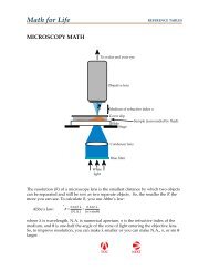
11 • Regulation of Gene Expression in Eukaryotes - W.H. Freeman
11 • Regulation of Gene Expression in Eukaryotes - W.H. Freeman
11 • Regulation of Gene Expression in Eukaryotes - W.H. Freeman
- No tags were found...

Create successful ePaper yourself
Turn your PDF publications into a flip-book with our unique Google optimized e-Paper software.
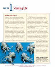
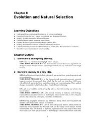
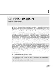
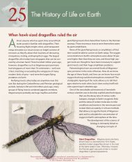
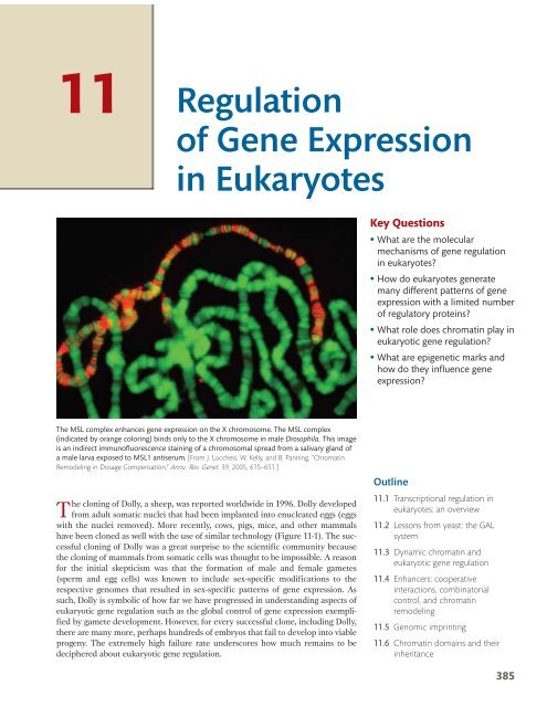
<strong>11</strong><strong>Regulation</strong><strong>of</strong> <strong>Gene</strong> <strong>Expression</strong><strong>in</strong> <strong>Eukaryotes</strong>Key Questions• What are the molecularmechanisms <strong>of</strong> gene regulation<strong>in</strong> eukaryotes?• How do eukaryotes generatemany different patterns <strong>of</strong> geneexpression with a limited number<strong>of</strong> regulatory prote<strong>in</strong>s?• What role does chromat<strong>in</strong> play <strong>in</strong>eukaryotic gene regulation?• What are epigenetic marks andhow do they <strong>in</strong>fluence geneexpression?The MSL complex enhances gene expression on the X chromosome. The MSL complex(<strong>in</strong>dicated by orange color<strong>in</strong>g) b<strong>in</strong>ds only to the X chromosome <strong>in</strong> male Drosophila. This imageis an <strong>in</strong>direct immun<strong>of</strong>luorescence sta<strong>in</strong><strong>in</strong>g <strong>of</strong> a chromosomal spread from a salivary gland <strong>of</strong>a male larva exposed to MSL1 antiserum. [From J. Lucchesi, W. Kelly, and B. Pann<strong>in</strong>g, “Chromat<strong>in</strong>Remodel<strong>in</strong>g <strong>in</strong> Dosage Compensation,” Annu. Rev. <strong>Gene</strong>t. 39, 2005, 615–651.]The clon<strong>in</strong>g <strong>of</strong> Dolly, a sheep, was reported worldwide <strong>in</strong> 1996. Dolly developedfrom adult somatic nuclei that had been implanted <strong>in</strong>to enucleated eggs (eggswith the nuclei removed). More recently, cows, pigs, mice, and other mammalshave been cloned as well with the use <strong>of</strong> similar technology (Figure <strong>11</strong>-1). The successfulclon<strong>in</strong>g <strong>of</strong> Dolly was a great surprise to the scientific community becausethe clon<strong>in</strong>g <strong>of</strong> mammals from somatic cells was thought to be impossible. A reasonfor the <strong>in</strong>itial skepticism was that the formation <strong>of</strong> male and female gametes(sperm and egg cells) was known to <strong>in</strong>clude sex-specific modifications to therespective genomes that resulted <strong>in</strong> sex-specific patterns <strong>of</strong> gene expression. Assuch, Dolly is symbolic <strong>of</strong> how far we have progressed <strong>in</strong> understand<strong>in</strong>g aspects <strong>of</strong>eukaryotic gene regulation such as the global control <strong>of</strong> gene expression exemplifiedby gamete development. However, for every successful clone, <strong>in</strong>clud<strong>in</strong>g Dolly,there are many more, perhaps hundreds <strong>of</strong> embryos that fail to develop <strong>in</strong>to viableprogeny. The extremely high failure rate underscores how much rema<strong>in</strong>s to bedeciphered about eukaryotic gene regulation.Outl<strong>in</strong>e<strong>11</strong>.1 Transcriptional regulation <strong>in</strong>eukaryotes: an overview<strong>11</strong>.2 Lessons from yeast: the GALsystem<strong>11</strong>.3 Dynamic chromat<strong>in</strong> andeukaryotic gene regulation<strong>11</strong>.4 Enhancers: cooperative<strong>in</strong>teractions, comb<strong>in</strong>atorialcontrol, and chromat<strong>in</strong>remodel<strong>in</strong>g<strong>11</strong>.5 Genomic impr<strong>in</strong>t<strong>in</strong>g<strong>11</strong>.6 Chromat<strong>in</strong> doma<strong>in</strong>s and their<strong>in</strong>heritance385
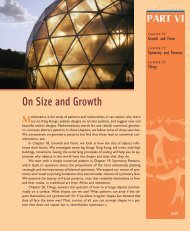
386 Chapter <strong>11</strong> • <strong>Regulation</strong> <strong>of</strong> <strong>Gene</strong> <strong>Expression</strong> <strong>in</strong> <strong>Eukaryotes</strong>The first cloned mammalFIGURE <strong>11</strong>-1 The first cloned mammalwas a sheep named Dolly. [PHOTOTAKE/Alamy.]In this chapter, we will exam<strong>in</strong>e gene regulation <strong>in</strong>eukaryotes. In many ways, our look at gene regulation will bea study <strong>of</strong> contrasts. In bacteria, you learned how the activities<strong>of</strong> genetic switches were <strong>of</strong>ten governed by s<strong>in</strong>gle activatoror repressor prote<strong>in</strong>s and how the control <strong>of</strong> sets <strong>of</strong> geneswas achieved by their organization <strong>in</strong>to operons or by theactivity <strong>of</strong> specific factors (see Chapter 10). Initial expectationswere that eukaryotic gene expression would be regulatedby similar means. In eukaryotes, however, most genesare not found <strong>in</strong> operons. Furthermore, we will see that theprote<strong>in</strong>s and DNA sequences participat<strong>in</strong>g <strong>in</strong> eukaryotic generegulation are more numerous. Often, many DNA-b<strong>in</strong>d<strong>in</strong>gprote<strong>in</strong>s act on a s<strong>in</strong>gle switch, with many separate switchesper gene, and the regulatory sequences <strong>of</strong> these switches are<strong>of</strong>ten located far from promoters. A key additional differencebetween bacteria and eukaryotes is that the access to eukaryoticgene promoters is restricted by chromat<strong>in</strong>. <strong>Gene</strong> regulation<strong>in</strong> eukaryotes requires the activity <strong>of</strong> large prote<strong>in</strong> complexesthat promote or restrict access to gene promoters by RNA polymerase. Thischapter will provide an essential foundation for understand<strong>in</strong>g the spatiotemporalregulation <strong>of</strong> gene expression that choreographs the process <strong>of</strong> developmentdescribed <strong>in</strong> Chapter 12.<strong>11</strong>.1 Transcriptional <strong>Regulation</strong><strong>in</strong> <strong>Eukaryotes</strong>: An OverviewThe biological properties <strong>of</strong> each eukaryotic cell type are largely determ<strong>in</strong>ed by theprote<strong>in</strong>s expressed with<strong>in</strong> it. This constellation <strong>of</strong> expressed prote<strong>in</strong>s determ<strong>in</strong>esmuch <strong>of</strong> the cell’s architecture, its enzymatic activities, its <strong>in</strong>teractions with itsenvironment, and many other physiological properties. However, at any given time<strong>in</strong> a cell’s life history, only a fraction <strong>of</strong> the RNAs and prote<strong>in</strong>s encoded <strong>in</strong> itsgenome are expressed. At different times, the pr<strong>of</strong>ile <strong>of</strong> expressed gene productscan differ dramatically, both <strong>in</strong> regard to which prote<strong>in</strong>s are expressed and at whatlevels. How are these specific pr<strong>of</strong>iles generated?As one might expect, if the f<strong>in</strong>al product is a prote<strong>in</strong>, regulation could beachieved by controll<strong>in</strong>g the transcription <strong>of</strong> DNA <strong>in</strong>to RNA or the translation <strong>of</strong>RNA <strong>in</strong>to prote<strong>in</strong>. In fact, gene regulation takes place at many levels, <strong>in</strong>clud<strong>in</strong>gat the mRNA level (through alterations <strong>in</strong> splic<strong>in</strong>g or the stability <strong>of</strong> the mRNA)and after translation (by modifications <strong>of</strong> prote<strong>in</strong>s). However, most regulation isthought to take place at the level <strong>of</strong> gene transcription; so, <strong>in</strong> this chapter, the primaryfocus is on the regulation <strong>of</strong> transcription. The basic mechanism at work isthat molecular signals from outside or <strong>in</strong>side the cell lead to the b<strong>in</strong>d<strong>in</strong>g <strong>of</strong> regulatoryprote<strong>in</strong>s to specific DNA sites outside <strong>of</strong> prote<strong>in</strong>-encod<strong>in</strong>g regions, and theb<strong>in</strong>d<strong>in</strong>g <strong>of</strong> these prote<strong>in</strong>s modulates the rate <strong>of</strong> transcription. These prote<strong>in</strong>s maydirectly or <strong>in</strong>directly assist RNA polymerase <strong>in</strong> b<strong>in</strong>d<strong>in</strong>g to its transcription <strong>in</strong>itiationsite—the promoter—or they may repress transcription by prevent<strong>in</strong>g the b<strong>in</strong>d<strong>in</strong>g<strong>of</strong> RNA polymerase.Although bacteria and eukaryotes have much <strong>of</strong> the logic <strong>of</strong> gene regulation <strong>in</strong>common, there are some fundamental differences <strong>in</strong> the underly<strong>in</strong>g mechanismsand mach<strong>in</strong>ery. Both use sequence-specific DNA-b<strong>in</strong>d<strong>in</strong>g prote<strong>in</strong>s to modulate thelevel <strong>of</strong> transcription. However, eukaryotic genomes are bigger and their range <strong>of</strong>properties is larger than those <strong>of</strong> bacteria. Inevitably their regulation is more complex,requir<strong>in</strong>g more types <strong>of</strong> regulatory prote<strong>in</strong>s and more types <strong>of</strong> <strong>in</strong>teractionswith the adjacent regulatory regions <strong>in</strong> DNA. The most important difference is
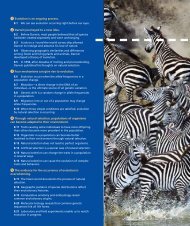
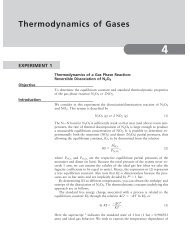
<strong>11</strong>.1 Transcriptional <strong>Regulation</strong> <strong>in</strong> <strong>Eukaryotes</strong>: An Overview387Activatorprote<strong>in</strong>BACTERIALRNA polPromoterOverview <strong>of</strong> transcriptional regulationOperatorGround state: onCod<strong>in</strong>gregionEUKARYOTICTATAGround state: <strong>of</strong>fFIGURE <strong>11</strong>-2 In bacteria, RNApolymerase can usually beg<strong>in</strong>transcription unless a repressorprote<strong>in</strong> blocks it. In eukaryotes,however, the packag<strong>in</strong>g <strong>of</strong> DNAwith nucleosomes preventstranscription unless otherregulatory prote<strong>in</strong>s are present.These regulatory prote<strong>in</strong>sexpose promoter sequences byalter<strong>in</strong>g nucleosome density orposition. They may also recruitRNA polymerase II more directlythrough b<strong>in</strong>d<strong>in</strong>g.TranscriptionfactorsEnhancerRepressorprote<strong>in</strong>RNA pol IITATARepressed state: <strong>of</strong>fActive state: onthat eukaryotic DNA is packaged <strong>in</strong>to nucleosomes, form<strong>in</strong>g chromat<strong>in</strong>, whereasbacterial DNA lacks nucleosomes. In eukaryotes, chromat<strong>in</strong> structure is dynamicand is an essential <strong>in</strong>gredient <strong>in</strong> gene regulation.In general, the ground state <strong>of</strong> a bacterial gene is “on.” Thus, RNA polymerasecan usually b<strong>in</strong>d to a promoter when no other regulatory prote<strong>in</strong>s are around tob<strong>in</strong>d to the DNA. In bacteria, transcription <strong>in</strong>itiation is prevented or reduced if theb<strong>in</strong>d<strong>in</strong>g <strong>of</strong> RNA polymerase is blocked, usually through the b<strong>in</strong>d<strong>in</strong>g <strong>of</strong> a repressorregulatory prote<strong>in</strong>. Activator regulatory prote<strong>in</strong>s <strong>in</strong>crease the b<strong>in</strong>d<strong>in</strong>g <strong>of</strong> RNApolymerase to promoters where a little help is needed. In contrast, the groundstate <strong>in</strong> eukaryotes is “<strong>of</strong>f.” Therefore, the transcriptional mach<strong>in</strong>ery (<strong>in</strong>clud<strong>in</strong>gRNA polymerase II and associated general transcription factors) cannot b<strong>in</strong>d tothe promoter <strong>in</strong> the absence <strong>of</strong> other regulatory prote<strong>in</strong>s (Figure <strong>11</strong>-2). In manycases, the b<strong>in</strong>d<strong>in</strong>g <strong>of</strong> the transcriptional apparatus is not possible, ow<strong>in</strong>g to theposition <strong>of</strong> nucleosomes near the promoter. Thus, chromat<strong>in</strong> structure usually hasto be changed to activate eukaryotic transcription. The structure <strong>of</strong> chromat<strong>in</strong>around activated or repressed genes with<strong>in</strong> cells can be quite stable and <strong>in</strong>heritedby daughter cells. The <strong>in</strong>heritance <strong>of</strong> chromat<strong>in</strong> structure is a form <strong>of</strong> <strong>in</strong>heritancethat does not directly entail DNA sequence.The unique features <strong>of</strong> eukaryotic transcriptional regulation are the focus <strong>of</strong>the rest <strong>of</strong> this chapter. Some differences from transcriptional regulation <strong>in</strong> bacteriawere already noted <strong>in</strong> Chapter 8:1. In bacteria, all genes are transcribed <strong>in</strong>to RNA by the same RNApolymerase, whereas three RNA polymerases function <strong>in</strong> eukaryotes. RNA
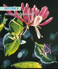
388 Chapter <strong>11</strong> • <strong>Regulation</strong> <strong>of</strong> <strong>Gene</strong> <strong>Expression</strong> <strong>in</strong> <strong>Eukaryotes</strong>Promoter-proximal elements precedethe promoter <strong>of</strong> a eukaryotic geneGC-richboxGGGCGG – 200 bpCCAAT–100 bpPromoter-proximalelementsFIGURE <strong>11</strong>-3 The region upstream <strong>of</strong>the transcription start site <strong>in</strong> highereukaryotes conta<strong>in</strong>s promoter-proximalelements and the promoter.polymerase II, which transcribes mRNAs, was the focus <strong>of</strong> Chapter 8 andwill be the only polymerase discussed <strong>in</strong> this chapter.2. RNA transcripts are extensively processed dur<strong>in</strong>g transcription <strong>in</strong>eukaryotes; the 5 and 3 ends are modified and <strong>in</strong>trons are spliced out.3. RNA polymerase II is much larger and more complex than its bacterialcounterpart. One reason for the added complexity is that RNA polymerase IImust synthesize RNA and coord<strong>in</strong>ate the special process<strong>in</strong>g events uniqueto eukaryotes.Multicellular eukaryotes may have as many as 25,000 genes, severalfold morethan the average bacterium. Moreover, patterns <strong>of</strong> eukaryotic gene expression canbe extraord<strong>in</strong>arily complex. That is, there is great variation among genes <strong>in</strong> when agene is on (transcribed) or <strong>of</strong>f (not transcribed) and <strong>in</strong> how much transcript needsto be made. For example, one gene may be transcribed only dur<strong>in</strong>g early developmentand another only <strong>in</strong> the presence <strong>of</strong> a viral <strong>in</strong>fection. F<strong>in</strong>ally, the majority <strong>of</strong>the genes <strong>in</strong> a eukaryotic cell are <strong>of</strong>f at any one time. On the basis <strong>of</strong> these considerationsalone, eukaryotic gene regulation must be able to1. ensure that the expression <strong>of</strong> most genes <strong>in</strong> the genome is <strong>of</strong>f at any onetime while activat<strong>in</strong>g a subset <strong>of</strong> genes; and2. generate thousands <strong>of</strong> patterns <strong>of</strong> gene expression.As you will see later <strong>in</strong> the chapter, mechanisms have evolved to ensure thatmost <strong>of</strong> the genes <strong>in</strong> a eukaryotic cell are not transcribed. Before consider<strong>in</strong>g howgenes are kept transcriptionally <strong>in</strong>active, we will focus on the second po<strong>in</strong>t: Howare eukaryotic genes able to exhibit an enormous number and diversity <strong>of</strong> expressionpatterns? The mach<strong>in</strong>ery required for generat<strong>in</strong>g so many patterns <strong>of</strong> genetranscription <strong>in</strong> vivo has many components, <strong>in</strong>clud<strong>in</strong>g both regulatory prote<strong>in</strong>sand cis-act<strong>in</strong>g regulatory sequences. The first set <strong>of</strong> prote<strong>in</strong>s comprises the largeRNA polymerase II complex and the general transcription factors that you learnedabout <strong>in</strong> Chapter 8. To <strong>in</strong>itiate transcription, these prote<strong>in</strong>s <strong>in</strong>teract with DNAsequences called promoter-proximal elements near the promoter <strong>of</strong> a gene. Thesecond group <strong>of</strong> prote<strong>in</strong> components consists <strong>of</strong> specific transcription factors thatb<strong>in</strong>d to cis-act<strong>in</strong>g regulatory sequences <strong>in</strong> the DNA called enhancers or upstreamactivat<strong>in</strong>g sequences (UAS’s). These regulatory sequences may be located aconsiderable distance from gene promoters. <strong>Gene</strong>rally speak<strong>in</strong>g, promoters andpromoter-proximal elements are bound by transcription factors that affect theexpression <strong>of</strong> many genes. Enhancers are the targets <strong>of</strong> more specific transcriptionfactors that control the regulation <strong>of</strong> smaller subsets <strong>of</strong> genes. Often, an enhancerwill act <strong>in</strong> only one or a few cell types <strong>in</strong> a multicellular eukaryote.TATA–30 bpPromotermRNAFor RNA polymerase II to transcribe DNA <strong>in</strong>to RNA ata maximum rate, multiple cis-act<strong>in</strong>g regulatory elementsmust play a part. The promoters, promoter-proximal elements,and enhancers are all targets for b<strong>in</strong>d<strong>in</strong>g by differenttrans-act<strong>in</strong>g DNA b<strong>in</strong>d<strong>in</strong>g prote<strong>in</strong>s. Figure <strong>11</strong>-3 is a schematicrepresentation <strong>of</strong> the promoter and promoter-proximal sequenceelements. The b<strong>in</strong>d<strong>in</strong>g <strong>of</strong> RNA polymerase II to thepromoter does not produce efficient transcription by itself.Transcription requires the b<strong>in</strong>d<strong>in</strong>g <strong>of</strong> general transcriptionfactors to additional promoter-proximal elements that arecommonly found with<strong>in</strong> 100 bp <strong>of</strong> the transcription <strong>in</strong>itiationsite <strong>of</strong> many (but not all) genes. One <strong>of</strong> these elements is the CCAAT box, and <strong>of</strong>tenanother is a GC-rich segment farther upstream. The general transcription factorsthat b<strong>in</strong>d to the promoter-proximal elements are expressed <strong>in</strong> most cells, and sothey are available to <strong>in</strong>itiate transcription at any time. Mutations <strong>in</strong> these sites can
<strong>11</strong>.1 Transcriptional <strong>Regulation</strong> <strong>in</strong> <strong>Eukaryotes</strong>: An Overview389Promoter-proximal elements are necessary for efficient transcription3.5Relative transcription level3.01.00GCCACACCCGGCCAATCATATAAFIGURE <strong>11</strong>-4 Po<strong>in</strong>t mutations <strong>in</strong> the promoter and promoter-proximal elements h<strong>in</strong>dertranscription <strong>of</strong> the β-glob<strong>in</strong> gene. Po<strong>in</strong>t mutations throughout the promoter region wereanalyzed for their effects on transcription rates. The height <strong>of</strong> each l<strong>in</strong>e represents thetranscription level relative to a wild-type promoter or promoter-proximal element (1.0).Only the base substitutions that lie with<strong>in</strong> the three elements shown change the level <strong>of</strong>transcription. Positions with black dots were not tested. [From T. Maniatis, S. Goodbourn, andJ. A. Fischer, “<strong>Regulation</strong> <strong>of</strong> Inducible and Tissue-Specific <strong>Gene</strong> <strong>Expression</strong>,” Science 236, 1987, 1237.]have a dramatic effect on transcription, demonstrat<strong>in</strong>g how important they are. Anexample <strong>of</strong> the consequences on transcription rates <strong>of</strong> mutat<strong>in</strong>g these sequenceelements is shown <strong>in</strong> Figure <strong>11</strong>-4.To modulate transcription, regulatory prote<strong>in</strong>s possess one or more <strong>of</strong> the follow<strong>in</strong>gfunctional doma<strong>in</strong>s:1. A doma<strong>in</strong> that recognizes a DNA regulatory sequence (the prote<strong>in</strong>’sDNA-b<strong>in</strong>d<strong>in</strong>g site)2. A doma<strong>in</strong> that <strong>in</strong>teracts with one or more prote<strong>in</strong>s <strong>of</strong> the transcriptionalapparatus (RNA polymerase or a prote<strong>in</strong> associated with RNA polymerase)3. A doma<strong>in</strong> that <strong>in</strong>teracts with prote<strong>in</strong>s bound to nearby regulatorysequences on DNA such that they can act cooperatively to regulatetranscription4. A doma<strong>in</strong> that <strong>in</strong>fluences chromat<strong>in</strong> condensation either directly or<strong>in</strong>directly5. A doma<strong>in</strong> that acts as a sensor <strong>of</strong> physiological conditions with<strong>in</strong> the cellMuch <strong>of</strong> the strategy <strong>of</strong> eukaryotic transcriptional control h<strong>in</strong>ges on how specifictranscription factors control the access <strong>of</strong> general transcription factors andRNA polymerase II. Eukaryotic gene regulatory mechanisms have been discoveredthrough both biochemical and genetic approaches. The latter has been advanced<strong>in</strong> particular by studies <strong>of</strong> the s<strong>in</strong>gle-celled yeast Saccharomyces cerevisiae (see theModel Organism box). Several decades <strong>of</strong> research have been a source <strong>of</strong> many<strong>in</strong>sights <strong>in</strong>to general pr<strong>in</strong>ciples <strong>of</strong> how eukaryotic transcriptional regulatory prote<strong>in</strong>swork and how different cell types are generated. We’ll exam<strong>in</strong>e two yeastgene regulatory systems <strong>in</strong> detail: the first concerns the galactose-utilization pathway;the second is the control <strong>of</strong> mat<strong>in</strong>g type.
390 Chapter <strong>11</strong> • <strong>Regulation</strong> <strong>of</strong> <strong>Gene</strong> <strong>Expression</strong> <strong>in</strong> <strong>Eukaryotes</strong>Model OrganismYeastSaccharomyces cerevisiae, or budd<strong>in</strong>g yeast, has emerged<strong>in</strong> recent years as the premier eukaryotic genetic system.Humans have grown yeast for centuries because it is anessential component <strong>of</strong> beer, bread, and w<strong>in</strong>e. Yeast hasmany features that make it an ideal model organism. As aunicellular eukaryote, it can be grown on agar plates and,with a life cycle <strong>of</strong> just 90 m<strong>in</strong>utes, large quantities can becultured <strong>in</strong> liquid media. It has a very compact genomewith only about 12 megabase pairs <strong>of</strong> DNA (comparedwith almost 3000 megabase pairs for humans) conta<strong>in</strong><strong>in</strong>gapproximately 6000 genes that are distributed on 16 chromosomes.It was the first eukaryote to have its genomesequenced.The yeast life cycle makes it very versatile for laboratorystudies. Cells can be grown as either diploid or haploid.In both cases, the mother cell produces a bud conta<strong>in</strong><strong>in</strong>gan identical daughter cell. Diploid cells either cont<strong>in</strong>ueto grow by budd<strong>in</strong>g or are <strong>in</strong>duced to undergo meiosis,which produces four haploid spores held together <strong>in</strong> anascus (also called a tetrad). Haploid spores <strong>of</strong> opposite mat<strong>in</strong>gtype (a or `) will fuse and form a diploid. Spores <strong>of</strong> thesame mat<strong>in</strong>g type will cont<strong>in</strong>ue growth by budd<strong>in</strong>g.Electron micrograph <strong>of</strong> budd<strong>in</strong>g yeast cells. [SciMAT/PhotoResearchers.]Mitosis(n)Culturecolony(n) Ascus(2n)a /a(n) (n)FusionMitosis+a /aa /aMeiosis(2n)(2n)a (n)a (n)(n) aThe life cycle <strong>of</strong> baker’s yeast. The nuclear alleles MATa and MATadeterm<strong>in</strong>e mat<strong>in</strong>g type.Yeast has been called the E. coli <strong>of</strong> eukaryotes because<strong>of</strong> the ease <strong>of</strong> forward and reverse mutant analysis. Toisolate mutants by us<strong>in</strong>g a forward genetic approach, haploidcells are mutagenized (with X rays, for example) andscreened on plates for mutant phenotypes. This procedureis usually done by first plat<strong>in</strong>g cells on a rich medium onwhich all cells grow and by copy<strong>in</strong>g, or replica plat<strong>in</strong>g, thecolonies from this master plate onto replica plates conta<strong>in</strong><strong>in</strong>gselective media or special growth conditions. (See alsoChapter 15.) For example, temperature-sensitive mutantswill grow on the master plate at the permissive temperaturebut not on a replica plate at a restrictive temperature.Comparison <strong>of</strong> the colonies on the master and replicaplates will reveal the temperature-sensitive mutants. Us<strong>in</strong>greverse genetics, scientists can also replace any yeast gene(<strong>of</strong> known or unknown function) with a mutant version(synthesized <strong>in</strong> a test tube) to understand the nature <strong>of</strong> thegene product.(n)CulturecolonyaMitosis<strong>11</strong>.2 Lessons from Yeast: The GAL SystemTo make use <strong>of</strong> extracellular galactose, yeast imports the sugar and converts it <strong>in</strong>toa form <strong>of</strong> glucose that can be metabolized. Several genes—GAL1, GAL2, GAL7, andGAL10—<strong>in</strong> the yeast genome encode enzymes that catalyze steps <strong>in</strong> the biochemicalpathway that converts galactose <strong>in</strong>to glucose (Figure <strong>11</strong>-5). Three additionalgenes—GAL3, GAL4, and GAL80—encode prote<strong>in</strong>s that regulate the expression <strong>of</strong>the enzyme genes. Just as <strong>in</strong> the lac system, the abundance <strong>of</strong> the sugar determ<strong>in</strong>es
<strong>11</strong>.2 Lesson from Yeast: The GAL System391the level <strong>of</strong> gene expression <strong>in</strong> the biochemical pathway. In yeast cells grow<strong>in</strong>g <strong>in</strong>media lack<strong>in</strong>g galactose, the GAL genes are largely silent. But, <strong>in</strong> the presence <strong>of</strong>galactose (and the absence <strong>of</strong> glucose), the GAL genes are <strong>in</strong>duced. Just as for thelac operon, genetic and molecular analyses <strong>of</strong> mutants have been key to understand<strong>in</strong>ghow the expression <strong>of</strong> the genes <strong>in</strong> the galactose pathway is controlled.The key regulator <strong>of</strong> GAL gene expression is the Gal4 prote<strong>in</strong>, a sequencespecificDNA-b<strong>in</strong>d<strong>in</strong>g prote<strong>in</strong>. Gal4 is perhaps the best-studied transcriptional regulatoryprote<strong>in</strong> <strong>in</strong> eukaryotes. The detailed dissection <strong>of</strong> its regulation and activityhas been a source <strong>of</strong> several key <strong>in</strong>sights <strong>in</strong>to the control <strong>of</strong> transcription <strong>in</strong>eukaryotes.Gal4 regulates multiple genes through upstream activat<strong>in</strong>gsequencesIn the presence <strong>of</strong> galactose, the GAL1, GAL2, GAL7, and GAL10 genes are <strong>in</strong>duced1000-fold or more. In GAL4 mutants, however, they rema<strong>in</strong> silent. Each <strong>of</strong> thesefour genes has two or more Gal4-b<strong>in</strong>d<strong>in</strong>g sites located 5 (upstream) <strong>of</strong> its promoter.Consider the GAL10 and GAL1 genes, which are adjacent to each other andtranscribed <strong>in</strong> opposite directions. Between the GAL1 transcription start site andthe GAL10 transcription start site is a s<strong>in</strong>gle <strong>11</strong>8-bp region that conta<strong>in</strong>s four Gal4-b<strong>in</strong>d<strong>in</strong>g sites (Figure <strong>11</strong>-6). Each Gal4-b<strong>in</strong>d<strong>in</strong>g site is 17 base pairs long and isbound by one Gal4 prote<strong>in</strong> dimer. There are two Gal4-b<strong>in</strong>d<strong>in</strong>g sites upstream <strong>of</strong>the GAL2 gene as well, and another two upstream <strong>of</strong> the GAL7 gene. These b<strong>in</strong>d<strong>in</strong>gsites are required for gene activation <strong>in</strong> vivo. If they are deleted, the genes aresilent, even <strong>in</strong> the presence <strong>of</strong> galactose. These regulatory sequences are enhancersthat are also referred to as upstream activat<strong>in</strong>g sequences. The presence <strong>of</strong> enhancerslocated at a considerable l<strong>in</strong>ear distance from a eukaryotic gene’s promoteris typical.Message The b<strong>in</strong>d<strong>in</strong>g <strong>of</strong> sequence-specific DNA-b<strong>in</strong>d<strong>in</strong>g prote<strong>in</strong>s to regionsoutside the promoters <strong>of</strong> target genes is a common feature <strong>of</strong> eukaryotic transcriptionalregulation.The Gal pathwayGalactose (extracellular)Gal2Galactose (<strong>in</strong>tracellular)Gal1Galactose-1-phosphateGal7UDP-galactoseGal10UDP-glucoseGal7Glucose-1-phosphateGlycosisFIGURE <strong>11</strong>-5 Galactose is converted<strong>in</strong>to glucose-1-phosphate <strong>in</strong> a series <strong>of</strong>steps. These steps are catalyzed byenzymes (Gal1, and so forth) encoded bythe structural genes GAL1, GAL2, GAL7,and GAL10.Transcriptional activator prote<strong>in</strong>s b<strong>in</strong>d to UAS elements <strong>in</strong> yeastGal4Chr II5′ GAL7GAL10 GAL1 3′ Chr XII 5′GAL2 3′UASUASUASFIGURE <strong>11</strong>-6 The Gal4 prote<strong>in</strong> activates target genes through upstream-activat<strong>in</strong>g-sequence(UAS) elements. The Gal4 prote<strong>in</strong> has two functional doma<strong>in</strong>s: a DNA-b<strong>in</strong>d<strong>in</strong>g doma<strong>in</strong> (redsquare) and an activation doma<strong>in</strong> (orange oval). The prote<strong>in</strong> b<strong>in</strong>ds to specific sequencesupstream <strong>of</strong> the promoters <strong>of</strong> Gal-pathway genes. Some <strong>of</strong> the GAL genes are adjacent (GAL1,GAL10), whereas others are on different chromosomes. The GAL1 UAS element conta<strong>in</strong>s fourGal4-b<strong>in</strong>d<strong>in</strong>g sites.
392 Chapter <strong>11</strong> • <strong>Regulation</strong> <strong>of</strong> <strong>Gene</strong> <strong>Expression</strong> <strong>in</strong> <strong>Eukaryotes</strong>(a) The complete Gal4 dimerGal4Activationdoma<strong>in</strong>DNA-b<strong>in</strong>d<strong>in</strong>gdoma<strong>in</strong>Gal4 site(b) Gal4 lack<strong>in</strong>g the activation doma<strong>in</strong>Gal4 site(c) LexA lack<strong>in</strong>g the activation doma<strong>in</strong>DNA-b<strong>in</strong>d<strong>in</strong>gdoma<strong>in</strong>(d) Gal4–LexA hybridTranscriptional activatorprote<strong>in</strong>s are modularGal4 activationdoma<strong>in</strong>LexADNA-b<strong>in</strong>d<strong>in</strong>gdoma<strong>in</strong>LexA siteLexA siteFIGURE <strong>11</strong>-7 Transcriptional activatorprote<strong>in</strong>s have multiple, separable doma<strong>in</strong>s.(a) The Gal4 prote<strong>in</strong> has two doma<strong>in</strong>s andforms a dimer. (b) The experimental removal<strong>of</strong> the activation doma<strong>in</strong> shows that DNAb<strong>in</strong>d<strong>in</strong>g is not sufficient for gene activation.(c) Similarly, the bacterial LexA prote<strong>in</strong> cannotactivate transcription on its own, but, whenfused to the Gal4 activation doma<strong>in</strong> (d), it canactivate transcription through LexA-b<strong>in</strong>d<strong>in</strong>gsites. [After J. Watson et al., Molecular Biology <strong>of</strong>the <strong>Gene</strong>, Fifth Edition, copyright © 2004,Benjam<strong>in</strong> Cumm<strong>in</strong>gs.]The Gal4 prote<strong>in</strong> has separable DNA-b<strong>in</strong>d<strong>in</strong>gand activation doma<strong>in</strong>sAfter Gal4 is bound to the UAS element, how is gene expression <strong>in</strong>duced? A dist<strong>in</strong>ctdoma<strong>in</strong> <strong>of</strong> the Gal4 prote<strong>in</strong>, the activation doma<strong>in</strong>, is required for regulatoryactivity. Thus, the Gal4 prote<strong>in</strong> has at least two doma<strong>in</strong>s: one for DNA b<strong>in</strong>d<strong>in</strong>gand another for activat<strong>in</strong>g transcription. A similar modular organization has beenfound to be a common feature <strong>of</strong> other DNA-b<strong>in</strong>d<strong>in</strong>g transcription factors as well.The modular organization <strong>of</strong> the Gal4 prote<strong>in</strong> was demonstrated<strong>in</strong> a series <strong>of</strong> simple, elegant experiments. The strategy waslacZlacZlacZlacZONOFFOFFONto test the DNA b<strong>in</strong>d<strong>in</strong>g and gene activation <strong>of</strong> mutant forms <strong>of</strong> theprote<strong>in</strong> <strong>in</strong> which parts had been either deleted or fused to otherprote<strong>in</strong>s. In this manner, whether a part <strong>of</strong> the prote<strong>in</strong> was necessaryfor a particular function could be determ<strong>in</strong>ed. To carry outthese studies, experimenters needed a simple means <strong>of</strong> assay<strong>in</strong>gthe expression <strong>of</strong> the enzymes encoded by the GAL genes. Theexpression <strong>of</strong> GAL genes and other targets <strong>of</strong> transcription factorsis typically monitored by us<strong>in</strong>g a reporter gene whose expressionis easily tracked. Often, the reporter gene is the lacZ gene <strong>of</strong> E. coli,which can act on substrates whose products are easily measured bytheir bright color or fluourescence. Another common reportergene is the gene that encodes the green fluorescent prote<strong>in</strong> <strong>of</strong> jellyfish,which, as its name suggests, is easily tracked by the light that itemits. The cod<strong>in</strong>g region <strong>of</strong> one <strong>of</strong> these reporter genes and a promoterare placed downstream <strong>of</strong> a UAS element from a GAL gene.Reporter expression is then a read-out <strong>of</strong> Gal4 activity <strong>in</strong> cells.When a form <strong>of</strong> the Gal4 prote<strong>in</strong> lack<strong>in</strong>g the activation doma<strong>in</strong>is expressed <strong>in</strong> yeast, the b<strong>in</strong>d<strong>in</strong>g sites <strong>of</strong> the UAS element are occupied,but no transcription is stimulated (Figure <strong>11</strong>-7). The same istrue when other regulatory prote<strong>in</strong>s lack<strong>in</strong>g activation doma<strong>in</strong>s,such as the bacterial repressor LexA, are expressed <strong>in</strong> cells bear<strong>in</strong>greporter genes with their respective b<strong>in</strong>d<strong>in</strong>g sites. The more <strong>in</strong>terest<strong>in</strong>gresult is obta<strong>in</strong>ed when a form <strong>of</strong> the Gal4 prote<strong>in</strong> lack<strong>in</strong>gthe DNA-b<strong>in</strong>d<strong>in</strong>g doma<strong>in</strong> is grafted to the LexA DNA-b<strong>in</strong>d<strong>in</strong>gdoma<strong>in</strong>; the hybrid prote<strong>in</strong> now activates transcription from LexAb<strong>in</strong>d<strong>in</strong>gsites (Figure <strong>11</strong>-7). Further “doma<strong>in</strong> swap” experimentshave revealed that the transcriptional activation function <strong>of</strong> theGal4 prote<strong>in</strong> resides <strong>in</strong> two small doma<strong>in</strong>s about 50 to 100 am<strong>in</strong>oacids <strong>in</strong> length. These doma<strong>in</strong>s are separable from those used <strong>in</strong>the dimerization <strong>of</strong> the prote<strong>in</strong>, DNA b<strong>in</strong>d<strong>in</strong>g, and <strong>in</strong>teractionwith the Gal80 prote<strong>in</strong> (see next). The activation doma<strong>in</strong> helpsrecruit the transcriptional mach<strong>in</strong>ery to the promoter, as we willsee <strong>in</strong> Section <strong>11</strong>.3. This highly modular arrangement <strong>of</strong> activityregulat<strong>in</strong>gdoma<strong>in</strong>s is found <strong>in</strong> many transcription factors.Message Many eukaryotic transcriptional regulatory prote<strong>in</strong>s are modular prote<strong>in</strong>s,with separable doma<strong>in</strong>s for DNA b<strong>in</strong>d<strong>in</strong>g, activation or repression, and <strong>in</strong>teraction withother prote<strong>in</strong>s.Gal4 activity is physiologically regulatedHow does Gal4 become active <strong>in</strong> the presence <strong>of</strong> galactose? Key clues came fromanalyses <strong>of</strong> mutations <strong>in</strong> the GAL80 and GAL3 genes. In GAL80 mutants, the GALstructural genes are active even <strong>in</strong> the absence <strong>of</strong> galactose. This result suggeststhat the normal function <strong>of</strong> the Gal80 prote<strong>in</strong> is to somehow <strong>in</strong>hibit GAL gene
<strong>11</strong>.2 Lesson from Yeast: The GAL System393expression. Conversely, <strong>in</strong> GAL3 mutants, the GAL structural genes arenot active <strong>in</strong> the presence <strong>of</strong> galactose, suggest<strong>in</strong>g that Gal3 normallypromotes expression <strong>of</strong> the GAL genes.Extensive biochemical analyses have revealed that the Gal80 prote<strong>in</strong>b<strong>in</strong>ds to the Gal4 prote<strong>in</strong> with high aff<strong>in</strong>ity and directly <strong>in</strong>hibitsGal4 activity. Specifically, Gal80 b<strong>in</strong>ds to a region with<strong>in</strong> one <strong>of</strong> theGal4 activation doma<strong>in</strong>s, block<strong>in</strong>g its ability to promote the transcription<strong>of</strong> target genes. The role <strong>of</strong> the Gal3 prote<strong>in</strong> is to release Gal4 fromits <strong>in</strong>hibition by Gal80 <strong>in</strong> the presence <strong>of</strong> galactose. Gal3 is a sensor and<strong>in</strong>ducer. When Gal3 b<strong>in</strong>ds galactose and ATP, it undergoes an allostericchange that promotes b<strong>in</strong>d<strong>in</strong>g to Gal80, which <strong>in</strong> turn causes Gal80 torelease Gal4, which is then able to activate transcription <strong>of</strong> its targetgenes. Thus, Gal3, Gal80, and Gal4 are all part <strong>of</strong> a switch whose state isdeterm<strong>in</strong>ed by the presence or absence <strong>of</strong> galactose (Figure <strong>11</strong>-8). Inthis switch, DNA b<strong>in</strong>d<strong>in</strong>g by the transcriptional regulator is not thephysiologically regulated step (as is the case <strong>in</strong> the lac operon and bacteriophage); rather, the activity <strong>of</strong> the activation doma<strong>in</strong> is regulated.Transcriptional activator prote<strong>in</strong>smay be activated by an <strong>in</strong>ducerInactiveGal4+ Galactose+ Gal3ActiveGal4UASGal80GAL1OFFMessage The activity <strong>of</strong> eukaryotic transcriptional regulatory prote<strong>in</strong>sis <strong>of</strong>ten controlled by <strong>in</strong>teractions with other prote<strong>in</strong>s.UASGAL1ONGal4 functions <strong>in</strong> most eukaryotesIn addition to its action <strong>in</strong> yeast cells, Gal4 has been shown to be able to activatetranscription <strong>in</strong> <strong>in</strong>sect cells, human cells, and many other eukaryotic species. Thisversatility suggests that biochemical mach<strong>in</strong>ery and mechanisms <strong>of</strong> gene activationare common to a broad array <strong>of</strong> eukaryotes and that features revealed <strong>in</strong> yeast aregenerally present <strong>in</strong> other eukaryotes, and vice versa. Furthermore, because <strong>of</strong> theirversatility, Gal4 and its UAS elements have become favored tools <strong>in</strong> genetic analysisfor manipulat<strong>in</strong>g gene expression and function <strong>in</strong> a wide variety <strong>of</strong> model systems.FIGURE <strong>11</strong>-8 Gal4 activity is regulatedby the Gal80 prote<strong>in</strong>. (Top) In the absence<strong>of</strong> galactose, the Gal4 prote<strong>in</strong> is <strong>in</strong>active,even though it can b<strong>in</strong>d to sites upstream<strong>of</strong> the GAL1 target gene. Gal4 activity issuppressed by the b<strong>in</strong>d<strong>in</strong>g <strong>of</strong> the Gal80prote<strong>in</strong>. (Bottom) In the presence <strong>of</strong>galactose and the Gal3 prote<strong>in</strong>, Gal80undergoes a conformational change andreleases the Gal4 activation doma<strong>in</strong>,permitt<strong>in</strong>g target gene transcription.Message The ability <strong>of</strong> Gal4, as well as other eukaryotic regulators, to function <strong>in</strong> avariety <strong>of</strong> eukaryotes <strong>in</strong>dicates that eukaryotes generally have the transcriptionalregulatory mach<strong>in</strong>ery and mechanisms <strong>in</strong> common.Now we look at how activators and other regulatory prote<strong>in</strong>s <strong>in</strong>teract with thetranscriptional mach<strong>in</strong>ery to control gene expression.Activators recruit the transcriptional mach<strong>in</strong>eryIn bacteria, activators commonly stimulate transcription by <strong>in</strong>teract<strong>in</strong>g directlywith DNA and with RNA polymerase. In eukaryotes, activators generally work <strong>in</strong>directlyto recruit RNA polymerase II to gene promoters through two major mechanisms.First, activators can <strong>in</strong>teract with subunits <strong>of</strong> the prote<strong>in</strong> complexes hav<strong>in</strong>groles <strong>in</strong> transcription <strong>in</strong>itiation. Second, activators can recruit prote<strong>in</strong>s that modifychromat<strong>in</strong> structure, allow<strong>in</strong>g RNA polymerase II and other prote<strong>in</strong>s access to theDNA. Many activators, <strong>in</strong>clud<strong>in</strong>g Gal4, have both activities. We’ll exam<strong>in</strong>e therecruitment <strong>of</strong> parts <strong>of</strong> the transcriptional <strong>in</strong>itiation complex first.Recall from Chapter 8 that the eukaryotic transcriptional mach<strong>in</strong>ery conta<strong>in</strong>smany prote<strong>in</strong>s that are parts <strong>of</strong> various subcomplexes with<strong>in</strong> the transcriptionalapparatus that is assembled on gene promoters. One subcomplex, transcription factorIID (TFIID), b<strong>in</strong>ds to the TATA box <strong>of</strong> eukaryotic promoters through the TATAb<strong>in</strong>d<strong>in</strong>gprote<strong>in</strong> (TBP; see Figure 8-12). Gal4 b<strong>in</strong>ds to TBP at a site <strong>in</strong> its activation
394 Chapter <strong>11</strong> • <strong>Regulation</strong> <strong>of</strong> <strong>Gene</strong> <strong>Expression</strong> <strong>in</strong> <strong>Eukaryotes</strong>Transcriptional activator prote<strong>in</strong>srecruit the transcriptional mach<strong>in</strong>eryTBPTFIIDGal4UASMediatorRNA polymerase IIdoma<strong>in</strong>, and, through this b<strong>in</strong>d<strong>in</strong>g, it recruits the TFIID complexand, <strong>in</strong> turn, RNA polymerase II to the promoter (Figure<strong>11</strong>-9). The aff<strong>in</strong>ity <strong>of</strong> this <strong>in</strong>teraction correlates well withGal4’s potency as an activator. Gal4 also <strong>in</strong>teracts with thelarge Mediator complex, which directly <strong>in</strong>teracts with RNApolymerase II to recruit it to gene promoters. The Mediatorcomplex is an example <strong>of</strong> a coactivator, a term applied to aprote<strong>in</strong> or prote<strong>in</strong> complex that facilitates gene activation bya transcription factor but that itself is neither part <strong>of</strong> the transcriptionalmach<strong>in</strong>ery nor a DNA-b<strong>in</strong>d<strong>in</strong>g prote<strong>in</strong>.The ability <strong>of</strong> activators to b<strong>in</strong>d to upstream DNAsequences and to <strong>in</strong>teract with prote<strong>in</strong>s that b<strong>in</strong>d directly or<strong>in</strong>directly to promoters helps to expla<strong>in</strong> how transcriptioncan be stimulated from more distant regulatory sequences(see Figure <strong>11</strong>-9).TATAFIGURE <strong>11</strong>-9 Gal4 recruits thetranscriptional mach<strong>in</strong>ery. The Gal4prote<strong>in</strong>, and many other transcriptionalactivators, b<strong>in</strong>ds to multiple prote<strong>in</strong>complexes, <strong>in</strong>clud<strong>in</strong>g the TFIID andMediator complexes, that recruit RNApolymerase II to gene promoters. The<strong>in</strong>teractions facilitate gene activationthrough b<strong>in</strong>d<strong>in</strong>g sites that are distant fromgene promoters. [After J. Watson et al.,Molecular Biology <strong>of</strong> the <strong>Gene</strong>, Fifth Edition,copyright © 2004, Benjam<strong>in</strong> Cumm<strong>in</strong>gs.]GAL genesMessage Eukaryotic transcriptional activators <strong>of</strong>ten workby recruit<strong>in</strong>g parts <strong>of</strong> the transcriptional mach<strong>in</strong>ery to genepromoters.<strong>11</strong>.3 Dynamic Chromat<strong>in</strong> andEukaryotic <strong>Gene</strong> <strong>Regulation</strong>A second mechanism for <strong>in</strong>fluenc<strong>in</strong>g gene transcription <strong>in</strong> eukaryotes modifies thelocal chromat<strong>in</strong> structure around gene regulatory sequences. To fully understandhow this mechanism works, we need to first review chromat<strong>in</strong> structure and thenconsider how it can change and how these changes affect gene expression.The recruitment <strong>of</strong> transcriptional mach<strong>in</strong>ery by activators may appear to besomewhat similar <strong>in</strong> eukaryotes and bacteria, with the major difference be<strong>in</strong>g <strong>in</strong>the number <strong>of</strong> <strong>in</strong>teract<strong>in</strong>g prote<strong>in</strong>s <strong>in</strong> the transcriptional mach<strong>in</strong>ery. Indeed, lessthan a decade ago, many biologists pictured eukaryotic regulation simply as a biochemicallymore complicated version <strong>of</strong> what had been discovered <strong>in</strong> bacteria.However, this view has changed dramatically as biologists have considered theeffect <strong>of</strong> the organization <strong>of</strong> genomic DNA <strong>in</strong> eukaryotes.Compared with eukaryotic DNA, bacterial DNA is relatively “naked,” mak<strong>in</strong>git readily accessible to RNA polymerase. In contrast, eukaryotic chromosomes arepackaged <strong>in</strong>to chromat<strong>in</strong>, which is composed <strong>of</strong> DNA and prote<strong>in</strong>s (mostly histones).As mentioned briefly <strong>in</strong> Chapter 2, the basic unit <strong>of</strong> chromat<strong>in</strong> is the nucleosome,conta<strong>in</strong><strong>in</strong>g about 150 bp <strong>of</strong> DNA wrapped twice around a histone octamer(Figure <strong>11</strong>-10). The histone octamer is composed <strong>of</strong> two subunits <strong>of</strong> each <strong>of</strong> thefour histones: histone 2A, 2B, 3, and 4. Nucleosomes can associate <strong>in</strong>to higherorderstructures that further condense the DNA. The packag<strong>in</strong>g <strong>of</strong> eukaryoticDNA <strong>in</strong>to chromat<strong>in</strong> means that much <strong>of</strong> the DNA is not readily accessible to regulatoryprote<strong>in</strong>s and the transcriptional apparatus. Thus, whereas prokaryoticgenes are generally accessible and “on” unless repressed, eukaryotic genes are <strong>in</strong>accessibleand “<strong>of</strong>f” unless activated. Therefore, the modification <strong>of</strong> chromat<strong>in</strong> structureis a dist<strong>in</strong>ctive feature <strong>of</strong> eukaryotic gene regulation.One can imag<strong>in</strong>e several ways to alter chromat<strong>in</strong> structure. For example, onemechanism might be to simply move the histone octamer along the DNA. In the1980s, biochemical techniques were developed that allowed researchers to determ<strong>in</strong>ethe position <strong>of</strong> nucleosomes <strong>in</strong> and around specific genes. In these studies,chromat<strong>in</strong> was isolated from tissues or cells <strong>in</strong> which a gene was on and comparedwith chromat<strong>in</strong> from tissue where the same gene was <strong>of</strong>f. The result for most genesanalyzed was that nucleosome positions changed, especially <strong>in</strong> a gene’s regulatoryregions. Thus, which DNA regions are wrapped up <strong>in</strong> nucleosomes can change:
<strong>11</strong>.3 Dynamic Chromat<strong>in</strong> and Eukaryotic <strong>Gene</strong> <strong>Regulation</strong>395The structure <strong>of</strong> chromat<strong>in</strong>(a)(b)Short region <strong>of</strong>DNA double helix2 nmNucleosomes:the basic unit<strong>of</strong> chromat<strong>in</strong><strong>11</strong> nmChromat<strong>in</strong> fiber<strong>of</strong> packednucleosomes30 nmFIGURE <strong>11</strong>-10 (a) The nucleosome <strong>in</strong> decondensed and condensed chromat<strong>in</strong>.(b) Chromat<strong>in</strong> structure varies along the length <strong>of</strong> a chromosome. The least-condensedchromat<strong>in</strong> (euchromat<strong>in</strong>) is shown <strong>in</strong> yellow, regions <strong>of</strong> <strong>in</strong>termediate condensation are <strong>in</strong>orange and blue, and heterochromat<strong>in</strong> coated with special prote<strong>in</strong>s (purple) is <strong>in</strong> red.[(b) From P. J. Horn and C. L. Peterson, “Chromat<strong>in</strong> Higher Order Fold<strong>in</strong>g: Wrapp<strong>in</strong>g Up Transcription,”Science 297, 2002, 1827, Fig. 3. Copyright 2002, AAAS.]nucleosome positions can shift on the DNA from cell to cell and over the life cycle<strong>of</strong> an organism. Transcription might be repressed when the promoter and flank<strong>in</strong>gsequences are wound up <strong>in</strong> a nucleosome and <strong>in</strong>accessible to RNA polymerase II.Activation <strong>of</strong> transcription would thus require the block<strong>in</strong>g nucleosome to be reorganizedby nudg<strong>in</strong>g the histones or remov<strong>in</strong>g them entirely. Conversely, when generepression is necessary, histone octamers may shift <strong>in</strong>to a position that preventstranscription. The chang<strong>in</strong>g <strong>of</strong> nucleosome position is referred to as chromat<strong>in</strong>remodel<strong>in</strong>g. Now, chromat<strong>in</strong> remodel<strong>in</strong>g is known to be an <strong>in</strong>tegral part <strong>of</strong>eukaryotic gene expression, and great advances are be<strong>in</strong>g made <strong>in</strong> determ<strong>in</strong><strong>in</strong>g theunderly<strong>in</strong>g mechanism(s) and the regulatory prote<strong>in</strong>s tak<strong>in</strong>g part. Here, aga<strong>in</strong>,genetic studies <strong>in</strong> yeast have been pivotal.Chromat<strong>in</strong>-remodel<strong>in</strong>g prote<strong>in</strong>s and gene activationTwo genetic screens <strong>in</strong> yeast for mutants <strong>in</strong> seem<strong>in</strong>gly unrelated processes led to thediscovery <strong>of</strong> the same gene whose product plays a key role <strong>in</strong> chromat<strong>in</strong> remodel<strong>in</strong>g.In both cases, yeast cells were treated with agents that would cause mutations.In one screen, these mutagenized yeast cells were screened for cells that could notgrow well on sucrose (sugar nonferment<strong>in</strong>g mutants, snf). In another screen, mutagenizedyeast cells were screened for mutants that were defective <strong>in</strong> switch<strong>in</strong>g theirmat<strong>in</strong>g type (switch mutants, swi; see Section <strong>11</strong>.4). Many mutants for different lociwere recovered <strong>in</strong> each screen, but one mutant gene was found to cause both phenotypes.Mutants at the so-called swi2/snf2 locus (“switch–sniff”) could neither utilizesucrose effectively nor switch mat<strong>in</strong>g type.What was the connection between the ability to utilize sugar and the abilityto switch mat<strong>in</strong>g types? The Snf2–Swi2 prote<strong>in</strong> was purified and discovered to bepart <strong>of</strong> a large, multisubunit complex called the SWI–SNF complex that can repositionnucleosomes <strong>in</strong> a test-tube assay if ATP is provided as an energy source(Figure <strong>11</strong>-<strong>11</strong>). In some situations, the multisubunit SWI–SNF complex activatestranscription by mov<strong>in</strong>g nucleosomes that are cover<strong>in</strong>g the TATA sequences and,<strong>in</strong> this way, facilitates the b<strong>in</strong>d<strong>in</strong>g <strong>of</strong> RNA polymerase II. The SWI–SNF complex isthus a coactivator.Chromat<strong>in</strong> remodel<strong>in</strong>gexposes regulatorysequencesNucleosomeremodel<strong>in</strong>gFIGURE <strong>11</strong>-<strong>11</strong> The histone octamerslides <strong>in</strong> response to chromat<strong>in</strong>remodel<strong>in</strong>gactivity (such as that <strong>of</strong> theSWI–SNF complex), <strong>in</strong> this caseexpos<strong>in</strong>g the DNA marked <strong>in</strong> red. (SeeFigure <strong>11</strong>-15 for details on howSWI–SNF is recruited to a particularDNA region). [After J. Watson et al.,Molecular Biology <strong>of</strong> the <strong>Gene</strong>, Fifth Edition,copyright © 2004, Benjam<strong>in</strong> Cumm<strong>in</strong>gs.]
396 Chapter <strong>11</strong> • <strong>Regulation</strong> <strong>of</strong> <strong>Gene</strong> <strong>Expression</strong> <strong>in</strong> <strong>Eukaryotes</strong>Gal4 also b<strong>in</strong>ds to the SWI–SNF complex and recruits the chromat<strong>in</strong>-remodel<strong>in</strong>gcomplex to activated promoters. Yeast stra<strong>in</strong>s conta<strong>in</strong><strong>in</strong>g a defective SWI–SNFcomplex show a reduced level <strong>of</strong> Gal4 activity. Why might an activator use multipleactivation mechanisms? There are at least two reasons understood at present. Thefirst is that the accessibility <strong>of</strong> target promoters may change at different stages <strong>of</strong>the cell cycle or <strong>in</strong> different cell types (<strong>in</strong> multicellular eukaryotes). For example,dur<strong>in</strong>g mitosis, when chromat<strong>in</strong> is more condensed, genes are less accessible. Atthat stage, Gal4 must recruit the chromat<strong>in</strong>-remodel<strong>in</strong>g complexes, whereas, atother times, such recruitment might not be required to activate gene expression.A second reason is that many transcription factors act <strong>in</strong> comb<strong>in</strong>ations to controlgene expression synergistically. We will see shortly that this comb<strong>in</strong>atorialsynergy is a result <strong>of</strong> the fact that chromat<strong>in</strong>-remodel<strong>in</strong>g complexes and the transcriptionalmach<strong>in</strong>ery are recruited more efficiently when multiple transcriptionfactors act together.Message Chromat<strong>in</strong> can be dynamic; nucleosomes are not necessarily <strong>in</strong> fixedpositions on the chromosome. Chromat<strong>in</strong> remodel<strong>in</strong>g changes nucleosome density orposition and is an <strong>in</strong>tegral part <strong>of</strong> eukaryotic gene regulation.AcetylationModified histone tails protrudefrom the nucleosomeMethylationH4H2AHistones and chromat<strong>in</strong> remodel<strong>in</strong>gLet’s look at the nucleosome more closely to see if anypart <strong>of</strong> this structure could carry the <strong>in</strong>formation necessaryto <strong>in</strong>fluence nucleosome position or nucleosomedensity or both.H3H2AH3H4FIGURE <strong>11</strong>-12 Nucleosome structureshow<strong>in</strong>g seven <strong>of</strong> the eight histones andmost but not all <strong>of</strong> their tails. The sites <strong>of</strong>posttranslational modifications such asacetylation and methylation are shownfor one histone tail. In fact, all the tailsconta<strong>in</strong> such sites.A histone code As already stated, most nucleosomes arecomposed <strong>of</strong> an octamer made up <strong>of</strong> two copies each <strong>of</strong>the four core histones. Histones are known to be the mostconserved prote<strong>in</strong>s <strong>in</strong> nature; that is, histones are almostidentical <strong>in</strong> all eukaryotic organisms from yeast to plantsto animals. This conservation contributed to the view thathistones could not take part <strong>in</strong> anyth<strong>in</strong>g more complicatedthan the packag<strong>in</strong>g <strong>of</strong> DNA to fit <strong>in</strong> the nucleus.However, recall that DNA with its four bases also was consideredtoo “dumb” a molecule to carry the bluepr<strong>in</strong>t forall organisms on Earth.Figure <strong>11</strong>-12 shows a model <strong>of</strong> nucleosome structurethat represents contributions from many studies. Of noteis that the histone prote<strong>in</strong>s are organized <strong>in</strong>to the coreoctamer with their am<strong>in</strong>o-term<strong>in</strong>al ends protrud<strong>in</strong>g fromthe nucleosome. These protrud<strong>in</strong>g ends are called histonetails. S<strong>in</strong>ce the early 1960s, specific lys<strong>in</strong>e residues <strong>in</strong> thehistone tails have been known to be able to be covalentlymodified by the attachment <strong>of</strong> acetyl and methyl groups.H2BThese reactions take place after the histone prote<strong>in</strong> hasbeen translated and even after the histone has been <strong>in</strong>corporated<strong>in</strong>to a nucleosome.There are now known to be at least 150 different histonemodifications that require a wide variety <strong>of</strong> molecules <strong>in</strong> addition to theacetyl and methyl groups already mentioned (for example, phosphorylation andubiquitylation).Histone acetylation, deacetylation, and gene expression The acetylation reactionis the best-characterized histone modification:
<strong>11</strong>.3 Dynamic Chromat<strong>in</strong> and Eukaryotic <strong>Gene</strong> <strong>Regulation</strong>397NH 3Am<strong>in</strong>o group at end<strong>of</strong> lys<strong>in</strong>e side cha<strong>in</strong> CoAOCS CH 3Acetyl CoANHOCAcetyl groupCH 3Note that the reaction is reversible, which means that acetyl groups can beadded and removed from the same histone residue. With 44 histone lys<strong>in</strong>e residuesavailable to accept acetyl groups, the presence or absence <strong>of</strong> these groups can carrya tremendous amount <strong>of</strong> <strong>in</strong>formation. For this reason, the covalent modification <strong>of</strong>histone tails is said to be a histone code. Scientists co<strong>in</strong>ed the expression histonecode because the covalent modification <strong>of</strong> histone tails is rem<strong>in</strong>iscent <strong>of</strong> the geneticcode. For the histone code, <strong>in</strong>formation is stored <strong>in</strong> the patterns <strong>of</strong> histone modificationrather than <strong>in</strong> the sequence <strong>of</strong> nucleotides. With more than 150 known histonemodifications, there are a huge number <strong>of</strong> possible patterns and their effectson chromat<strong>in</strong> structure and transcriptional regulation are just beg<strong>in</strong>n<strong>in</strong>g to be deciphered.To add to this complexity, the code is likely not <strong>in</strong>terpreted <strong>in</strong> precisely thesame way <strong>in</strong> all organisms. For now, let’s see how the acetylation <strong>of</strong> histone am<strong>in</strong>oacids <strong>in</strong>fluences chromat<strong>in</strong> structure and gene expression.Evidence had been accumulat<strong>in</strong>g for years that the histones associated with thenucleosomes <strong>of</strong> active genes are rich <strong>in</strong> acetyl groups (said to be hyperacetylated),whereas <strong>in</strong>active genes are underacetylated (hypoacetylated). The enzyme responsiblefor add<strong>in</strong>g acetyl groups, histone acetyltransferase (HAT), proved verydifficult to isolate. When it was f<strong>in</strong>ally isolated and its prote<strong>in</strong> sequence deduced, itwas found to be an ortholog <strong>of</strong> a yeast transcriptional activator called GCN5 (mean<strong>in</strong>gthat it was encoded by the same gene <strong>in</strong> a different organism). Thus, the conclusionwas that GCN5 is a histone acetyltransferase. It b<strong>in</strong>ds to the DNA <strong>in</strong> the regulatoryregions <strong>of</strong> some genes and activates transcription by acetylat<strong>in</strong>g nearbyhistones. Various prote<strong>in</strong> complexes that are recruited by transcriptional activatorsare now understood to possess a HAT activity.How does histone acetylation facilitate changes <strong>in</strong> gene expression? Thereappear to be at least two mechanisms for do<strong>in</strong>g so. First, the addition <strong>of</strong> acetylgroups to specific histone residues can alter the <strong>in</strong>teraction <strong>in</strong> a nucleosomebetween the DNA and a histone octamer so that the octamer ismore likely to slide along the DNA to a new position. Second,histone acetylation, <strong>in</strong> conjunction with other histone modifications,<strong>in</strong>fluences the b<strong>in</strong>d<strong>in</strong>g <strong>of</strong> regulatory prote<strong>in</strong>s to the DNA.The bound regulatory prote<strong>in</strong> may take part <strong>in</strong> one <strong>of</strong> severalfunctions that either directly or <strong>in</strong>directly <strong>in</strong>crease the frequency<strong>of</strong> transcription <strong>in</strong>itiation.Like other histone modifications, acetylation is reversible,and histone deacetylases (HDAT’s) also have been identified.Such prote<strong>in</strong>s play key roles <strong>in</strong> gene repression. For example, <strong>in</strong>the presence <strong>of</strong> galactose and glucose, the activation <strong>of</strong> GALgenes is prevented by the Mig1 prote<strong>in</strong>. Mig1 is a sequence-specific DNA-b<strong>in</strong>d<strong>in</strong>grepressor that b<strong>in</strong>ds to a site between the UAS element and the promoter <strong>of</strong> theGAL1 gene (Figure <strong>11</strong>-13). Mig1 recruits a prote<strong>in</strong> complex called Tup1 that conta<strong>in</strong>sa histone deacetylase and that represses gene transcription. The Tup1 complexis an example <strong>of</strong> a corepressor, which faciliates gene repression but is notitself a DNA-b<strong>in</strong>d<strong>in</strong>g repressor. The Tup1 complex is also recruited by other yeastrepressors, such as MATα2 (see page 400), and counterparts <strong>of</strong> this complex arefound <strong>in</strong> all eukaryotes.Gal4Message In most cases exam<strong>in</strong>ed, histone acetylation and deacetylation promoteand repress gene transcription, respectively. These activities are recruited to genes bysequence-specific activators and repressors.UASHistone deacetylation can turn<strong>of</strong>f gene transcriptionTup1Mig1Mig1siteGAL1OFFFIGURE <strong>11</strong>-13 Recruitment <strong>of</strong> arepress<strong>in</strong>g complex leads to repression<strong>of</strong> transcription. In the presence <strong>of</strong>glucose, GAL1 transcription is repressedby the Mig1 prote<strong>in</strong>, which b<strong>in</strong>ds to asite between the UAS and the promoter<strong>of</strong> the GAL1 gene. Mig1 recruits the Tup1repress<strong>in</strong>g complex, which recruits ahistone deacetylase, turn<strong>in</strong>g genetranscription <strong>of</strong>f. [After J. Watson et al.,Molecular Biology <strong>of</strong> the <strong>Gene</strong>, Fifth Edition,copyright © 2004, Benjam<strong>in</strong> Cumm<strong>in</strong>gs.]
398 Chapter <strong>11</strong> • <strong>Regulation</strong> <strong>of</strong> <strong>Gene</strong> <strong>Expression</strong> <strong>in</strong> <strong>Eukaryotes</strong><strong>11</strong>.4 Mechanism <strong>of</strong> Enhancer ActionDNAbend<strong>in</strong>gprote<strong>in</strong>sEnhanceosomes help recruitthe transcriptional mach<strong>in</strong>eryCBPRNA pol IIThe development <strong>of</strong> a complex organism requires that transcription levels be regulatedover a wide range. Th<strong>in</strong>k <strong>of</strong> a regulation mechanism as more like a rheostatthan an on-or-<strong>of</strong>f switch. In eukaryotes, transcription levels are made f<strong>in</strong>elyadjustable by the cluster<strong>in</strong>g <strong>of</strong> b<strong>in</strong>d<strong>in</strong>g sites <strong>in</strong>to enhancers. Several different transcriptionfactors or several molecules <strong>of</strong> the same transcription factor may b<strong>in</strong>d toadjacent sites. The b<strong>in</strong>d<strong>in</strong>g <strong>of</strong> these factors to sites that arethe correct distance apart leads to an amplified, or superadditive,effect on activat<strong>in</strong>g transcription. When an effectis greater than additive, it is said to be synergistic.The b<strong>in</strong>d<strong>in</strong>g <strong>of</strong> multiple regulatory prote<strong>in</strong>s to themultiple b<strong>in</strong>d<strong>in</strong>g sites <strong>in</strong> an enhancer can catalyze the formation<strong>of</strong> an enhanceosome, a large prote<strong>in</strong> complex thatacts synergistically to activate transcription. In Figure <strong>11</strong>-14,you can see how architectural prote<strong>in</strong>s bend the DNA topromote cooperative <strong>in</strong>teractions between the other DNAb<strong>in</strong>d<strong>in</strong>gprote<strong>in</strong>s. In this mode <strong>of</strong> enhanceosome action,transcription is activated to very high levels only when allthe prote<strong>in</strong>s are present and touch<strong>in</strong>g one another <strong>in</strong> justthe right way.To better understand what an enhanceosome is andhow it acts synergistically, let’s look at a specific example.FIGURE <strong>11</strong>-14 The β-<strong>in</strong>terferonenhanceosome. In this case, thetranscription factors recruit acoactivator (CBP), which b<strong>in</strong>ds both tothe transcription factors and to RNApolymerase II, <strong>in</strong>itiat<strong>in</strong>g transcription.[After A. J. Courey, “Cooperativity <strong>in</strong>Transcriptional Control,” Curr. Biol. 7, 2001,R250–R253, Fig. 1.]The a-<strong>in</strong>terferon enhanceosomeThe human β-<strong>in</strong>terferon gene, which encodes the antiviral prote<strong>in</strong> <strong>in</strong>terferon, isone <strong>of</strong> the best-characterized genes <strong>in</strong> eukaryotes. It is normally switched <strong>of</strong>f butis activated to very high levels <strong>of</strong> transcription on viral <strong>in</strong>fection. The key to theactivation <strong>of</strong> this gene is the assembly <strong>of</strong> transcription factors <strong>in</strong>to an enhanceosomeabout 100 bp upstream <strong>of</strong> the TATA box and transcription start site. Theregulatory prote<strong>in</strong>s <strong>of</strong> the β-<strong>in</strong>terferon enhanceosome all b<strong>in</strong>d to the same face <strong>of</strong>the DNA double helix. B<strong>in</strong>d<strong>in</strong>g to the other side <strong>of</strong> the helix are several architecturalprote<strong>in</strong>s that bend the DNA and allow the different regulatory prote<strong>in</strong>s totouch one another and form an activated complex. When all <strong>of</strong> the regulatoryprote<strong>in</strong>s are bound and <strong>in</strong>teract<strong>in</strong>g correctly, they form a “land<strong>in</strong>g pad,” a highaff<strong>in</strong>ityb<strong>in</strong>d<strong>in</strong>g site for the prote<strong>in</strong> CBP, a coactivator prote<strong>in</strong> that also recruitsthe transcriptional mach<strong>in</strong>ery. The large CBP prote<strong>in</strong> also conta<strong>in</strong>s an <strong>in</strong>tr<strong>in</strong>sichistone acetylase activity that modifies nucleosomes and facilitates high levels <strong>of</strong>transcription.Although the β-<strong>in</strong>terferon promoter is shown without nucleosomes <strong>in</strong> Figure<strong>11</strong>-14, the enhanceosome is actually surrounded by two nucleosomes, called nuc 1and nuc 2 <strong>in</strong> Figure <strong>11</strong>-15. One <strong>of</strong> them, nuc 2, is strategically positioned over theTATA box and transcription start site. However, the b<strong>in</strong>d<strong>in</strong>g <strong>of</strong> GCN5, another coactivator,is now known to actually precede CBP b<strong>in</strong>d<strong>in</strong>g. GCN5 acetylates the twonucleosomes. After acetylation, the activat<strong>in</strong>g transcription factors recruit the coactivatorCBP, the RNA pol II holoenzyme, and the SWI–SNF chromat<strong>in</strong>-remodel<strong>in</strong>gcomplex. SWI–SNF is then positioned to nudge the nucleosome 37 bp <strong>of</strong>f the TATAbox, mak<strong>in</strong>g the TATA box accessible to the TATA-b<strong>in</strong>d<strong>in</strong>g prote<strong>in</strong> and allow<strong>in</strong>gtranscription to be <strong>in</strong>itiated.Cooperative <strong>in</strong>teractions help to expla<strong>in</strong> several perplex<strong>in</strong>g observations aboutenhancers. For example, they expla<strong>in</strong> why mutat<strong>in</strong>g any one transcription factor orb<strong>in</strong>d<strong>in</strong>g site dramatically reduces enhancer activity. They also expla<strong>in</strong> why the distancebetween b<strong>in</strong>d<strong>in</strong>g sites with<strong>in</strong> the enhancer is such a critical feature. Furthermore,enhancers do not have to be close to the start site <strong>of</strong> transcription, as is the
<strong>11</strong>.4 Enhancers: Cooperative Interactions, Comb<strong>in</strong>atorial Control, and Chromat<strong>in</strong> Remodel<strong>in</strong>g399example shown <strong>in</strong> Figure <strong>11</strong>-15. One characteristic <strong>of</strong> enhancersis that they can activate transcription when they arelocated at great distances from the promoter (>50 kb), eitherupstream or downstream from a gene or even <strong>in</strong> an <strong>in</strong>tron.Message Eukaryotic enhancers can act at great distances tomodulate the activity <strong>of</strong> the transcriptional apparatus.Enhancers conta<strong>in</strong> b<strong>in</strong>d<strong>in</strong>g sites for many transcription factors,which b<strong>in</strong>d and <strong>in</strong>teract cooperatively. These <strong>in</strong>teractions result<strong>in</strong> a variety <strong>of</strong> responses, <strong>in</strong>clud<strong>in</strong>g the recruitment <strong>of</strong>additional coactivators and the remodel<strong>in</strong>g <strong>of</strong> chromat<strong>in</strong>.Enhanceosomes recruit chromat<strong>in</strong> remodelersEnhanceosomenuc 1 nuc 2GCN5The enhanceosome formsa b<strong>in</strong>d<strong>in</strong>g site for GCN5, whichb<strong>in</strong>ds and adds acetylgroups to nuc 1, 2.The control <strong>of</strong> yeast mat<strong>in</strong>g type:Comb<strong>in</strong>atorial <strong>in</strong>teractionsThus far, we have focused <strong>in</strong> this chapter on the regulation<strong>of</strong> s<strong>in</strong>gle genes or a few genes <strong>in</strong> one pathway. In multicellularorganisms, dist<strong>in</strong>ct cell types differ <strong>in</strong> the expression <strong>of</strong>hundreds <strong>of</strong> genes. The expression or repression <strong>of</strong> sets <strong>of</strong>genes must therefore be coord<strong>in</strong>ated <strong>in</strong> the mak<strong>in</strong>g <strong>of</strong> particularcell types. One <strong>of</strong> the best-understood examples <strong>of</strong>cell-type regulation <strong>in</strong> eukaryotes is the regulation <strong>of</strong> mat<strong>in</strong>gtype <strong>in</strong> yeast. This regulatory system has been dissectedby an elegant comb<strong>in</strong>ation <strong>of</strong> genetics, molecular biology,and biochemistry. Mat<strong>in</strong>g type serves as an excellent modelfor understand<strong>in</strong>g the logic <strong>of</strong> gene regulation <strong>in</strong> multicellularanimals.The yeast Saccharomyces cerevisiae can exist <strong>in</strong> any <strong>of</strong>three different cell types known as a, `, and a/` (see Chapter2). The two cell types a and ` are haploid and conta<strong>in</strong>only one copy <strong>of</strong> each chromosome. Although the two haploidcell types cannot be dist<strong>in</strong>guished by their appearance<strong>in</strong> the microscope, they can be differentiated by a number <strong>of</strong>specific cellular characteristics, pr<strong>in</strong>cipally their mat<strong>in</strong>g type(see the Model Organism box on page 390). An ` cell matesonly with an a cell and secretes an oligopeptide pheromone,or sex hormone, called ` factor that arrests a cells <strong>in</strong> the cellcycle. A cell <strong>of</strong> the a type mates only with an ` cell andsecretes a pheromone, called a factor, that arrests ` cells.The diploid a/` cell does not mate, is larger than the ` anda cells, and does not respond to the mat<strong>in</strong>g hormones.<strong>Gene</strong>tic analysis <strong>of</strong> mutants defective <strong>in</strong> mat<strong>in</strong>g hasshown that cell type is controlled by a s<strong>in</strong>gle genetic locus,the mat<strong>in</strong>g-type locus, MAT. There are two alleles <strong>of</strong> theSWI–SNFMAT locus: haploid a cells have the MATa allele, haploid ` cells have the MATaallele, and the a/` diploid has both alleles. Although mat<strong>in</strong>g type is under geneticcontrol, certa<strong>in</strong> stra<strong>in</strong>s switch their mat<strong>in</strong>g type, sometimes as frequently as everycell division. We will exam<strong>in</strong>e the basis <strong>of</strong> switch<strong>in</strong>g later <strong>in</strong> this chapter, but, first,let’s see how each cell type expresses the right set <strong>of</strong> genes.+GCN5CBPGCN5complexRNA pol IICBPRNA pol IICBPThe coactivator CBP b<strong>in</strong>ds,recruit<strong>in</strong>g RNA pol II.SWI–SNF nudges aside nuc 2.SWI–SNFThe TATA-b<strong>in</strong>d<strong>in</strong>g prote<strong>in</strong> (TBP)b<strong>in</strong>ds to the newly exposed TATAbox, allow<strong>in</strong>g transcription to beg<strong>in</strong>.TBPFIGURE <strong>11</strong>-15 The β-<strong>in</strong>terferonenhanceosome acts to movenucleosomes by recruit<strong>in</strong>g the SWI–SNFcomplex.DNA-b<strong>in</strong>d<strong>in</strong>g prote<strong>in</strong>s comb<strong>in</strong>atorially regulate the expression<strong>of</strong> cell-type-specific genesHow does the MAT locus control cell type? <strong>Gene</strong>tic analyses <strong>of</strong> mutants that cannotmate have identified a number <strong>of</strong> structural genes that are separate from the
400 Chapter <strong>11</strong> • <strong>Regulation</strong> <strong>of</strong> <strong>Gene</strong> <strong>Expression</strong> <strong>in</strong> <strong>Eukaryotes</strong>MAT locus, but their prote<strong>in</strong> products are required for mat<strong>in</strong>g. One group <strong>of</strong> structuralgenes is expressed only <strong>in</strong> the ` cell type (`-specific genes), and another set isexpressed only <strong>in</strong> the a cell type (a-specific genes). The different alleles <strong>of</strong> theMAT locus encode different regulatory prote<strong>in</strong>s that control which <strong>of</strong> these sets <strong>of</strong>structural genes is expressed <strong>in</strong> each cell type. In addition, a regulatory prote<strong>in</strong> notencoded by the MAT locus, called MCM1, plays a key role <strong>in</strong> regulat<strong>in</strong>g cell type.The simplest case is the a cell type (Figure <strong>11</strong>-16a). The MATa locus encodes as<strong>in</strong>gle regulatory prote<strong>in</strong>, a1. However, this regulatory prote<strong>in</strong> has no effect <strong>in</strong> haploidcells, only <strong>in</strong> diploid cells. In a haploid a cell, the regulatory prote<strong>in</strong> MCM1turns on the expression <strong>of</strong> the structural genes needed by an a cell, by b<strong>in</strong>d<strong>in</strong>g toregulatory sequences with<strong>in</strong> a-specific gene promoters.Comb<strong>in</strong>ations <strong>of</strong> regulatory prote<strong>in</strong>s control cell typesMAT locusa1a1α2 α1 α2 α1Expressedregulatoryprote<strong>in</strong>sMCM1a1α2a1α1MCM1MCM1α2a-specificgenesMCM1ONα2MCM1α2OFFα2MCM1α2OFFα-specificgenesOFFMCM1α1ONOFFHaploid-specificgenesONONa1α2OFF(a) a cell(b) α cell(c) a/α cellFIGURE <strong>11</strong>-16 Control <strong>of</strong> cell-typespecificgene expression <strong>in</strong> yeast. Thethree cell types <strong>of</strong> S. cerevisiae aredeterm<strong>in</strong>ed by the regulatory prote<strong>in</strong>sa1, α1, and α2, which regulate differentsubsets <strong>of</strong> target genes. The MCM1prote<strong>in</strong> acts <strong>in</strong> all three cell types and<strong>in</strong>teracts with α1 and α2.In an ` cell, the `-specific structural genes must be transcribed, but, <strong>in</strong> addition,the MCM1 prote<strong>in</strong> must be prevented from activat<strong>in</strong>g the a-specific genes.The DNA sequence <strong>of</strong> the MATa allele encodes two prote<strong>in</strong>s, α1 and α2, that areproduced by separate transcription units. These two prote<strong>in</strong>s have different regulatoryroles <strong>in</strong> the ` cell, as can be demonstrated by analyz<strong>in</strong>g their DNA-b<strong>in</strong>d<strong>in</strong>gproperties <strong>in</strong> vitro (Figure <strong>11</strong>-16b). The α1 prote<strong>in</strong> b<strong>in</strong>ds <strong>in</strong> concert with the MCM1prote<strong>in</strong> to a discrete DNA sequence controll<strong>in</strong>g several `-specific genes. Thus, α1 isan activator <strong>of</strong> `-specific gene expression. The α2 prote<strong>in</strong> represses transcription <strong>of</strong>the a-specific genes. It b<strong>in</strong>ds as a dimer, with MCM1, to sites <strong>in</strong> DNA sequenceslocated 5 <strong>of</strong> a group <strong>of</strong> a-specific genes and acts as a repressor.In a diploid yeast cell, all three regulatory prote<strong>in</strong>s encoded by the MAT locusare expressed (Figure <strong>11</strong>-16c). What is the result? The a1 prote<strong>in</strong> encoded by MATahas a part to play at last. The a1 prote<strong>in</strong> can b<strong>in</strong>d to α2 and alter its b<strong>in</strong>d<strong>in</strong>g specificitysuch that the a1–α2 complex does not b<strong>in</strong>d to a-specific genes. Rather, thea1–α2 complex b<strong>in</strong>ds to a different sequence found upstream <strong>of</strong> another set <strong>of</strong>genes, called haploid specific, that are expressed <strong>in</strong> haploid cells but not diploidcells. In diploid cells, then, α2 exists <strong>in</strong> two forms: (1) as an α2–MCM1 complex thatrepresses a-specific genes and (2) <strong>in</strong> a complex with a1 that represses haploid-spe-
<strong>11</strong>.4 Enhancers: Cooperative Interactions, Comb<strong>in</strong>atorial Control, and Chromat<strong>in</strong> Remodel<strong>in</strong>g401Enhancer-block<strong>in</strong>g <strong>in</strong>sulators prevent enhancer activation×FIGURE <strong>11</strong>-17 Enhancer-block<strong>in</strong>g<strong>in</strong>sulators prevent gene activation whenplaced between an enhancer and apromoter. [After M. Gaszner and G. Felsenfeld,“Insulators: Exploit<strong>in</strong>g Transcriptional andEpigenetic Mechanisms,” Nat. Rev. <strong>Gene</strong>t. 7,2006, 703–713.]ONPromoter 2 Enhancer Enhancerblock<strong>in</strong>gPromoter 1<strong>in</strong>sulatorOFFcific genes. The different b<strong>in</strong>d<strong>in</strong>g partners determ<strong>in</strong>e which specific DNA sequencesare bound and which genes are regulated by each α2-conta<strong>in</strong><strong>in</strong>g complex.The regulation <strong>of</strong> different sets <strong>of</strong> target genes by the association <strong>of</strong> the same transcriptionfactor with different b<strong>in</strong>d<strong>in</strong>g partners plays a major role <strong>in</strong> the generation<strong>of</strong> different patterns <strong>of</strong> gene expression <strong>in</strong> different cell types with<strong>in</strong> multicellulareukaryotes.Message In yeast and <strong>in</strong> multicellular eukaryotes, cell-type-specific patterns <strong>of</strong> geneexpression are governed by comb<strong>in</strong>ations <strong>of</strong> <strong>in</strong>teract<strong>in</strong>g transcription factors.Enhancer-block<strong>in</strong>g <strong>in</strong>sulatorsA regulatory element, such as an enhancer, that can act over tens <strong>of</strong> thousands <strong>of</strong>base pairs could <strong>in</strong>terfere with the regulation <strong>of</strong> nearby genes. To prevent suchpromiscuous activation, regulatory elements called enhancer-block<strong>in</strong>g <strong>in</strong>sulatorshave evolved. When positioned between an enhancer and a promoter, enhancerblock<strong>in</strong>g<strong>in</strong>sulators prevent the enhancer from activat<strong>in</strong>g transcription at that promoter.Such <strong>in</strong>sulators have no effect on the activation <strong>of</strong> other promoters that arenot separated from their enhancers by the <strong>in</strong>sulator (Figure <strong>11</strong>-17). Several modelshave been proposed to expla<strong>in</strong> how an <strong>in</strong>sulator could block enhancer activity onlywhen placed between an enhancer and a promoter. Many <strong>of</strong> the models, like theone shown <strong>in</strong> Figure <strong>11</strong>-18, propose that the DNA is organized <strong>in</strong>to loops conta<strong>in</strong><strong>in</strong>gModel for how enhancer-block<strong>in</strong>g <strong>in</strong>sulators might workONPromoter 1Promoter 2×OFFEnhancerEnhancer-block<strong>in</strong>g<strong>in</strong>sulatorFIGURE <strong>11</strong>-18 The proposal is thatenhancer-block<strong>in</strong>g <strong>in</strong>sulators (EB) createnew loops that physically separate apromoter from its enhancer (E).[After M. Gaszner and G. Felsenfeld,“Insulators: Exploit<strong>in</strong>g Transcriptional andEpigenetic Mechanisms,” Nat. Rev. <strong>Gene</strong>t. 7,2006, 703–713.]
402 Chapter <strong>11</strong> • <strong>Regulation</strong> <strong>of</strong> <strong>Gene</strong> <strong>Expression</strong> <strong>in</strong> <strong>Eukaryotes</strong>active genes. Accord<strong>in</strong>g to this model, <strong>in</strong>sulators act by mov<strong>in</strong>g a promoter <strong>in</strong>to anew loop, where it is shielded from the enhancer.As you will see next, enhancer-block<strong>in</strong>g <strong>in</strong>sulators are a fundamental component<strong>of</strong> a phenomenon called genomic impr<strong>in</strong>t<strong>in</strong>g.The phenomenon <strong>of</strong> genomic impr<strong>in</strong>t<strong>in</strong>g was discovered almost 20 years ago <strong>in</strong>mammals. In genomic impr<strong>in</strong>t<strong>in</strong>g, certa<strong>in</strong> autosomal genes have unusual <strong>in</strong>heritancepatterns. For example, an igf2 allele is expressed <strong>in</strong> a mouse only if it is<strong>in</strong>herited from the mouse’s father—an example <strong>of</strong> maternal impr<strong>in</strong>t<strong>in</strong>g becausea copy <strong>of</strong> the gene derived from the mother is <strong>in</strong>active. Conversely, a mouse H19allele is expressed only if it is <strong>in</strong>herited from the mother; H19 is an example <strong>of</strong>paternal impr<strong>in</strong>t<strong>in</strong>g because the paternal copy is <strong>in</strong>active. The consequence <strong>of</strong>parental impr<strong>in</strong>t<strong>in</strong>g is that impr<strong>in</strong>ted genes are expressed as if there were onlyone copy <strong>of</strong> the gene present <strong>in</strong> the cell even though there are two. Hence,impr<strong>in</strong>t<strong>in</strong>g is an example <strong>of</strong> monoallelic <strong>in</strong>heritance. Importantly, no changesare observed <strong>in</strong> the DNA sequences <strong>of</strong> impr<strong>in</strong>ted genes; that is, the identical genecan be active or <strong>in</strong>active <strong>in</strong> the progeny, depend<strong>in</strong>g on whether it was <strong>in</strong>heritedfrom mom or dad.If the DNA sequence <strong>of</strong> the gene does not correlate with activity, what does?The answer is that that the DNA <strong>in</strong> the regulatory regions <strong>of</strong> impr<strong>in</strong>ted genes ismethylated <strong>in</strong> a sex-specific manner <strong>in</strong> the development <strong>of</strong> gametes. DNA methylationusually results from the enzymatic addition <strong>of</strong> methyl groups to the carbon-5position <strong>of</strong> a specific cytos<strong>in</strong>e residue.Methyl groupGenomic impr<strong>in</strong>t<strong>in</strong>g requires <strong>in</strong>sulatorsNH 2 NH 2CC CH Maternal allele3Igf2OFF >50 Kb ICR ON Paternal alleleIgf2CTCFM M M MON >50 Kb ICR OFFFIGURE <strong>11</strong>-19 Genomic impr<strong>in</strong>t<strong>in</strong>g <strong>in</strong>the mouse. The impr<strong>in</strong>t<strong>in</strong>g controlregion (ICR) is unmethylated <strong>in</strong> femalegametes and can b<strong>in</strong>d a CTCF dimer,form<strong>in</strong>g an <strong>in</strong>sulator that blocksenhancer activation <strong>of</strong> Igf2. Methylation(M) <strong>of</strong> the ICR <strong>in</strong> male germ cellsprevents CTCF b<strong>in</strong>d<strong>in</strong>g, but it alsoprevents the b<strong>in</strong>d<strong>in</strong>g <strong>of</strong> other prote<strong>in</strong>sto the H19 promoter.<strong>11</strong>.5 Genomic Impr<strong>in</strong>t<strong>in</strong>gON3C261 CNCytos<strong>in</strong>e45 C MethyltransferaseC261 CNH19Both DNA methylation marks and histone modificationmarks can be stably <strong>in</strong>heritable from one cell generation toEnhancerthe next. We will see later <strong>in</strong> the chapter how such marks arethought to be duplicated <strong>in</strong> the course <strong>of</strong> DNA replication.For now, suffice it to say that such heritable alteration, <strong>in</strong>which the DNA sequence itself is unchanged, is called epigenetic<strong>in</strong>heritance, and the alterations (<strong>in</strong>clud<strong>in</strong>g bothDNA methylation and histone modifications) are called epigeneticmarks.Let’s turn aga<strong>in</strong> to the mouse ifg2 and H19 genes to seehow impr<strong>in</strong>t<strong>in</strong>g works at the molecular level. These twoH19genes are located <strong>in</strong> a cluster <strong>of</strong> impr<strong>in</strong>ted genes on mouseEnhancer chromosome 7. There are an estimated 100 impr<strong>in</strong>ted genes<strong>in</strong> the mouse, and most are found <strong>in</strong> clusters compris<strong>in</strong>gfrom 3 to <strong>11</strong> impr<strong>in</strong>ted genes. (Humans have most <strong>of</strong> the same clustered impr<strong>in</strong>tedgenes as those <strong>in</strong> the mouse.) In all cases exam<strong>in</strong>ed, there is a specific DNA methylationpattern for each gene copy <strong>of</strong> an impr<strong>in</strong>ted gene. For the ifg2–H19 cluster, aspecific region <strong>of</strong> DNA ly<strong>in</strong>g between the two genes (Figure <strong>11</strong>-19) is methylated <strong>in</strong>male germ cells and unmethylated <strong>in</strong> female germ cells. This region is called theimpr<strong>in</strong>t<strong>in</strong>g control region (ICR). Only the unmethylated (female) ICR can b<strong>in</strong>d aregulatory prote<strong>in</strong> called CTCF. When bound, CTCF acts as an enhancer-block<strong>in</strong>g<strong>in</strong>sulator that prevents enhancer activation <strong>of</strong> Igf2 transcription. However, theenhancer <strong>in</strong> females can still activate H19 transcription. In males, CTCF cannotON345 C
<strong>11</strong>.5 Genomic Impr<strong>in</strong>t<strong>in</strong>g403b<strong>in</strong>d to the ICR and the enhancer can activate Igf2 transcription (recall thatenhancers can act at great distances). The enhancer cannot activate H19, however,because the methylated region extends <strong>in</strong>to the H19 promoter. The methylatedpromoter cannot b<strong>in</strong>d prote<strong>in</strong>s needed for the transcription <strong>of</strong> H19.Thus, we see how an enhancer-block<strong>in</strong>g <strong>in</strong>sulator (<strong>in</strong> this case, CTCF bound topart <strong>of</strong> the ICR) prevents the enhancer from activat<strong>in</strong>g a distant gene (<strong>in</strong> this case,Igf2). Furthermore, we see that the CTCF-b<strong>in</strong>d<strong>in</strong>g site is methylated only <strong>in</strong> chromosomesderived from the male parent. The methylation <strong>of</strong> the CTCF-b<strong>in</strong>d<strong>in</strong>g siteprevents CTCF b<strong>in</strong>d<strong>in</strong>g <strong>in</strong> males and permits the enhancer to activate Igf2.Note that parental impr<strong>in</strong>t<strong>in</strong>g can greatly affect pedigree analysis. Because the<strong>in</strong>herited allele from one parent is <strong>in</strong>active, a mutation <strong>in</strong> the allele <strong>in</strong>herited fromthe other parent will appear to be dom<strong>in</strong>ant, whereas, <strong>in</strong> fact, the allele is expressedbecause only one <strong>of</strong> the two homologs is active for this gene. Figure <strong>11</strong>-20shows how a mutation <strong>in</strong> an impr<strong>in</strong>ted gene can have different outcomes on thephenotype <strong>of</strong> the organism if <strong>in</strong>herited from the male or from the female parent.Many steps are required for impr<strong>in</strong>t<strong>in</strong>g (Figure <strong>11</strong>-21). Soon after fertilization,mammals set aside cells that will become their germ cells. Impr<strong>in</strong>ts areremoved or erased before the germ cells form. Without their dist<strong>in</strong>guish<strong>in</strong>g mark<strong>of</strong> DNA methylation, these genes are now said to be epigenetically equivalent. Asthese primordial germ cells become fully formed gametes, impr<strong>in</strong>ted genes receivethe sex-specific mark that will determ<strong>in</strong>e whether the gene will be active orsilent after fertilization.Unusual <strong>in</strong>heritance<strong>of</strong> impr<strong>in</strong>ted genesNo mutations A BM A BICRMutation <strong>in</strong> impr<strong>in</strong>ted gene A BM A BICROUTCOME UNAFFECTEDHomologouschromosomesSteps required for impr<strong>in</strong>t<strong>in</strong>gMaleTwo l<strong>in</strong>ked genes,one active, one silentFemale A BM A BICROUTCOME AFFECTEDPrimordialgerm cellsIgf2 H19 Igf2 H191Impr<strong>in</strong>tserasedFIGURE <strong>11</strong>-20 A mutation (represented byan orange star) <strong>in</strong> gene A will have no effectif <strong>in</strong>herited from the male. Abbreviations:M, methylation; ICR, impr<strong>in</strong>t<strong>in</strong>g controlregion. [After S. T. da Rocha and A. C. Ferguson-Smith, “Genomic Impr<strong>in</strong>t<strong>in</strong>g,” Curr. Biol. 14, 2004,R646–R649.]Primordialgerm cells2Impr<strong>in</strong>ts<strong>in</strong>itiatedGametesSperm3Propagation<strong>of</strong> impr<strong>in</strong>tsOocyteFertilization anddevelopmentSilent alleleActive alleleFIGURE <strong>11</strong>-21 How Igf2 and H19 aredifferentially impr<strong>in</strong>ted <strong>in</strong> males and females.
404 Chapter <strong>11</strong> • <strong>Regulation</strong> <strong>of</strong> <strong>Gene</strong> <strong>Expression</strong> <strong>in</strong> <strong>Eukaryotes</strong>But what about Dolly and other cloned mammals?Genomic impr<strong>in</strong>t<strong>in</strong>g leads to what many thought would be a requirement for theparticipation <strong>of</strong> male and female germ cells <strong>in</strong> mammalian embryo development.That is, male and female gametes conta<strong>in</strong> different subsets <strong>of</strong> impr<strong>in</strong>ted genes sothat the embryo will have a full complement <strong>of</strong> active impr<strong>in</strong>ted genes. Why thenare mammals such as Dolly and, more recently, cloned pigs, cats, dogs, and cowsthat were derived from somatic nuclei able to survive and even flourish? After all,as already noted, the mutation <strong>of</strong> even a s<strong>in</strong>gle impr<strong>in</strong>ted gene can be lethal or canlead to serious disease.At this po<strong>in</strong>t, scientists do not understand why the clon<strong>in</strong>g <strong>of</strong> many mammalianspecies has been successful. However, despite these successes, clon<strong>in</strong>g isextremely <strong>in</strong>efficient <strong>in</strong> all species tested. For most experiments, a successful cloneis an exceed<strong>in</strong>gly rare event, requir<strong>in</strong>g hundreds, even thousands, <strong>of</strong> attempts. Onecould argue that the failure <strong>of</strong> most cloned embryos to develop <strong>in</strong>to viable organismsis a testament to the importance <strong>of</strong> the epigenetic mechanisms <strong>of</strong> gene regulation<strong>in</strong> eukaryotes. As such, it illustrates how knowledge <strong>of</strong> the complete DNAsequence <strong>of</strong> all genes <strong>in</strong> an organism is only a first step <strong>in</strong> understand<strong>in</strong>g howeukaryotic genes are regulated.<strong>11</strong>.6 Chromat<strong>in</strong> Doma<strong>in</strong>sand Their InheritanceThus far, we have looked at how genes are activated <strong>in</strong> a chromat<strong>in</strong> environment.However, as stated at the beg<strong>in</strong>n<strong>in</strong>g <strong>of</strong> this chapter, most <strong>of</strong> the genes <strong>in</strong> eukaryoticgenomes are <strong>of</strong>f at any one time. Let’s now turn to those vast regions <strong>of</strong> thegenome that are transcriptionally <strong>in</strong>active. One <strong>of</strong> the most useful models forunderstand<strong>in</strong>g mechanisms that ma<strong>in</strong>ta<strong>in</strong> the <strong>in</strong>activity <strong>of</strong> genes concerns the control<strong>of</strong> yeast mat<strong>in</strong>g type and mat<strong>in</strong>g-type switch<strong>in</strong>g.Mat<strong>in</strong>g-type switch<strong>in</strong>g and gene silenc<strong>in</strong>gHaploid yeast cells are able to switch their mat<strong>in</strong>g type. <strong>Gene</strong>tic analyses <strong>of</strong> certa<strong>in</strong>mutants that either could not switch or could not mate (they were sterile) weresources <strong>of</strong> key <strong>in</strong>sights <strong>in</strong>to mat<strong>in</strong>g-type switch<strong>in</strong>g. Among the switch mutantswere several mutant loci <strong>in</strong>clud<strong>in</strong>g the HO gene and the HMRa and HMLa genes.Further study revealed that the HO gene encodes an endonuclease, an enzymethat cleaves DNA, required for the <strong>in</strong>itiation <strong>of</strong> switch<strong>in</strong>g. It was also found thatthe HMRa and HMLa loci conta<strong>in</strong> “cassettes” <strong>of</strong> unexpressed genetic <strong>in</strong>formationfor the MATa and MATα mat<strong>in</strong>g types, respectively. The HMR and HML loci arethus referred to as “silent” cassettes.The HO endonuclease <strong>in</strong>itiates the mat<strong>in</strong>g-type switch by <strong>in</strong>sert<strong>in</strong>g a doublestrandbreak at the MAT locus. The <strong>in</strong>terconversion <strong>of</strong> mat<strong>in</strong>g type then takesplace by a type <strong>of</strong> recomb<strong>in</strong>ation between the segment <strong>of</strong> DNA (a cassette) fromone <strong>of</strong> the two unexpressed loci and the MAT locus. The result is the replacement<strong>of</strong> the old cassette at the MAT locus with a new cassette. The result<strong>in</strong>g mat<strong>in</strong>g typeis either the MATa or the MATα type, depend<strong>in</strong>g on which cassette is at the MATlocus (Figure <strong>11</strong>-22). The <strong>in</strong>serted cassette is actually copied from the HML orHMR locus. In this manner, the switch is reversible because the <strong>in</strong>formation for thea and a cassettes is always present at the HMR and HML loci and never lost.Normally, the HMR and HML cassettes are “silent.” However, <strong>in</strong> SIR mutants(silent <strong>in</strong>formation regulators), silenc<strong>in</strong>g is compromised such that both a and a<strong>in</strong>formation is expressed. The result<strong>in</strong>g mutants are sterile. The Sir2, Sir3, and Sir4prote<strong>in</strong>s form a complex that plays a key role <strong>in</strong> gene silenc<strong>in</strong>g. Sir2 is a histonedeacetylase that facilitates the condensation <strong>of</strong> chromat<strong>in</strong> and helps lock up HMRand HML <strong>in</strong> chromat<strong>in</strong> doma<strong>in</strong>s that are <strong>in</strong>accessible to transcriptional activators.
<strong>11</strong>.6 Chromat<strong>in</strong> Doma<strong>in</strong>s and Their Inheritance405(a)Mat<strong>in</strong>g-type switch<strong>in</strong>g is controlledby recomb<strong>in</strong>ation <strong>of</strong> DNA cassettesHMLα MATa HMRaSilentActiveSilentFIGURE <strong>11</strong>-22 S. cerevisiae chromosome IIIencodes three mat<strong>in</strong>g-type loci, but only thegenes at the MAT locus are expressed. HMLencodes a silent cassette <strong>of</strong> the ` genes, andHMR encodes a silent cassette <strong>of</strong> the a genes.Copy<strong>in</strong>g <strong>of</strong> a silent cassette and <strong>in</strong>sertionthrough recomb<strong>in</strong>ation at the MAT locusswitches mat<strong>in</strong>g type.a mat<strong>in</strong>g type(b) HMLα is copied <strong>in</strong>to the MAT locusHMLα MATα HMRaSilenta mat<strong>in</strong>g type(c) HMRa is copied <strong>in</strong>to the MAT locusHMLα MATa HMRaSilenta mat<strong>in</strong>g type<strong>Gene</strong> silenc<strong>in</strong>g is a very different process from gene repression; silenc<strong>in</strong>g is a positioneffect that depends on the neighborhood <strong>in</strong> which genetic <strong>in</strong>formation islocated. You will learn more about position effects later, <strong>in</strong> the section on positioneffectvariegation <strong>in</strong> the fruit fly Drosophila melanogaster.In summary, there are two dist<strong>in</strong>ct levels <strong>in</strong> the control <strong>of</strong> yeast mat<strong>in</strong>g type.First, the regulation <strong>of</strong> a DNA rearrangement controls the array <strong>of</strong> regulatoryproducts synthesized with<strong>in</strong> the cell. Second, the DNA-b<strong>in</strong>d<strong>in</strong>g activities <strong>of</strong> theseregulatory prote<strong>in</strong>s (a1, α1, and α2) control the batteries <strong>of</strong> structural genesexpressed with<strong>in</strong> each cell type. These two levels form a hierarchy: the genes <strong>of</strong> thefirst level control the activation <strong>of</strong> genes on the second level, which <strong>in</strong> turn controlthe activation <strong>of</strong> the structural genes. These structural genes encode the prote<strong>in</strong>shav<strong>in</strong>g roles <strong>in</strong> the actual mat<strong>in</strong>g process and the biology <strong>of</strong> each cell type. As weshall see <strong>in</strong> regard to animals <strong>in</strong> Chapter 12, the genetic control <strong>of</strong> developmentalprocesses is <strong>of</strong>ten hierarchical: networks <strong>of</strong> regulatory genes set up the cell- andtissue-specific expression <strong>of</strong> prote<strong>in</strong>s that mediate cell behavior and function.Heterochromat<strong>in</strong> and euchromat<strong>in</strong> comparedLet’s return to the silent cassettes HML and HMR to understand why gene silenc<strong>in</strong>gis a very different process from gene repression and what is meant by a genomicneighborhood. To do so, it is important to note that chromat<strong>in</strong> is not uniform overall chromosomes; certa<strong>in</strong> doma<strong>in</strong>s <strong>of</strong> chromosomes are bundled <strong>in</strong> highly condensedchromat<strong>in</strong> called heterochromat<strong>in</strong>. Other doma<strong>in</strong>s are packaged <strong>in</strong> lesscondensedchromat<strong>in</strong> called euchromat<strong>in</strong> (see Figure <strong>11</strong>-10b). Chromat<strong>in</strong> condensationalso changes <strong>in</strong> the course <strong>of</strong> the cell cycle. The chromat<strong>in</strong> <strong>of</strong> cells enter<strong>in</strong>gmitosis becomes highly condensed as the chromosomes align <strong>in</strong> preparation for celldivision. After cell division, regions form<strong>in</strong>g heterochromat<strong>in</strong> rema<strong>in</strong> condensed
406 Chapter <strong>11</strong> • <strong>Regulation</strong> <strong>of</strong> <strong>Gene</strong> <strong>Expression</strong> <strong>in</strong> <strong>Eukaryotes</strong>especially around the centromeres and telomeres (called constitutive heterochromat<strong>in</strong>),whereas the regions form<strong>in</strong>g euchromat<strong>in</strong> become less condensed.<strong>Gene</strong>ticists first suspected a limited role for the <strong>in</strong>fluence <strong>of</strong> chromat<strong>in</strong> structureon gene regulation early <strong>in</strong> the history <strong>of</strong> genetics. At that time, they noticedthat heterochromatic DNA conta<strong>in</strong>ed few genes, whereas euchromat<strong>in</strong> was rich<strong>in</strong> genes. But what is heterochromat<strong>in</strong> if not genes? Most <strong>of</strong> the eukaryoticgenome is composed <strong>of</strong> repetitive sequences that do not make prote<strong>in</strong> or structuralRNA—sometimes called junk DNA (see Chapter 14). Thus, the denselypacked nucleosomes <strong>of</strong> heterochromat<strong>in</strong> were said to form a “closed” structurethat was <strong>in</strong>accessible to regulatory prote<strong>in</strong>s and <strong>in</strong>hospitable to gene activity. Incontrast, euchromat<strong>in</strong>, with its more widely spaced nucleosomes, was proposed toassume an “open” structure that permitted transcription. The existence <strong>of</strong> openand closed regions <strong>of</strong> chromat<strong>in</strong> was also suggested as a reason that recomb<strong>in</strong>ationfrequencies are 100- to 1000-fold higher <strong>in</strong> euchromat<strong>in</strong> compared with heterochromat<strong>in</strong>.Euchromat<strong>in</strong>, with its more open conformation, was hypothesized tobe more accessible to prote<strong>in</strong>s needed for DNA recomb<strong>in</strong>ation.Message The chromat<strong>in</strong> <strong>of</strong> eukaryotes is not uniform. Highly condensedheterochromatic regions have fewer genes and lower recomb<strong>in</strong>ation frequencies thando the less-condensed euchromatic regions.Position-effect variegation <strong>in</strong> Drosophila reveals genomicneighborhoodsThe geneticist Hermann Muller first discovered an <strong>in</strong>terest<strong>in</strong>g genetic phenomenonwhile study<strong>in</strong>g Drosophila: there exist chromosomal neighborhoods that cansilence genes that are experimentally “relocated” to adjacent regions <strong>of</strong> the chromosome.In these experiments, flies were irradiated with X rays to <strong>in</strong>duce mutations<strong>in</strong> their germ cells. The progeny <strong>of</strong> the irradiated flies were screened forunusual phenotypes. A mutation <strong>in</strong> the white gene, near the tip <strong>of</strong> the X chromosome,will result <strong>in</strong> progeny with white eyes <strong>in</strong>stead <strong>of</strong> the wild-type red color.Some <strong>of</strong> the progeny had very unusual eyes with patches <strong>of</strong> white and red color.Cytological exam<strong>in</strong>ation revealed a chromosomal rearrangement <strong>in</strong> the mutantflies: present <strong>in</strong> the X chromosome was an <strong>in</strong>version <strong>of</strong> a piece <strong>of</strong> the chromosomecarry<strong>in</strong>g the white gene (Figure <strong>11</strong>-23). Inversions and other chromosomal rearrangementswill be discussed <strong>in</strong> Chapter 16. In this rearrangement, the white gene,which is normally located <strong>in</strong> a euchromatic region <strong>of</strong> the X chromosome, nowf<strong>in</strong>ds itself near the heterochromatic centromere. In some cells, the heterochromat<strong>in</strong>can “spread” to the neighbor<strong>in</strong>g euchromat<strong>in</strong> and silences the white gene.Patches <strong>of</strong> white tissue <strong>in</strong> the eye are derived from the descendants <strong>of</strong> a s<strong>in</strong>gle cell<strong>in</strong> which the white gene has been epigenetically silenced and rema<strong>in</strong>s silencedthrough future cell divisions. In contrast, the red patches arise from cells <strong>in</strong> whichheterochromat<strong>in</strong> has not spread to the white gene, and so this gene rema<strong>in</strong>s active<strong>in</strong> all its descendants. The existence <strong>of</strong> red and white patches <strong>of</strong> cells <strong>in</strong> the eye <strong>of</strong>a s<strong>in</strong>gle organism dramatically illustrates two features <strong>of</strong> epigenetic silenc<strong>in</strong>g. First,that the expression <strong>of</strong> a gene can be repressed by virtue <strong>of</strong> its position <strong>in</strong> the chromosomerather than by a mutation <strong>in</strong> its DNA sequence. Second, that epigeneticsilenc<strong>in</strong>g can be <strong>in</strong>herited from one cell generation to the next.F<strong>in</strong>d<strong>in</strong>gs from subsequent studies <strong>in</strong> Drosophila and yeast demonstrated thatmany active genes are silenced <strong>in</strong> this mosaic fashion when they are relocated toneighborhoods (near centromeres or telomeres) that are heterochromatic. Thus,the ability <strong>of</strong> heterochromat<strong>in</strong> to spread <strong>in</strong>to euchromat<strong>in</strong> and silence genes is afeature common to many organisms. This phenomenon has been called positioneffectvariegation (PEV). It provides powerful evidence that chromat<strong>in</strong> structure
<strong>11</strong>.6 Chromat<strong>in</strong> Doma<strong>in</strong>s and Their Inheritance407<strong>Gene</strong> silenc<strong>in</strong>g is caused by the spread <strong>of</strong> heterochromat<strong>in</strong>Chromosome white +TelomereCentromerewhite + geneexpressedWild-type eyeInversion places white +close to heterochromat<strong>in</strong>.white +Red facetwhite + geneexpressedwhite +Heterochromat<strong>in</strong> spreadsWhite facetwhite + genesilentEye is a mixture<strong>of</strong> red and whitefacets.is able to regulate the expression <strong>of</strong> genes—<strong>in</strong> this case, determ<strong>in</strong><strong>in</strong>g whether geneswith identical DNA sequence will be active or silenced.Message Active genes that are relocated to genomic neighborhoods that areheterochromatic may be silenced if the heterochromat<strong>in</strong> spreads to the genes.<strong>Gene</strong>tic analysis <strong>of</strong> PEV reveals prote<strong>in</strong>s necessaryfor heterochromat<strong>in</strong> formationTo f<strong>in</strong>d out what prote<strong>in</strong>s might be implicated <strong>in</strong> the establishment <strong>of</strong> heterochromat<strong>in</strong>,geneticists isolated mutations at a second chromosomal locus that eithersuppressed or enhanced the variegated pattern (Figure <strong>11</strong>-24). Suppressors <strong>of</strong> variegation[called Su(var)] are genes that, when mutated, reduce the spread <strong>of</strong> heterochromat<strong>in</strong>,mean<strong>in</strong>g that the wild-type products <strong>of</strong> these genes are required forspread<strong>in</strong>g. In fact, the Su(var) alleles have proved to be a treasure trove for scientists<strong>in</strong>terested <strong>in</strong> the prote<strong>in</strong>s that are required to establish and ma<strong>in</strong>ta<strong>in</strong> the <strong>in</strong>active,heterochromatic state. Among more than 50 Drosophila gene products identified bythese screens was heterochromat<strong>in</strong> prote<strong>in</strong>-1 (HP-1), which had previously beenfound associated with the heterochromatic telomeres and centromeres. Thus, itmakes sense that a mutation <strong>in</strong> the gene encod<strong>in</strong>g HP-1 will show up as a Su(var)allele because the prote<strong>in</strong> is required <strong>in</strong> some way to produce or ma<strong>in</strong>ta<strong>in</strong> heterochromat<strong>in</strong>.Another Su(var) gene was found to encode a methyltransferase thatFIGURE <strong>11</strong>-23 Chromosomalrearrangement produces position-effectvariegation. Chromosomal <strong>in</strong>versionplaces the wild-type white allele close toheterochromat<strong>in</strong>. The spread <strong>of</strong>heterochromat<strong>in</strong> silences the allele. Eyefacets are white <strong>in</strong>stead <strong>of</strong> the wild-typered wherever the allele has been silenced.[After J. C. Eissenberg and S. Elg<strong>in</strong>,Encyclopedia <strong>of</strong> Life Sciences. NaturePublish<strong>in</strong>g Group, 2001, p. 3, Fig. 1.]
408 Chapter <strong>11</strong> • <strong>Regulation</strong> <strong>of</strong> <strong>Gene</strong> <strong>Expression</strong> <strong>in</strong> <strong>Eukaryotes</strong>FIGURE <strong>11</strong>-24 Mutations were used toidentify genes that suppress, Su(var), orenhance, E(var), position-effect variegation.[After J. C. Eissenberg and S. Elg<strong>in</strong>, Encyclopedia<strong>of</strong> Life Sciences. Nature Publish<strong>in</strong>g Group, 2001,p. 3, Fig. 1.]Some genes enhance or suppressthe spread <strong>of</strong> heterochromat<strong>in</strong>Second-sitemutations that affectthe spread<strong>in</strong>g <strong>of</strong>heterochromat<strong>in</strong>Drosophila eye(translocated white + )E(var)Spread<strong>in</strong>genhanced.More white +are silenced.Su(var)Spread<strong>in</strong>gsuppressed.Fewer white +are silenced.adds methyl groups to a specific am<strong>in</strong>o acid residue <strong>in</strong> the tail <strong>of</strong> histone H3(called histone H3 methyltransferase or HMTase). One <strong>of</strong> the reactions catalyzedby HMTase is shown here:COO –COO –COO –COO –+ H 3 NC H(CH 2) 4NH +3HMTase+ H 3 NH 3CC H(CH 2) 4N +HHHMTase+ H 3 N C H(CH 2) 4N +H 3C CH 3HHMTase+ H 3 NCH(CH 2) 4N +H 3C CH 3CH 3Lys<strong>in</strong>eMonomethyl lys<strong>in</strong>eDimethyl lys<strong>in</strong>eTrimethyl lys<strong>in</strong>eProte<strong>in</strong>s similar to HP-1 and HMTase have been isolated <strong>in</strong> diverse taxa, suggest<strong>in</strong>gthe conservation <strong>of</strong> an important eukaryotic function.We have seen that actively transcribed regions are associated with nucleosomeswhose histone tails are hyperacetylated and that transcriptional activatorssuch as GCN5 encode a histone acetytransferase activity. As heret<strong>of</strong>ore discussed,acetyl marks can also be removed from histones by histone deacetylases. Similarly,chromat<strong>in</strong> made up <strong>of</strong> nucleosomes that are methylated at lys<strong>in</strong>e 9 <strong>of</strong> H3 (calledH3meK9) and bound up with HP-1 prote<strong>in</strong> conta<strong>in</strong> epigenetic marks that are associatedwith heterochromat<strong>in</strong>. Scientists are now able to separate heterochromat<strong>in</strong>and euchromat<strong>in</strong> and analyze differences <strong>in</strong> histone modifications and bound pro-
<strong>11</strong>.6 Chromat<strong>in</strong> Doma<strong>in</strong>s and Their Inheritance409te<strong>in</strong>s. The procedure used, chromat<strong>in</strong> immunoprecipitation (ChIP), isdescribed <strong>in</strong> Chapter 20.Figure <strong>11</strong>-25 illustrates how, <strong>in</strong> the absence <strong>of</strong> any barriers, heterochromat<strong>in</strong>might spread <strong>in</strong>to adjo<strong>in</strong><strong>in</strong>g regions <strong>in</strong> some cells butnot <strong>in</strong> others and <strong>in</strong>activate genes. It could be what is happen<strong>in</strong>g tothe white gene <strong>of</strong> Drosophila when it is translocated near the doma<strong>in</strong><strong>of</strong> heterochromat<strong>in</strong> associated with the chromosome ends. But canthe spread <strong>of</strong> heterochromat<strong>in</strong> be stopped? One can imag<strong>in</strong>e that thespread<strong>in</strong>g <strong>of</strong> heterochromat<strong>in</strong> <strong>in</strong>to active gene regions could be disastrousfor an organism because active genes would be silenced as theyare converted <strong>in</strong>to heterochromat<strong>in</strong>. To avert this potential disaster,the genome conta<strong>in</strong>s DNA elements called barrier <strong>in</strong>sulators thatprevent the spread<strong>in</strong>g <strong>of</strong> heterochromat<strong>in</strong> by creat<strong>in</strong>g a local environmentthat is not favorable to heterochromat<strong>in</strong> formation. Forexample, a barrier <strong>in</strong>sulator could b<strong>in</strong>d HATs and, <strong>in</strong> do<strong>in</strong>g so, makesure that the adjacent histones are hyperacetlyated. A model for howa barrier <strong>in</strong>sulator might act to “protect” a region <strong>of</strong> euchromat<strong>in</strong>from be<strong>in</strong>g converted <strong>in</strong>to heterochromat<strong>in</strong> is shown <strong>in</strong> Figure <strong>11</strong>-26.Heterochromat<strong>in</strong> may spreadfarther <strong>in</strong> some cells than <strong>in</strong> othersOFFOFFOFFONOFFOFFOFFSilenc<strong>in</strong>g an entire chromosome: X-chromosome<strong>in</strong>activationThe epigenetic phenomenon called X-chromosome <strong>in</strong>activation has<strong>in</strong>trigued scientists for decades. In Chapter 16, you will learn about the effects <strong>of</strong>gene copy number on the phenotype <strong>of</strong> an organism. For now, it is sufficient toknow that the number <strong>of</strong> transcripts produced by a gene is usually proportional tothe number <strong>of</strong> copies <strong>of</strong> that gene <strong>in</strong> a cell. Mammals, for example, are diploid andhave two copies <strong>of</strong> each gene located on their autosomes. For the vast majority <strong>of</strong>genes, both alleles are expressed. However, this is not possible for the sex chromosomes.As discussed <strong>in</strong> Chapter 2, the number <strong>of</strong> the X and Y sex chromosomes differsbetween the sexes, with female mammals hav<strong>in</strong>g two X chromosomes andmales hav<strong>in</strong>g only one. The mammalian X chromosome is thought to conta<strong>in</strong>about 1000 genes. Females have twice as many copies <strong>of</strong> these X-l<strong>in</strong>ked genes andwould otherwise express twice as much transcript from these genes as males do ifthere were not a mechanism to correct this imbalance. (Not hav<strong>in</strong>g a Y chromosomeis not a problem for females, because the very few genes on this chromosomeare required only for the development <strong>of</strong> males.) This dosage imbalance is correctedby a process called dosage compensation, which makes the amount <strong>of</strong>most gene products from the two copies <strong>of</strong> the X chromosome <strong>in</strong> females equivalentto the s<strong>in</strong>gle dose <strong>of</strong> the X chromosome <strong>in</strong> males. In mammals, this equivalencyis accomplished by random <strong>in</strong>activation <strong>of</strong> one <strong>of</strong> the two X chromosomes <strong>in</strong>each cell at an early stage <strong>in</strong> development. This <strong>in</strong>active state is then propagated toONFIGURE <strong>11</strong>-25 The spread <strong>of</strong>heterochromat<strong>in</strong> <strong>in</strong>to adjacenteuchromat<strong>in</strong> is variable. In four geneticallyidentical diploid cells, heterochromat<strong>in</strong>spread enough to knock out a gene <strong>in</strong>some chromosomes but not others.Heterochromat<strong>in</strong> and euchromat<strong>in</strong> arerepresented by orange and green spheres,respectively. [After M. Gaszner and G.Felsenfeld, “Insulators: Exploit<strong>in</strong>g Transcriptionaland Epigenetic Mechanisms,” Nat. Rev. <strong>Gene</strong>t. 7,2006, 703–713.]Barrier <strong>in</strong>sulators stop the spread <strong>of</strong> heterochromat<strong>in</strong>HP-1 HMTaseM M M MHeterochromat<strong>in</strong>AcHATBarrier<strong>in</strong>sulatorAcAcEuchromat<strong>in</strong>AcFIGURE <strong>11</strong>-26 In this model, barrier<strong>in</strong>sulators recruit enzymatic activities suchas histone acetyltransferase (HAT) thatpromote euchromat<strong>in</strong> formation. The letter“M” stands for methylation and the letters“Ac” for acetylation. [After M. Gaszner andG. Felsenfeld, “Insulators: Exploit<strong>in</strong>gTranscriptional and Epigenetic Mechanisms,”Nat. Rev. <strong>Gene</strong>t. 7, 2006, 703–713.]
410 Chapter <strong>11</strong> • <strong>Regulation</strong> <strong>of</strong> <strong>Gene</strong> <strong>Expression</strong> <strong>in</strong> <strong>Eukaryotes</strong>Different mechanisms <strong>of</strong>dosage compensationXFemaleMaleHypertranscription (Drosophila)1 + 1 = 2X12X12+ = 1X YX <strong>in</strong>activation (mammals)X X Y1 = 1Hypotranscription (C. elegans)X X No YXFIGURE <strong>11</strong>-27 Dosage compensation canbe achieved by doubl<strong>in</strong>g the expression <strong>of</strong>the male X chromosome (hypertranscription),by X <strong>in</strong>activation, or by halv<strong>in</strong>g theexpression <strong>of</strong> both female X chromosomes(hypotranscription).all progeny cells. (In the germ l<strong>in</strong>e, the second X chromosome becomes reactivated<strong>in</strong> oogenesis). The <strong>in</strong>activated chromosome, called a Barr body, can be seen <strong>in</strong> thenucleus as a darkly sta<strong>in</strong><strong>in</strong>g, highly condensed, heterochromatic structure.Two aspects <strong>of</strong> X-chromosome <strong>in</strong>activation are relevant to a discussion <strong>of</strong>chromat<strong>in</strong> and the regulation <strong>of</strong> gene expression. First, most <strong>of</strong> the genes on the<strong>in</strong>activated X chromosome are silenced, and the chromosome has epigeneticmarks associated with heterochromat<strong>in</strong> <strong>in</strong>clud<strong>in</strong>g methylation <strong>of</strong> H3 at lys<strong>in</strong>e 9and hypermethylation <strong>of</strong> its DNA. Second, genes on the <strong>in</strong>activated chromosomerema<strong>in</strong> <strong>in</strong>active <strong>in</strong> all descendants <strong>of</strong> these cells. Because the DNA sequence itselfis unchanged, this heritable alteration is an example <strong>of</strong> epigenetic <strong>in</strong>heritance.Interest<strong>in</strong>gly, although diverse taxa exhibit dosage compensation, the compensationmechanism can differ dramatically. For example, <strong>in</strong> fruit flies, the expression<strong>of</strong> genes on the X chromosome is compensated not by <strong>in</strong>activat<strong>in</strong>g one <strong>of</strong> thetwo X’s <strong>in</strong> females, but <strong>in</strong>stead, by doubl<strong>in</strong>g the expression <strong>of</strong> the genes on the oneX <strong>in</strong> the male (Figure <strong>11</strong>-27). This mechanism is characterized by the b<strong>in</strong>d<strong>in</strong>g <strong>of</strong> aRNA–prote<strong>in</strong> complex, called MSL, along the entire length <strong>of</strong> the X chromosome<strong>in</strong> males (see illustration on page 385). One <strong>of</strong> the components <strong>of</strong> the MSL complexis a histone acetyltransferase. Recall that acetylated histones are a ma<strong>in</strong> feature<strong>of</strong> active chromat<strong>in</strong>. Thus, the function <strong>of</strong> the MSL complex appears to be toadd acetyl groups to histones. MSL stands for male-specific lethal, and the complexwas so named because genetic screens for mutations lethal to males identified itscomponents.Message For most diploid organisms, both alleles <strong>of</strong> a gene are expressed<strong>in</strong>dependently. X <strong>in</strong>activation and genomic impr<strong>in</strong>t<strong>in</strong>g are examples <strong>of</strong> monoallelicexpression. In these cases, epigenetic mechanisms silence one copy <strong>of</strong> an entirechromosome or <strong>of</strong> a s<strong>in</strong>gle chromosomal locus, respectively.The <strong>in</strong>heritance <strong>of</strong> epigenetic marks and chromat<strong>in</strong> structureEpigenetic <strong>in</strong>heritance can be def<strong>in</strong>ed operationally as the <strong>in</strong>heritance <strong>of</strong> chromat<strong>in</strong>states from one cell generation to the next. What this <strong>in</strong>heritance means isthat, <strong>in</strong> DNA replication, both the DNA sequence and the chromat<strong>in</strong> structure arefaithfully passed on to the next cell generation. However, unlike the sequence <strong>of</strong>DNA, chromat<strong>in</strong> structure can change <strong>in</strong> the course <strong>of</strong> the cell cycle when, forexample, transcription factors modify the histone code, caus<strong>in</strong>g local changes <strong>in</strong>nucleosome position or nucleosome density or both.As mentioned <strong>in</strong> Chapter 7, the replisome not only copies the parental strandsbut also disassembles the nucleosomes <strong>in</strong> the parental strands and reassemblesInheritance <strong>of</strong> chromat<strong>in</strong> statesNucleosomeFIGURE <strong>11</strong>-28 In replication,old histones (purple) with theirhistone codes are distributedrandomly to the daughterstrands, where they direct thecod<strong>in</strong>g <strong>of</strong> adjacent newlyassembled histones (orange) t<strong>of</strong>orm complete nucleosomes.ReplicationNewly synthesizedhistones, no histone codeHistones with code
Summary4<strong>11</strong>them <strong>in</strong> both the parental and the daughter strands. This process is accomplishedby the random distribution <strong>of</strong> the old histones from exist<strong>in</strong>g nucleosomes todaughter strands and the delivery <strong>of</strong> new histones to the replisome. In this way, theold histones with their modified tails and the new histones with unmodified tailsare assembled <strong>in</strong>to nucleosomes that become associated with both daughterstrands. The code carried by the old histones most likely guides the modification <strong>of</strong>the new histones (Figure <strong>11</strong>-28).The <strong>in</strong>heritance <strong>of</strong> DNA methylation is better understood. Semiconservativereplication generates daughter helices that are methylated on one <strong>of</strong> their twostrands (the parental strand). The unmethylated strands are methylated by DNAmethyltransferases that have a high aff<strong>in</strong>ity for these so-called hemimethylatedsubstrates and are guided by the methylation pattern on the parental strand (Figure<strong>11</strong>-29). Thus, the <strong>in</strong>formation <strong>in</strong>herent <strong>in</strong> the histone code and the exist<strong>in</strong>gDNA methylation patterns serve to reconstitute the local chromat<strong>in</strong> structure thatexisted before DNA synthesis and mitosis.A model for the <strong>in</strong>heritance <strong>of</strong> DNA methylationMethylatedM CpGGpC MDNAreplicationM CpGGpCCpGGpC MDNAmethyltransferaseMCpGGpC MMCpGGpC MFIGURE <strong>11</strong>-29 After replication, thehemimethylated d<strong>in</strong>ucleotide CG (shown as CpG)residues are fully methylated. The parental strandsare black, and the daughter strand is red. Theletter “M” represents the methyl group on theC nucleotide. [After Y. H. Jiang, J. Bressler, and A. L.Beaudet, “Epigenetics and Human Disease,” Annu. Rev.Genomics Hum. <strong>Gene</strong>t. 5, 2004, 479–510.]Message Chromat<strong>in</strong> structure is <strong>in</strong>herited from cell generation to cell generationbecause mechanisms exist to replicate the DNA along with the associated epigeneticmarks.SummaryMany aspects <strong>of</strong> eukaryotic gene regulation resemble the regulation<strong>of</strong> bacterial operons. Both operate largely at the level<strong>of</strong> transcription, and both rely on trans-act<strong>in</strong>g prote<strong>in</strong>s thatb<strong>in</strong>d to cis-act<strong>in</strong>g regulatory target sequences on the DNAmolecule. These regulatory prote<strong>in</strong>s determ<strong>in</strong>e the level <strong>of</strong>transcription from a gene by controll<strong>in</strong>g the b<strong>in</strong>d<strong>in</strong>g <strong>of</strong> RNApolymerase to the gene’s promoter.There are three major dist<strong>in</strong>guish<strong>in</strong>g features <strong>of</strong> the control<strong>of</strong> transcription <strong>in</strong> eukaryotes. First, eukaryotic genes possessenhancers, which are cis-act<strong>in</strong>g regulatory elements locatedat sometimes great l<strong>in</strong>ear distances from the promoter.Many genes possess multiple enhancers. Second, these enhancersare <strong>of</strong>ten bound by more transcription factors thanare bacterial operons. Multicellular eukaryotes must generatethousands <strong>of</strong> patterns <strong>of</strong> gene expression with a limited number<strong>of</strong> regulatory prote<strong>in</strong>s (transcription factors). They do sothrough comb<strong>in</strong>atorial <strong>in</strong>teractions among transcription factors.Enhanceosomes are complexes <strong>of</strong> regulatory prote<strong>in</strong>sthat <strong>in</strong>teract <strong>in</strong> a cooperative and synergistic fashion to promotehigh levels <strong>of</strong> transcription through the recruitment <strong>of</strong>RNA polymerase II to the transcription start site.Third, eukaryotic genes are packaged <strong>in</strong> chromat<strong>in</strong>.<strong>Gene</strong> activation and repression require specific modificationsto chromat<strong>in</strong>. The vast majority <strong>of</strong> the tens <strong>of</strong> thousands<strong>of</strong> genes <strong>in</strong> a typical eukaryotic genome are turned <strong>of</strong>fat any one time. <strong>Gene</strong>s are ma<strong>in</strong>ta<strong>in</strong>ed <strong>in</strong> a transcriptionally<strong>in</strong>active state through the participation <strong>of</strong> nucleosomes,which serve to compact the chromat<strong>in</strong> and prevent the b<strong>in</strong>d<strong>in</strong>g<strong>of</strong> RNA polymerase II. The position <strong>of</strong> nucleosomes andthe extent <strong>of</strong> chromat<strong>in</strong> condensation are <strong>in</strong>structed by thehistone code, the pattern <strong>of</strong> posttranslational modifications<strong>of</strong> the histone tails. The histone code is an epigenetic markthat, along with the methylation <strong>of</strong> cytos<strong>in</strong>e bases, can bealtered by transcription factors. These factors b<strong>in</strong>d to regulatoryregions and recruit prote<strong>in</strong> complexes that enzymaticallymodify adjacent nucleosomes. These large multisubunitprote<strong>in</strong> complexes use the energy <strong>of</strong> ATP hydrolysis to movenucleosomes and remodel chromat<strong>in</strong>.
412 Chapter <strong>11</strong> • <strong>Regulation</strong> <strong>of</strong> <strong>Gene</strong> <strong>Expression</strong> <strong>in</strong> <strong>Eukaryotes</strong>The existence <strong>of</strong> epigenetic phenomena such as geneticimpr<strong>in</strong>t<strong>in</strong>g and X-chromosome <strong>in</strong>activation demonstratesthat eukaryotic gene expression can be silenced withoutchang<strong>in</strong>g the DNA sequence <strong>of</strong> the gene. Another epigeneticphenomenon, position-effect variegation, revealed theexistence <strong>of</strong> repressive heterochromatic doma<strong>in</strong>s that areassociated with highly condensed nucleosomes and conta<strong>in</strong>few genes. Barrier <strong>in</strong>sulators ma<strong>in</strong>ta<strong>in</strong> the <strong>in</strong>tegrity <strong>of</strong> thegenome by prevent<strong>in</strong>g the conversion <strong>of</strong> euchromat<strong>in</strong> <strong>in</strong>toheterochromat<strong>in</strong>.DNA replication faithfully copies both the DNA sequenceand the chromat<strong>in</strong> structure from parent to daughtercells. Newly formed cells <strong>in</strong>herit both genetic <strong>in</strong>formation,<strong>in</strong>herent <strong>in</strong> the nucleotide sequence <strong>of</strong> DNA, andepigenetic <strong>in</strong>formation, which is <strong>in</strong> the histone code andthe pattern <strong>of</strong> DNA methylation.Key Termsactivation doma<strong>in</strong> (p. 392)Barr body (p. 410)barrier <strong>in</strong>sulator (p. 409)chromat<strong>in</strong> remodel<strong>in</strong>g (p. 395)coactivator (p. 394)constitutive heterochromat<strong>in</strong> (p. 406)corepressor (p. 397)DNA methylation (p. 402)dosage compensation (p. 409)enhanceosome (p. 398)enhancer (p. 388)enhancer-block<strong>in</strong>g <strong>in</strong>sulator (p. 401)epigenetic <strong>in</strong>heritance (p. 402)epigenetic mark (p. 402)epigenetic silenc<strong>in</strong>g (p. 406)euchromat<strong>in</strong> (p. 405)gene silenc<strong>in</strong>g (p. 405)genomic impr<strong>in</strong>t<strong>in</strong>g (p. 402)hemimethylation (p. 4<strong>11</strong>)heterochromat<strong>in</strong> (p. 405)heterochromat<strong>in</strong> prote<strong>in</strong>-1 (HP-1)(p. 407)histone code (p. 397)histone deacetylase (HDAT) (p. 397)histone tail (p. 396)hyperacetylation (p. 397)hypoacetylation (p. 397)maternal impr<strong>in</strong>t<strong>in</strong>g (p. 402)Mediator complex (p. 394)monoallelic <strong>in</strong>heritance (p. 402)paternal impr<strong>in</strong>t<strong>in</strong>g (p. 402)pheromone (p. 399)position-effect variegation (PEV)(p. 406)promoter-proximal element (p. 388)reporter gene (p. 392)synergistic effect (p. 398)upstream activat<strong>in</strong>g sequence (UAS)(p. 388)ProblemsBASIC PROBLEMS1. What analogies can you draw between transcriptionaltrans-act<strong>in</strong>g factors that activate gene expression <strong>in</strong>eukaryotes and the correspond<strong>in</strong>g factors <strong>in</strong> bacteria?Give an example.2. Contrast the states <strong>of</strong> genes <strong>in</strong> bacteria and eukaryoteswith respect to gene activation.3. Predict and expla<strong>in</strong> the effect on GAL1 transcription,<strong>in</strong> the presence <strong>of</strong> galactose alone, <strong>of</strong> the follow<strong>in</strong>gmutations:a. Deletion <strong>of</strong> one Gal4-b<strong>in</strong>d<strong>in</strong>g site <strong>in</strong> the GAL1 UASelement.b. Deletion <strong>of</strong> all four Gal4-b<strong>in</strong>d<strong>in</strong>g sites <strong>in</strong> the GAL1UAS element.c. Deletion <strong>of</strong> the Mig1-b<strong>in</strong>d<strong>in</strong>g site upstream <strong>of</strong> GAL1.d. Deletion <strong>of</strong> the Gal4 activation doma<strong>in</strong>.e. Deletion <strong>of</strong> the GAL80 gene.f. Deletion <strong>of</strong> the GAL1 promoter.g. Deletion <strong>of</strong> the GAL3 gene.4. How is the activation <strong>of</strong> the GAL1 gene prevented <strong>in</strong>the presence <strong>of</strong> galactose and glucose?5. What are the roles <strong>of</strong> histone deacetylation and histoneacetylation <strong>in</strong> gene regulation, respectively?6. An ` stra<strong>in</strong> <strong>of</strong> yeast that cannot switch mat<strong>in</strong>g type isisolated. What mutations might it carry that wouldexpla<strong>in</strong> this phenotype?7. What genes are regulated by the α1 and α2 prote<strong>in</strong>s <strong>in</strong>an ` cell?8. What are Sir prote<strong>in</strong>s? How do mutations <strong>in</strong> SIR genesaffect the expression <strong>of</strong> mat<strong>in</strong>g-type cassettes?9. What is meant by the term epigenetic <strong>in</strong>heritance? Whatare two examples <strong>of</strong> such <strong>in</strong>heritance?
Problems41310. What is an enhanceosome? Why could a mutation <strong>in</strong>any one <strong>of</strong> the enhanceosome prote<strong>in</strong>s severely reducethe transcription rate?<strong>11</strong>. Why are mutations <strong>in</strong> impr<strong>in</strong>ted genes usually dom<strong>in</strong>ant?12. What features dist<strong>in</strong>guish an epigenetically silencedgene from a gene that is not expressed, ow<strong>in</strong>g to analteration <strong>in</strong> its DNA sequence?13. What mechanisms are thought to be responsible forthe <strong>in</strong>heritance <strong>of</strong> epigenetic <strong>in</strong>formation?14. What is the fundamental difference <strong>in</strong> how bacterialand eukaryotic genes are regulated?15. Why is it said that transcriptional regulation <strong>in</strong> eukaryotesis characterized by comb<strong>in</strong>atorial <strong>in</strong>teractions?16. The follow<strong>in</strong>g diagram represents the structure <strong>of</strong> a gene<strong>in</strong> Drosophila melanogaster; blue segments are exons, andyellow segments are <strong>in</strong>trons.AB C D E F G H I J K LEnhancer PromoterEnhancera. Which segments <strong>of</strong> the gene will be represented <strong>in</strong>the <strong>in</strong>itial RNA transcript?b. Which segments <strong>of</strong> the gene will be removed byRNA splic<strong>in</strong>g?c. Which segments would most likely b<strong>in</strong>d prote<strong>in</strong>sthat <strong>in</strong>teract with RNA polymerase?CHALLENGING PROBLEMS17. The transcription <strong>of</strong> a gene called YFG (your favoritegene) is activated when three transcription factors (TFA,TFB, TFC) <strong>in</strong>teract to recruit the coactivator CRX. TFA,TFB, TFC, and CRX and their respective b<strong>in</strong>d<strong>in</strong>g sitesconstitute an enhanceosome located 10 kb from thetranscription start site. Draw a diagram show<strong>in</strong>g howyou th<strong>in</strong>k the enhanceosome functions to recruit RNApolymerase to the promoter <strong>of</strong> YFG.22. You receive four stra<strong>in</strong>s <strong>of</strong> yeast <strong>in</strong> the mail and theaccompany<strong>in</strong>g <strong>in</strong>structions state that each stra<strong>in</strong> conta<strong>in</strong>sa s<strong>in</strong>gle copy <strong>of</strong> transgene A. You grow the fourstra<strong>in</strong>s and determ<strong>in</strong>e that only three stra<strong>in</strong>s express theprote<strong>in</strong> product <strong>of</strong> transgene A. Further analysis revealsthat transgene A is located at a different position <strong>in</strong> theyeast genome <strong>in</strong> each <strong>of</strong> the four stra<strong>in</strong>s. Provide anhypothesis to expla<strong>in</strong> this result.23. In Neurospora, all mutants affect<strong>in</strong>g the enzymes carbamylphosphate synthetase and aspartate transcarbamylasemap at the pyr-3 locus. If you <strong>in</strong>duce pyr-3mutations by ICR-170 (a chemical mutagen), you f<strong>in</strong>dthat either both enzyme functions are lack<strong>in</strong>g or onlythe transcarbamylase function is lack<strong>in</strong>g; <strong>in</strong> no case isthe synthetase activity lack<strong>in</strong>g when the transcarbamylaseactivity is present. (ICR-170 is assumed to <strong>in</strong>duceframeshifts.) Interpret these results <strong>in</strong> regard to a possibleoperon.24. You wish to f<strong>in</strong>d the cis-act<strong>in</strong>g regulatory DNA elementsresponsible for the transcriptional responses <strong>of</strong>two genes, c-fos and glob<strong>in</strong>. Transcription <strong>of</strong> the c-fosgene is activated <strong>in</strong> response to fibroblast growth factor(FGF), but it is <strong>in</strong>hibited by cortisol (Cort). On theother hand, transcription <strong>of</strong> the glob<strong>in</strong> gene is notaffected by either FGF or cortisol, but it is stimulatedby the hormone erythropoiet<strong>in</strong> (EP). To f<strong>in</strong>d the cisact<strong>in</strong>gregulatory DNA elements responsible for thesetranscriptional responses, you use the follow<strong>in</strong>g clones<strong>of</strong> the c-fos and glob<strong>in</strong> genes, as well as two “hybrid”comb<strong>in</strong>ations (fusion genes), as shown <strong>in</strong> diagram 1.The letter A represents the <strong>in</strong>tact c-fos gene, D representsthe <strong>in</strong>tact glob<strong>in</strong> gene, and B and C represent thec-fos–glob<strong>in</strong> gene fusions. The c-fos and glob<strong>in</strong> exons (E)and <strong>in</strong>trons (I) are numbered. For example, E3(f) is thethird exon <strong>of</strong> the c-fos gene and I2(g) is the second<strong>in</strong>tron <strong>of</strong> the glob<strong>in</strong> gene. (These labels are provided tohelp you make your answer clear.) The transcriptionstart sites (black arrows) and polyadenylation sites (redarrows) are <strong>in</strong>dicated.18. A s<strong>in</strong>gle mutation <strong>in</strong> one <strong>of</strong> the transcription factors <strong>in</strong>Problem 17 results <strong>in</strong> a drastic reduction <strong>in</strong> YFG transcription.Diagram what this mutant <strong>in</strong>teraction mightlook like.19. Diagram the effect <strong>of</strong> a mutation <strong>in</strong> the b<strong>in</strong>d<strong>in</strong>g site forone <strong>of</strong> the transcription factors <strong>in</strong> Problem 17.20. How does an epigenetically silenced gene differ from amutant gene (a null allele <strong>of</strong> the same gene)?21. What are epigenetic marks? Which are associated withheterochromat<strong>in</strong>? How are epigenetic marks thoughtto be <strong>in</strong>terpreted <strong>in</strong>to chromat<strong>in</strong> structure?ABCDp E1(f)I1(f)ppp I1(g)E1(g) E2(g)Diagram 1.E2(f)I2(g)I2(f)E3(f)ppE3(g)pp
414 Chapter <strong>11</strong> • <strong>Regulation</strong> <strong>of</strong> <strong>Gene</strong> <strong>Expression</strong> <strong>in</strong> <strong>Eukaryotes</strong>You <strong>in</strong>troduce all four <strong>of</strong> these clones simultaneously<strong>in</strong>to tissue-culture cells and then stimulate <strong>in</strong>dividualaliquots <strong>of</strong> these cells with one <strong>of</strong> the three factors. Gelanalysis <strong>of</strong> the RNA isolated from the cells gives thefollow<strong>in</strong>g results. The levels <strong>of</strong> transcripts producedNotreatment FGF Cort EPDiagram 2.CloneABCDfrom the <strong>in</strong>troduced genes <strong>in</strong> response to various treatmentsare shown; the <strong>in</strong>tensity <strong>of</strong> these bands is proportionalto the amount <strong>of</strong> transcript made from a particularclone. (The failure <strong>of</strong> a band to appear <strong>in</strong>dicatesthat the level <strong>of</strong> transcript is undetectable.)a. Where is the DNA element that permits activationby FGF?b. Where is the DNA element that permits repressionby Cort?c. Where is the DNA element that permits <strong>in</strong>ductionby EP? Expla<strong>in</strong> your answer.