An Anatomico-Clinical Overview - Advances in Clinical ...
An Anatomico-Clinical Overview - Advances in Clinical ...
An Anatomico-Clinical Overview - Advances in Clinical ...

Create successful ePaper yourself
Turn your PDF publications into a flip-book with our unique Google optimized e-Paper software.
The project falls <strong>in</strong>to three phases. Dur<strong>in</strong>g the first and<br />
second we will be develop<strong>in</strong>g a control system that, <strong>in</strong> the<br />
third, will be tested us<strong>in</strong>g conventional outcome measures<br />
– the Action Research Arm Test and the Fugl-Meyer<br />
(upper limb section). Throughout the study the effect of<br />
stimulation on muscle force, motor control, antagonist<br />
co-activation dur<strong>in</strong>g active flexion and extension and<br />
response to passive stretch<strong>in</strong>g will be assessed <strong>in</strong> a specially<br />
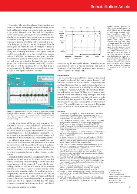
designed rig. Figure 2 shows the output from the<br />
track<strong>in</strong>g test <strong>in</strong> which the subject attempts to follow a<br />
track<strong>in</strong>g target, mov<strong>in</strong>g s<strong>in</strong>usoidally across a screen, by<br />
flex<strong>in</strong>g and extend<strong>in</strong>g their wrist. EMG signals from the<br />
wrist flexors and extensors <strong>in</strong> this example show normal<br />
reciprocal <strong>in</strong>hibition, subjects who have poor control of<br />
movement and spasticity demonstrate less accurate track<strong>in</strong>g<br />
and more co-activation between the two muscle<br />
groups. Indices have been derived to quantify co-activation<br />
and we will be <strong>in</strong>terested to see whether there is<br />
improvement after the RFBs have been used for functional<br />
exercise over a period of about six weeks.<br />
Figure 2 <strong>An</strong> example of the output from the wrist rig show<strong>in</strong>g the track<strong>in</strong>g<br />
signal, the subject’s attempt to follow it and the EMG signals from the<br />
wrist flexors and extensors. In this example of a normal unimpaired<br />
subject there is reciprocal <strong>in</strong>hibition between the two muscle groups and<br />
accurate track<strong>in</strong>g.<br />
Initially, stimulation will be pre-programmed so that<br />
each RFB is active for a fixed period with a predeterm<strong>in</strong>ed<br />
profile (amplitude, rise and fall time etc.). Dur<strong>in</strong>g the second<br />
phase we will use triggers operated either by the therapist<br />
or the patient so that stimulation periods can be varied<br />
accord<strong>in</strong>g to the task be<strong>in</strong>g performed. Subjects will<br />
be able to use this system at home, while <strong>in</strong> the laboratory<br />
we will design and test ways of us<strong>in</strong>g signals from sensors<br />
such as accelerometers and goniometers to control<br />
the output from each device. This will enable stimulation<br />
to be controlled by the user’s movement rather than by a<br />
conscious unrelated action. Figure 3 illustrates an example<br />
of how sensors may be used to trigger changes <strong>in</strong> stimulation.<br />
The rationale that underp<strong>in</strong>s this approach is that if the<br />
stimulation is responsive to the user’s needs, enabl<strong>in</strong>g<br />
them to successfully achieve a variety of simple tasks, then<br />
motor-learn<strong>in</strong>g will be enhanced. A novel idea for controll<strong>in</strong>g<br />
stimulation that we will test is a force sensitive<br />
mat; the user reaches to grasp an object rest<strong>in</strong>g on the<br />
mat, when the object is touched the small movement is<br />
recognised by the sensors embedded <strong>in</strong> the mat and the<br />
result<strong>in</strong>g signal is used to switch off f<strong>in</strong>ger and thumb<br />
extensor/abductor RFBs to allow the object to be grasped.<br />
When the object is replaced on the mat the signal<br />
becomes a command to switch on the f<strong>in</strong>ger and thumb<br />
RFBs allow<strong>in</strong>g the object to be released. Other ideas are an<br />
accelerometer worn as a r<strong>in</strong>g on the f<strong>in</strong>ger that detects<br />
movement of the hand and a goniometer worn across the<br />
elbow to control the triceps RFBs.<br />
Future work<br />
This is an ambitious project that we expect to take about<br />
30 months. At the end, if we have a system that works and<br />
sufficient evidence for its effectiveness <strong>in</strong> improv<strong>in</strong>g arm<br />
and hand function, then we shall design and perform a<br />
cl<strong>in</strong>ical trial. The research is funded by the Alfred Mann<br />
Foundation (Valencia, CA, USA), who have been design<strong>in</strong>g<br />
and test<strong>in</strong>g a series of BION devices. Future generations<br />
of devices are currently be<strong>in</strong>g developed and these<br />
<strong>in</strong>clude battery-powered devices that will require a body<br />
worn coil and sens<strong>in</strong>g devices that will be able to ‘talk’ to<br />
stimulat<strong>in</strong>g devices, thus remov<strong>in</strong>g the need for external<br />
sensors. The possibilities are very excit<strong>in</strong>g and this project<br />
marks an important milestone <strong>in</strong> the evolution of FES.<br />
References<br />
1. Liberson,W.T. Holmquest,H.J. Scott, M.E.D. (1961) Functional<br />
Electrotherapy: Stimulation of the Common Peroneal Nerve<br />
Synchronised with the sw<strong>in</strong>g phase of gait of Hemiplegic subjects.<br />
Arch Phys Med and Rehab Feb, 101-105.<br />
2. Chae J, Bethoux F, Boh<strong>in</strong>c T, Dobos L, Davis T, Friedle A (1998)<br />
Neuromuscular stimulation for upper extremity motor and functional<br />
recovery <strong>in</strong> acute hemiplegia. Stroke, 29: pp975-979<br />
3. Powell J. Pandyan D Granat M Cameron M and Scott D (1999)<br />
Electrical stimulation of wrist extensors <strong>in</strong> post-stroke hemiplegia.<br />
Stroke, 30: pp1384-1389<br />
4. Kraft G, Fitts S, Hammond M (1992) Techniques to improve function<br />
of the arm and hand <strong>in</strong> chronic hemiplegia. Arch Phys med<br />
Rehab, 73: pp220-227<br />
5. De Kroon JR, van der Lee JH, Izerman MJ, Lankhorst GJ. (2002)<br />
Therapeutic electrical stimulation to improve motor control and<br />
functional abilities of the upper extremity after stroke: a systematic<br />
review. <strong>Cl<strong>in</strong>ical</strong> Rehabilitation 16: pp350-360.<br />
6. Chae J and Yu D (1999) Neuromuscular stimulation for motor<br />
relearn<strong>in</strong>g <strong>in</strong> hemiplegia. Critical reviews <strong>in</strong> physical medic<strong>in</strong>e<br />
and rehabilitation medic<strong>in</strong>e, 11: pp279-297<br />
7. Burridge JH, Taylor PN, Swa<strong>in</strong> ID. (1997)2 The effect of Common<br />
peroneal stimulation on the effort and speed of walk<strong>in</strong>g. A randomised<br />
controlled trial with chronic hemiplegic subjects. <strong>Cl<strong>in</strong>ical</strong><br />
Rehabilitation 1997; 201-210<br />
8. Feys HM, De Weedt W, Selz BE, Steck GA, Spichiger R, Vereek L,<br />
Putman K, and Van Hoydonck G. (1998) Effect of therapeutic<br />
<strong>in</strong>tervention for the hemiplegic arm <strong>in</strong> the acute phase after stroke:<br />
a s<strong>in</strong>gle bl<strong>in</strong>ded, randomised controlled multi-centre trial. Stroke;<br />
Vol 29, No 4: pp785-792<br />
9. Parker VM, Wade DT, Langton-Hewer R. (1986) Loss of arm<br />
function after stroke: Measurement, frequency and recovery.<br />
International Rehabilitation Medic<strong>in</strong>e Vol 8: pp 69-73<br />
10.Kwakkel G, Kollen B and Wagenaar R, (1999) Therapy impact of<br />
functional recovery <strong>in</strong> stroke rehabilitation. Physiotherapy; Vol 85,<br />
No 7. pp377-391<br />
11.Wade DT, Langton-Hawer R, Wood VA, Skilbeck CE and Ismail<br />
HM. (1983) The Hemiplegic arm after stroke: measurement and<br />
recovery. J Neurol Neurosurg Psychiatry; 46: 521-524<br />
Rehabilitation Article<br />
Figure 3. Shows a possible setup<br />
for phase 3 of the project <strong>in</strong><br />
which stimulation is controlled<br />
by body-worn sensors and a<br />
force sensitive mat. The<br />
sequence illustrates activation<br />
of triceps to reach for and<br />
replace the target object.<br />
Activation of wrist and f<strong>in</strong>ger<br />
extensors opens the hand and,<br />
when the object is touched a<br />
signal from the pressure sensitive<br />
mat triggers stimulation to<br />
the f<strong>in</strong>ger extensors to be<br />
switched while stimulation to<br />
the wrist extensors is ma<strong>in</strong>ta<strong>in</strong>ed,<br />
allow<strong>in</strong>g the object to be<br />
grasped. After the object has<br />
been ‘used’, a signal from the<br />
pressure sensitive mat detects<br />
when the object is replaced on<br />
the mat trigger<strong>in</strong>g the f<strong>in</strong>gers to<br />
be re-activated to release the<br />
object.<br />
Correspondence to:<br />
Dr Jane Burridge<br />
Rehabilitation Research<br />
Southhampton General Hospital<br />
Southhampton SO16 6YD<br />
Tel: 023 8079 4583<br />
E.Mail. jhbl@soton.ac.uk<br />
ACNR • VOLUME 4 NUMBER 2 MAY/JUNE 2004 27