An Anatomico-Clinical Overview - Advances in Clinical ...
An Anatomico-Clinical Overview - Advances in Clinical ...
An Anatomico-Clinical Overview - Advances in Clinical ...

You also want an ePaper? Increase the reach of your titles
YUMPU automatically turns print PDFs into web optimized ePapers that Google loves.
Review Article<br />
Vascular Disorders of the Posterior Circulation –<br />
<strong>An</strong> <strong>An</strong>atomico-<strong>Cl<strong>in</strong>ical</strong> <strong>Overview</strong><br />
INTRODUCTION<br />
The posterior cerebral circulation consists of the vertebrobasilar<br />
system (vertebral arteries, basilar artery, posterior<br />
<strong>in</strong>ferior cerebellar arteries, anterior <strong>in</strong>ferior cerebellar<br />
arteries, superior cerebellar arteries, posterior<br />
cerebral arteries) (Fig. 1-3) and supplies the follow<strong>in</strong>g<br />
anatomical structures: upper cervical sp<strong>in</strong>al cord,<br />
medulla oblongata, pons, cerebellum, mesencephalon,<br />
thalami, occipital lobes, and parts of the temporal and<br />
parietal lobes (Fig. 4). Strokes <strong>in</strong> the posterior circulation<br />
comprise approximately 10% to 15% of all strokes 1<br />
and are more common <strong>in</strong> men than <strong>in</strong> women 2 . As <strong>in</strong><br />
supratentorial strokes, the most common etiology is<br />
ischemic (approximately 80% of cases), with the proportion<br />
of haemorrhagic strokes be<strong>in</strong>g similar to that<br />
seen <strong>in</strong> the anterior circulation. Although <strong>in</strong>traparenchymatous<br />
haemorrhage is slightly more frequent,<br />
while subarachnoid haemorrhage seems to be<br />
less common <strong>in</strong> the posterior territory 3 .<br />
AETIOLOGY<br />
The risk factors for posterior circulation <strong>in</strong>farction do not<br />
differ from those for anterior circulation strokes.<br />
However, a history of prior stroke, but not transient<br />
ischaemic attacks, may be more frequently encountered<br />
<strong>in</strong> patients with posterior circulation strokes 2 . The structures<br />
most commonly affected by <strong>in</strong>farction are the bra<strong>in</strong><br />
stem (60%) and cerebellum (50%) and so are caused by<br />
lesions of basilar and/or vertebral arteries (up to 50%) 4 .<br />
The most common cause is basilar artery (BA) stenosis or<br />
occlusion (approximately 40% of patients) with the proximal<br />
and middle segments of BA be<strong>in</strong>g the most frequent<br />
site of occlusion 5 . Some patients may have stenosis and/or<br />
occlusion of the extracranial or <strong>in</strong>tracranial part of the<br />
vertebral artery, or posterior cerebral arteries lesions. In<br />
rare <strong>in</strong>stances, a dolichoectasy of basilar artery or vertebral<br />
artery may be encountered 4 .<br />
Small artery disease is a presumed cause of stroke <strong>in</strong><br />
15% of patients, whereas cardiac embolism (e.g. from<br />
thrombus associated with an ak<strong>in</strong>etic left ventricle, atrial<br />
fibrillation, or paradoxical embolism through a PFO) is a<br />
causative factor of stroke <strong>in</strong> approximately 13% of<br />
BA<br />
Figure 1. A schematic<br />
draw<strong>in</strong>g of the posterior<br />
circulation arteries. VA:<br />
vertebral artery; PICA:<br />
posterior <strong>in</strong>ferior<br />
cerebellar artery; AICA:<br />
anterior <strong>in</strong>ferior<br />
cerebellar artery; BA:<br />
basilar artery; SCA:<br />
superior cerebellar artery;<br />
PCA: posterior cerebral<br />
artery.<br />
patients. In an equal proportion of patients (i.e. 13%)<br />
there may be more than one possible cause of stroke<br />
<strong>in</strong>clud<strong>in</strong>g arterial stenosis and/or occlusion, lacunar<br />
lesions or a potential cardiogenic source of embolism,<br />
whilst <strong>in</strong> 10% of cases no potential cause of stroke can be<br />
identified.<br />
However, there are two types of <strong>in</strong>farction <strong>in</strong> the posterior<br />
territory which are highly suggestive of a particular<br />
etiology. Isolated unilateral or bilateral bra<strong>in</strong>stem <strong>in</strong>farcts<br />
(<strong>in</strong>volv<strong>in</strong>g midbra<strong>in</strong> and/or pons) are associated with<br />
basilar stenosis, while isolated cerebellar <strong>in</strong>farctions are<br />
associated with cardioembolism 4 .<br />
Other etiologies of stroke such as for example, dissection<br />
of basilar artery or vertebral arteries (Figure 5), are<br />
rarely reported <strong>in</strong> patients with posterior circulation disorders.<br />
Cerebral venous and/or s<strong>in</strong>us thrombosis limited<br />
to the posterior circulation teritory are casuistic.<br />
CLINICAL FEATURES<br />
In posterior circulation <strong>in</strong>farctions severe headache and<br />
vomit<strong>in</strong>g are more frequently seen than <strong>in</strong> anterior circulation<br />
strokes 2 . The common signs and symptoms <strong>in</strong>clude<br />
bulbar or pseudobulbar palsy, vertigo and dizz<strong>in</strong>ess,<br />
hemiparesis, tetraparesis, cerebellar ataxia, eye movement<br />
disorders, changes <strong>in</strong> levels of consciousness and neuropsychological<br />
dysfunction. Indeed the organisation of<br />
the bra<strong>in</strong>stem relative to its vascular supply leads to a<br />
number of well-recognised syndromes. So ischemic<br />
lesions <strong>in</strong> the territory supplied by posterior <strong>in</strong>ferior cerebellar<br />
artery may give the lateral medullary syndrome<br />
(Wallenberg’s syndrome) [characterised by ipsilateral<br />
facial sensory disturbances, nystagmus, dysphagia,<br />
dysarthria, Horner’s syndrome with contralateral hemibody<br />
dissociated sensory disturbances]. The symptoms of<br />
anterior <strong>in</strong>ferior cerebellar artery occlusion are similar to<br />
those of Wallenberg’s syndrome, but can additionally be<br />
differentiated by the presence of limb and trunk ataxia,<br />
t<strong>in</strong>nitus, deafness, and facial nerve <strong>in</strong>volvement. Superior<br />
cerebellar artery occlusion can be dist<strong>in</strong>guished by the<br />
predom<strong>in</strong>ance of cerebellar symptoms and a trochlear<br />
nerve palsy 6 .<br />
Basilar artery occlusion may give a large spectrum of<br />
neurological signs and symptoms of which the most<br />
Section<br />
Dr Bartomiej<br />
Piechowski-Jówiak, is a<br />
Stroke fellow at the<br />
Department of<br />
Neurology, CHUV,<br />
Lausanne, Switzerland.<br />
His research <strong>in</strong>terests<br />
<strong>in</strong>clude cerebrovascular<br />
disorders and<br />
neurosonology.<br />
Professor Julien<br />
Boggouslavsky is<br />
Professor and Chairman<br />
of the University<br />
Department of<br />
Neurology, Lausanne,<br />
Switzerland. He is also<br />
Professor of cerebrovascular<br />
disease and Chief<br />
of the Lausanne Stroke<br />
Program, and Chief of<br />
the Neurology Service,<br />
University Hospital,<br />
Lausanne.<br />
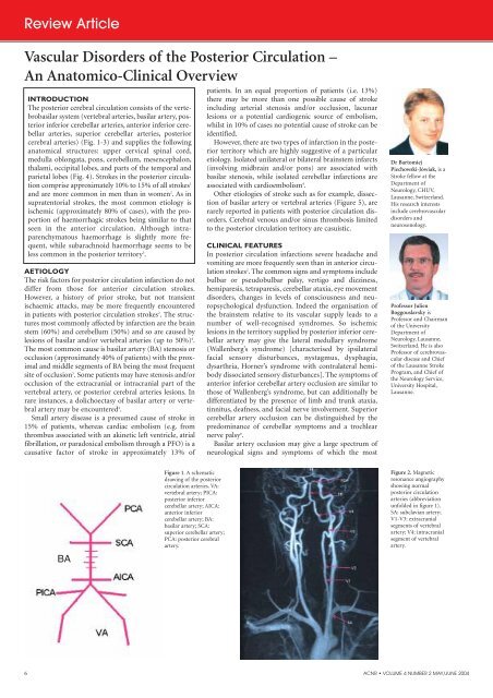
Figure 2. Magnetic<br />
resonance angiography<br />
show<strong>in</strong>g normal<br />
posterior circulation<br />
arteries (abbreviation<br />
unfolded <strong>in</strong> figure 1).<br />
SA: subclavian artery;<br />
V1-V3: extracranial<br />
segments of vertebral<br />
artery; V4: <strong>in</strong>tracranial<br />
segment of vertebral<br />
artery.<br />
6 ACNR • VOLUME 4 NUMBER 2 MAY/JUNE 2004