

2007 Winter Meeting - London - The Pathological Society of Great ...
2007 Winter Meeting - London - The Pathological Society of Great ...
2007 Winter Meeting - London - The Pathological Society of Great ...
- No tags were found...

Create successful ePaper yourself
Turn your PDF publications into a flip-book with our unique Google optimized e-Paper software.
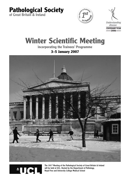
<strong>Pathological</strong> <strong>Society</strong><strong>of</strong> <strong>Great</strong> Britain & Ireland<strong>Winter</strong> Scientific <strong>Meeting</strong>incorporating the Trainees’ Programme3–5 January <strong>2007</strong><strong>The</strong> 191 st <strong>Meeting</strong> <strong>of</strong> the <strong>Pathological</strong> <strong>Society</strong> <strong>of</strong> <strong>Great</strong> Britain & Irelandwill be held at UCL. Hosted by the Department <strong>of</strong> Pathology,Royal Free and University College Medical School
CONTENTSProgramme Synopsis and Timetable . . . . . . . . . . . . . . . . . . . . . . 3Scientific Sessions Information . . . . . . . . . . . . . . . . . . . . . . . . . . . . . . 5General Arrangements . . . . . . . . . . . . . . . . . . . . . . . . . . . . . . . . . . . . . . . . 7Social Activities . . . . . . . . . . . . . . . . . . . . . . . . . . . . . . . . . . . . . . . . . . . . . . . . . . 9CPD . . . . . . . . . . . . . . . . . . . . . . . . . . . . . . . . . . . . . . . . . . . . . . . . . . . . . . . . . . . . . . . . 9Future <strong>Meeting</strong>s . . . . . . . . . . . . . . . . . . . . . . . . . . . . . . . . . . . . . . . . . . . . . . . . 10Detailed ProgrammeWednesday 3 January . . . . . . . . . . . . . . . . . . . . . . . . . . . . . . . . . . . . 11Thursday 4 January . . . . . . . . . . . . . . . . . . . . . . . . . . . . . . . . . . . . . . . 14Friday 5 January . . . . . . . . . . . . . . . . . . . . . . . . . . . . . . . . . . . . . . . . . . . 17Trade Exhibition/Sponsoring Companies . . . . . . . . . . . . . . . . . 21AbstractsPlenary . . . . . . . . . . . . . . . . . . . . . . . . . . . . . . . . . . . . . . . . . . . . . . . . . . . . . . . 23Posters . . . . . . . . . . . . . . . . . . . . . . . . . . . . . . . . . . . . . . . . . . . . . . . . . . . . . . . . 27Oral . . . . . . . . . . . . . . . . . . . . . . . . . . . . . . . . . . . . . . . . . . . . . . . . . . . . . . . . . . . 49Speakers . . . . . . . . . . . . . . . . . . . . . . . . . . . . . . . . . . . . . . . . . . . . . . . . . . . . . 57Abstract Reviewers . . . . . . . . . . . . . . . . . . . . . . . . . . . . . . . . . . . . . . . . . . . . . 63Maps . ......................................................Inside front &back coversPROGRAMME ACKNOWLEDGEMENTSPublished by<strong>The</strong> <strong>Pathological</strong> <strong>Society</strong> <strong>of</strong> <strong>Great</strong> Britain & Ireland© 2006COVER PHOTOGRAPH<strong>The</strong> cover photograph is reproduced with permissionProgramme is typeset in Times and ITC Stone SansThis programme was designed, produced and printed in England byByte & Type Limited · Birmingham · 0121 772 3828 · info@bytetype.co.uk2 <strong>Winter</strong> <strong>Meeting</strong> (191 st ) 3–5 January <strong>2007</strong> Scientific Programme
PROGRAMME SYNPOSIS AND TIMETABLE(Showing times, sessions and venues) CRUCIFORM BUILDING (BASEMENT), GOWER ST. UCL MAIN BUILDING, GOWER ST. ROCKEFELLER BUILDING, UNIVERSITY ST.WEDNESDAY 3 JANUARY <strong>2007</strong>TIME BUILDING & VENUE SESSION08.00 –NORTH CLOISTERS Registration and C<strong>of</strong>fee09.00–12.00 –LECTURE THEATRE 1 Symposium: Urological Pathology09.00–17.00 –CLUSTER ROOM Slide Seminar Competition Case Viewing: LBC innon-gynaecological cytology (Win a case <strong>of</strong> champagne!)10.30–12.00 –LECTURE THEATRE 2 Sino-British Pathology Forum10.30–11.00 –THE CLOISTERS C<strong>of</strong>fee12.00–12.15 –LECTURE THEATRE 1 Welcome Address: Pr<strong>of</strong> G Williams, UCL12.15–13.15 –LECTURE THEATRE 1 Academic Pathology Forum13.15–14.15 –THE CLOISTERS Lunch14.00–15.00 –THE CLOISTERS Poster Viewing (Abstracts P1–P39)14.00–15.00 –THE CLOISTERS Trade Exhibition15.00–18.00 –LECTURE THEATRE 1 Symposium: Undergraduate Education15.00–17.00 –LECTURE THEATRE 2 Oral Communications (Abstracts O1–O6)15.30–16.00 –THE CLOISTERS Tea (Oral Communications)16.30–17.00 –THE CLOISTERS Tea (Symposium: Undergraduate Education)18.00–19.30 –JEREMY BENTHAM ROOM Welcome ReceptionTHURSDAY 4 JANUARY <strong>2007</strong>TIME BUILDING & VENUE SESSION08.00 –NORTH CLOISTERS Registration and C<strong>of</strong>fee09.00–15.00 –CLUSTER ROOM Slide Seminar Competition Case Viewing: LBC in nongynaecologicalcytology (Win a case <strong>of</strong> champagne!)09.00–10.30 –LECTURE THEATRE 1 Plenary Oral Presentations (Abstracts PL1–PL6)10.30–11.00 –THE CLOISTERS C<strong>of</strong>fee11.00–13.00 –LECTURE THEATRE 1 Trainees Forum13.00–14.15 –THE CLOISTERS Lunch13.00–14.00 –JF SMITH SEMINAR ROOM Meet the Experts: Post Mortem Histology(3RD FLOOR)13.30–14.30 –THE CLOISTERS Poster Viewing (Abstracts P40–P82)13.30–14.30 –THE CLOISTERS Trade Exhibition14.30–17.30 –LECTURE THEATRE 1 Symposium: Advances in Lymphomas since the advent <strong>of</strong> theWHO Classification16.00–16.30 –THE CLOISTERS Tea17.30–18.15 –LECTURE THEATRE 1 <strong>Pathological</strong> <strong>Society</strong>’s 3 rd Goudie Lecture: Our changing view<strong>of</strong> the genome: implications for Pathology. Pr<strong>of</strong> PA Hall, Belfast19.30–20.00 –THE FLAXMAN GALLERY <strong>Society</strong> Dinner Reception20.00–22.30 –THE OLD REFECTORY <strong>Society</strong> Dinner<strong>Winter</strong> <strong>Meeting</strong> (191 st ) 3–5 January <strong>2007</strong> Scientific Programme3
CRUCIFORM BUILDING (BASEMENT), GOWER ST. UCL MAIN BUILDING, GOWER ST. ROCKEFELLER BUILDING, UNIVERSITY ST.FRIDAY 5 JANUARY <strong>2007</strong>TIME BUILDING & VENUE SESSION08.00 –NORTH CLOISTERS Registration and C<strong>of</strong>fee09.00–12.45 –LECTURE THEATRE 2 Oral Communications (Abstracts O7–O20)09.30–16.30 –GUSTAVE TUCK UK NEQAS for Immunocyotchemistry and FISH TestingLECTURE THEATRE09.30–12.00 –LECTURE THEATRE 1 Slide Seminar Follow-up Symposium: LBC innon-gynaecological cytology10.30–11.00 –THE CLOISTERS C<strong>of</strong>fee12.45–14.00 –THE CLOISTERS Lunch13.00–14.00 –JF SMITH SEMINAR ROOM Meet the Experts: Getting started in Research(3RD FLOOR)13.00–14.00 –THE CLOISTERS Trade Exhibition14.00–16.30 –LECTURE THEATRE 2 Trainee Oral Communications (Abstracts O21–O28)15.00–15.30 –THE CLOISTERS Tea4<strong>Winter</strong> <strong>Meeting</strong> (191 st ) 3–5 January <strong>2007</strong> Scientific Programme
SCIENTIFIC SESSIONS INFORMATION CRUCIFORM BUILDING (BASEMENT), GOWER ST. UCL MAIN BUILDING, GOWER ST. ROCKEFELLER BUILDING, UNIVERSITY ST.PLENARY ORAL SESSION –LECTURE THEATRE 1<strong>The</strong> plenary oral session, in which the 6 highest-ranked submitted oral abstracts will be presented, will beheld on Thursday 4 January, 09.00–10.30 hrs.Prize: A prize for the best presentation, donated by the Journal <strong>of</strong> Pathology will be presented at the<strong>Society</strong> Dinner.ORAL COMMUNICATIONS –LECTURE THEATRE 2Oral communication sessions will be held as follows:Wednesday 3 January, 15.00–17.00Friday 5 January, 09.00–12.45Note to presenters: Speakers are reminded that no communication may exceed the time allocated on theprogramme without the consent <strong>of</strong> the meeting, obtained through the Chairman.POSTER VIEWING & ROUNDS –THE CLOISTERSPosters will be displayed throughout the meeting, but dedicated viewing sessions will be on:Wednesday 3 January, 14.00–15.00Thursday 4 January, 13.30–14.30Poster Rounds: Chairmen will review posters during Wednesday 3 January and Thursday 4 January.Ideally, posters should be in place by 12.00 hrs on Wednesday 3 January and removed by 15.00 hrs onFriday 5 January. At least one <strong>of</strong> the contributors must be in attendance during the viewing period, asindicated in the programme synopsis.Prizes: Prizes are awarded for the three best posters: <strong>The</strong> Sir Alastair Currie Prize, Second and Thirdprizes will be presented at the <strong>Society</strong> Dinner on Thursday 4 January.SYMPOSIAFour symposia will be held:Wednesday 3 January –LECTURE THEATRE 109.00–12.00 Urological Pathology15.00–18.00 Undergraduate EducationThursday 4 January –LECTURE THEATRE 114.30–17.30 Advances in lymphomas since the advent <strong>of</strong> the WHO ClassificationFriday 5 January –LECTURE THEATRE 109.30–12.00 LBC in non-gynaecological cytology – Slide Seminar Follow-up<strong>Winter</strong> <strong>Meeting</strong> (191 st ) 3–5 January <strong>2007</strong> Scientific Programme5
CRUCIFORM BUILDING (BASEMENT), GOWER ST. UCL MAIN BUILDING, GOWER ST. ROCKEFELLER BUILDING, UNIVERSITY ST.FORAWednesday 3 January10.30–12.00 Sino-British Pathology Forum –LECTURE THEATRE 212.15–13.15 Academic Pathology Forum –LECTURE THEATRE 1TRAINEES PROGRAMMEThursday 4 January11.00–13.00 Trainees Forum –LECTURE THEATRE 113.00–14.00 Meet the Experts – Post Mortem Histology –JF SMITH SEMINAR ROOM (3RD FLOOR)Expert: Dr PJ Gallagher, SouthamptonFriday 5 January13.00–14.00 Meet the Experts – Getting started in research –JF SMITH SEMINAR ROOM (3RD FLOOR)Experts: Pr<strong>of</strong> NR Lemoine, <strong>London</strong> and Pr<strong>of</strong> I Tomlinson, <strong>London</strong>14.00–16.30 Trainee Oral Communications –LECTURE THEATRE 2SLIDE SEMINAR – LBC in non-gynaecological cytology –CLUSTER ROOMCompetition: <strong>The</strong>re will be a slide seminar competition and digital images <strong>of</strong> all cases will be available forpreview during the course <strong>of</strong> the meeting. <strong>The</strong> images will also become available on the <strong>Society</strong>’s websitein due course.<strong>The</strong> images will be available for viewing on:Wednesday 3 January, 09.00–17.00Thursday 4 January, 09.00–15.00Prize: A case <strong>of</strong> champagne will be awarded at the <strong>Society</strong> Dinner on Thursday 4 January.Discussion Session: Friday 5 January, 09.30–12.00 –LECTURE THEATRE 1SOCIETY LECTURE –LECTURE THEATRE 1Thursday 4 January, 17.30–18.15<strong>The</strong> <strong>Pathological</strong> <strong>Society</strong> <strong>of</strong> <strong>Great</strong> Britain & Ireland’s 3 rd Goudie Lecture entitled: Our changing view <strong>of</strong>the genome: implications for pathology, will be given by Pr<strong>of</strong> PA Hall, Centre for Cancer Research andCell Biology, Division <strong>of</strong> Pathology, Queen’s University, Befast.COMPANION MEETING –GUSTAVE TUCK LECTURE THEATREFriday 5 January09.30–16.30 UK NEQAS for Immunocytochemistry and FISH TestingTRADE EXHIBITION –THE CLOISTERSDelegates are encouraged to visit the Trade Exhibition and are requested to support the companiesrepresented there.6 <strong>Winter</strong> <strong>Meeting</strong> (191 st ) 3–5 January <strong>2007</strong> Scientific Programme
GENERAL ARRANGEMENTS CRUCIFORM BUILDING (BASEMENT), GOWER ST. UCL MAIN BUILDING, GOWER ST. ROCKEFELLER BUILDING, UNIVERSITY ST.REGISTRATIONRegistration is via the links on the <strong>Society</strong>’s website: www.pathsoc.org.uk<strong>The</strong> registration system will issue an automated email acknowledgement. All tickets will be issued onarrival at the Delegate Reception Desk at UCL.FEES (Fees include refreshments and lunch)Non-concessions• Up to and including 27 November 2006:<strong>Society</strong> MembersWhole meeting £180, or £80 per day (or part day)Non-membersWhole meeting £270, or £120 per day (or part day)• After 27 November 2006:<strong>Society</strong> MembersNon-membersWhole meeting £270, or £120 per day (or part day)Whole meeting £405, or £180 per day (or part day)Concessions – Qualifying CategoriesGroup A – <strong>Society</strong> MembersHonorary or Senior Members, Biomedical Scientists/Technicians (all grades), PhD Students,Post-Doctoral Fellows, Trainees.Group B – Non-MembersBiomedical Scientists/Technicians (all grades), PhD Students, Post-Doctoral Fellows, Trainees.Group C – Non-Members (at <strong>Society</strong> Member rates)Undergraduates.Groups B & C: Concessionary delegates must provide documentary evidence to qualify for theconcessionary Fees. Please send confirmation <strong>of</strong> your status, signed by your head <strong>of</strong> training,stating National Training Numbers, where applicable. This must be emailed or faxed to:(Email) julie@pathsoc.org.uk (Fax) +44(0)20 7976 1267Concessions• Up to and including 27 November 2006:<strong>Society</strong> MembersWhole meeting £50, or £25 per day (or part day)Non-membersWhole meeting £75, or £40 per day (or part day)• After 27 November 2006:<strong>Society</strong> MembersNon-members<strong>Society</strong> Dinner• £50 per ticketWhole meeting £75, or £40 per day (or part day)Whole meeting £105, or £60 per day (or part day)ADVANCE REGISTRATION – CLOSING DATEAdvance registration will close on Monday 18 December 2006.After this deadline registration will only be accepted on-site at the meeting.CANCELLATIONSPlease note that the <strong>Society</strong> is unable to refund registration fees for cancellations received afterMonday 11 December 2006.<strong>Winter</strong> <strong>Meeting</strong> (191 st ) 3–5 January <strong>2007</strong> Scientific Programme7
CRUCIFORM BUILDING (BASEMENT), GOWER ST. UCL MAIN BUILDING, GOWER ST. ROCKEFELLER BUILDING, UNIVERSITY ST.DELEGATE ENROLMENT (AT THE MEETING)Enrolment at the Delegate Reception Desk will take place at –NORTH CLOISTERS as follows:Wednesday 3 – Friday 5 January <strong>2007</strong>, from 08.00PRESENTATION CHECKING AND PREVIEWThis will be available at –CLUSTER ROOMORAL PRESENTATIONS AND LECTURESElectronic presentations must be in Micros<strong>of</strong>t PowerPoint (PC or Mac). Versions earlier than 2000 are notacceptable. Presentations should be submitted in advance <strong>of</strong> the meeting to arrive no later than 18December 2006.Files may be emailed or a CD posted to Bridgette Smith (telephone: +44(0)20 7679 6304) as follows:Email:Post:bridgette.smith@ucl.ac.uk (maximum attachment size is 5 megabytes)Bridgette A SmithPA/Office Manager, Department <strong>of</strong> Pathology, University College <strong>London</strong>,Rockefeller Building, University Street, <strong>London</strong> WC1E 6JJPlease label the files or CDs with:• Date and time <strong>of</strong> your lecture• Lecture theatre• Your nameIMPORTANT:Please bring another copy <strong>of</strong> your presentation with you to the meeting.MESSAGESDuring the meeting, messages for delegates may be left on telephone: +44(0)20 7679 2000 extn 5283.<strong>The</strong>re will also be a message board located beside the Delegate Reception Desk.REFRESHMENTSAll refreshments will be served at –THE CLOISTERS.BADGESDelegates are requested to wear their badges at all times.SMOKINGSmoking is prohibited at all meetings and social events.DISCLAIMER<strong>The</strong> <strong>Pathological</strong> <strong>Society</strong> <strong>of</strong> <strong>Great</strong> Britain & Ireland cannot be held responsible for any injury or losssustained during the <strong>Meeting</strong>.8 <strong>Winter</strong> <strong>Meeting</strong> (191 st ) 3–5 January <strong>2007</strong> Scientific Programme
CRUCIFORM BUILDING (BASEMENT), GOWER ST. UCL MAIN BUILDING, GOWER ST. ROCKEFELLER BUILDING, UNIVERSITY ST.TRAVELTrain – Mainline stationsKing’s Cross, St. Pancras, EustonTrain – Underground stationsWarren Street (Northern Line; Victoria Line)Euston Square (Hammersmith & City Line; Metropolitan Line; Circle Line)CarParking is almost impossible! NCP car parks are nearby but expensive. Daily Conjestion Charge <strong>of</strong> £8 willapply.AirFrom <strong>London</strong> Heathrow take the Piccadilly Line to Green Park, change to the Victoria Line and travel toWarren Street.ACCOMMODATIONFor a list <strong>of</strong> local hotels, at discounted “UCL rates,” see the <strong>Society</strong>’s website: www.pathsoc.org.ukSOCIAL ACTIVITIESWednesday 3 January, 18.00–19.30, at –THE JEREMY BENTHAM ROOMWelcome ReceptionTickets are free – to reserve your ticket please tick the relevant box when registering.Thursday 4 January, 19.30 for 20.00, at –THE OLD REFECTORY<strong>Society</strong> DinnerTickets are £50 each. To reserve your ticket please tick the relevant box when registering.Local Places <strong>of</strong> InterestPlease refer to the Internet for information.CONTINUING PROFESSIONAL DEVELOPMENT (CPD)This meeting has been approved by <strong>The</strong> Royal College <strong>of</strong> Pathologists for the purposes <strong>of</strong> ContinuingPr<strong>of</strong>essional Development. Credits can be accrued as follows:For each full day: 7 pointsFor each half day: 3 pointsCertificatesDelegates who are eligible for CPD points should complete the CPD Certificate Request Form which willbe provided in their delegate pack at the meeting.<strong>Winter</strong> <strong>Meeting</strong> (191 st ) 3–5 January <strong>2007</strong> Scientific Programme9
CRUCIFORM BUILDING (BASEMENT), GOWER ST. UCL MAIN BUILDING, GOWER ST. ROCKEFELLER BUILDING, UNIVERSITY ST.FUTURE MEETINGS<strong>2007</strong> 3–6 July Glasgow Pathology <strong>2007</strong>: 4 th Joint <strong>Meeting</strong> <strong>of</strong> the <strong>Pathological</strong><strong>Society</strong> and the British Division <strong>of</strong> the IAP2008 7–9 January Oxford1–4 July Leeds2009 7–9 January GKT, <strong>London</strong>30 June–3 July Cardiff Pathology 2009:5 th Joint <strong>Meeting</strong> <strong>of</strong> the <strong>Pathological</strong><strong>Society</strong> and the British Division <strong>of</strong> the IAP2010 January Imperial College, <strong>London</strong>29 June–2 July St AndrewsENQUIRIES BEFORE THE MEETING<strong>Pathological</strong> <strong>Society</strong> <strong>of</strong> <strong>Great</strong> Britain & Ireland2 Carlton House Terrace, <strong>London</strong> SW1Y 5AFTelephone +44 (0)20 7976 1260Fax +44 (0)20 7976 1267Emailadmin@pathsoc.org.uk10<strong>Winter</strong> <strong>Meeting</strong> (191 st ) 3–5 January <strong>2007</strong> Scientific Programme
DETAILED PROGRAMME – Wednesday 3 January <strong>2007</strong>Presenter = {P} Abstract numbers are shown in bold type and in square brackets eg [S123] CRUCIFORM BUILDING (BASEMENT), GOWER ST. UCL MAIN BUILDING, GOWER ST. ROCKEFELLER BUILDING, UNIVERSITY ST.08.00 –North CloistersREGISTRATION09.00–17.00 –Cluster RoomSLIDE SEMINAR COMPETITION CASE VIEWING: LBC in non-gynaecologicalcytology (WIN A CASE OF CHAMPAGNE!)09.00–12.00 –Lecture <strong>The</strong>atre 1SYMPOSIUM: Urological PathologyChair: Dr A Freeman, UCLHPr<strong>of</strong> S Fleming, University <strong>of</strong> Dundee09.00–09.30 Histopathology and Genetics <strong>of</strong> Sporadic and Familial Renal CarcinomaPr<strong>of</strong> H Moch, University Hospital, Zurich09.30–10.00 [S7] Proteomics and Prostate CarcinomaDr L Egevad, International Agency for Research on Cancer, Lyon, France10.00–10.30 [S8] <strong>The</strong> value <strong>of</strong> tissue microarrays in urological pathologyDr DM Berney, Barts & <strong>The</strong> <strong>London</strong>, Queen Mary School <strong>of</strong> Medicine &Dentistry, <strong>London</strong>10.30–11.00 COFFEE –THE CLOISTERS11.00–11.30 [S9] Evolving a 21 st Century Uropathology EQA scheme: from slides to imagesDr P Harnden, Leeds Teaching Hospitals, Leeds11.30–12.00 Growth factors and cell signalling in prostate cancerPr<strong>of</strong> S Fleming, University <strong>of</strong> Dundee10.30–12.00 –Lecture <strong>The</strong>atre 2SINO-BRITISH PATHOLOGY FORUMChair: Pr<strong>of</strong> PA Hall, Queen’s University, Belfast10.30–11.10 Molecular pathology <strong>of</strong> pancreatic cancer10.30–10.50 Pr<strong>of</strong> C Jie, Peking Union Medical College Hospital, CAMS & PUMC andDeputy President <strong>of</strong> Peking Union Medical College Hospital, President <strong>of</strong>Chinese <strong>Society</strong> <strong>of</strong> Pathology10.50–11.10 Pr<strong>of</strong> NR Lemoine, Institute <strong>of</strong> Cancer & CR-UK Clinical Centre, Bart’s &<strong>The</strong> <strong>London</strong> School <strong>of</strong> Medicine (QMUL)11.10–11.50 Gastrointestinal stromal tumours11.10–11.30 Pr<strong>of</strong> X-z Zhu, Cancer Hospital, Fudan University, Shanghai, China11.30–11.50 Dr BF Warren, John Radcliffe Hospital, Oxford11.50–12.00 Final discussion12.00–12.15 –Lecture <strong>The</strong>atre 1WELCOME ADDRESSPr<strong>of</strong> G Williams, UCL<strong>Winter</strong> <strong>Meeting</strong> (191 st ) 3–5 January <strong>2007</strong> Scientific Programme11
DETAILED PROGRAMME – Wednesday 3 January <strong>2007</strong>Presenter = {P} Abstract numbers are shown in bold type and in square brackets eg [S123] CRUCIFORM BUILDING (BASEMENT), GOWER ST. UCL MAIN BUILDING, GOWER ST. ROCKEFELLER BUILDING, UNIVERSITY ST.12.15–13.15 –Lecture <strong>The</strong>atre 1ACADEMIC PATHOLOGY FORUMChair: Pr<strong>of</strong> Sir NA Wright, Warden, Barts & <strong>The</strong> <strong>London</strong> School <strong>of</strong> Medicine andDentistry<strong>The</strong> Forum programme will be available on the <strong>Society</strong>’s website in due course.13.15–14.15 –<strong>The</strong> CloistersLUNCH14.00–15.00 –<strong>The</strong> CloistersPOSTER PRESENTATIONS, POSTER ROUNDS & TRADE EXHIBITIONCATEGORIESAutopsy & Forensic [P1–P4]Cellular/Molecular [P5–P12]Education & Audit [P13–P17]Experimental Tumour Pathology [P18]Gastrointestinal [P19–P26] (Note: P24 withdrawn)Genitourinary/Renal [P28–P32] (Note: P27 withdrawn)Head & Neck [P33]Hepatobiliary/Pancreas [P34]Skin [P35–P37]Technical Advances [P38–P39]Poster Round Chairs:Categories: Gastrointestinal; Head & Neck; Hepatobiliary/Pancreas; SkinDr MJ Arends, CambridgePr<strong>of</strong> Sir NA Wright, <strong>London</strong>Categories: Autopsy/Forensic; Education & Audit; Genitourinary/RenalDr SS Cross, SheffieldPr<strong>of</strong> AJ Howie, <strong>London</strong>Categories: Cellular/Molecular; Experimental Tumour Pathology; Technical AdvancesPr<strong>of</strong> CS Herrington, St. AndrewsPr<strong>of</strong> M Ilyas, Nottingham15.00–18.00 –Lecture <strong>The</strong>atre 1SYMPOSIUM: Undergraduate EducationChair: Pr<strong>of</strong> P Domizio, Barts & <strong>The</strong> <strong>London</strong> School <strong>of</strong> Medicine and Dentistry,Queen Mary, University <strong>of</strong> <strong>London</strong>15.00–15.30 What pathology should medical students know?Dr A Jubb, Medical Student, Leeds15.30–16.00 [S1] Pathology in a PBL CurriculumDr RFT McMahon, Division <strong>of</strong> Laboratory & Regenerative Medicine,<strong>The</strong> University <strong>of</strong> Manchester16.00–16.30 [S2] Assessment in pathologyDr J Chow, St George’s, University <strong>of</strong> <strong>London</strong>16.30–17.00 TEA –THE CLOISTERS12<strong>Winter</strong> <strong>Meeting</strong> (191 st ) 3–5 January <strong>2007</strong> Scientific Programme
DETAILED PROGRAMME – Wednesday 3 January <strong>2007</strong>Presenter = {P} Abstract numbers are shown in bold type and in square brackets eg [S123] CRUCIFORM BUILDING (BASEMENT), GOWER ST. UCL MAIN BUILDING, GOWER ST. ROCKEFELLER BUILDING, UNIVERSITY ST.17.00–18.00 DEBATE: Medical students DON’T need to learn pathologyProponent:Dr R Arnott, Dean <strong>of</strong> Medicine, University <strong>of</strong> BirminghamOpponent:[S3] Pr<strong>of</strong> AD Burt, Dean <strong>of</strong> Clinical Medicine, University <strong>of</strong> Newcastle-upon-Tyne15.00–16.00 –Lecture <strong>The</strong>atre 2ORAL COMMUNICATIONSCategories: Experimental Tumour Pathology; SkinChair: Dr A Ramsay, UCLPr<strong>of</strong> AH Wyllie, University <strong>of</strong> Cambridge15.00 [O1] Apc +/1310T Mice Develop Early-Onset, Severe Gastrointestinal Adenomas{P} MG Deheragoda, P Pollard, E Nye, P Seedhar, NA Wright, I Tomlinson15.15 [O2] Expression <strong>of</strong> CK17, CK15, CA15.3, ERα,ERβ‚1 and ERβ‚2 in cutaneous adnexaltumours{P} Y Alizadeh, IH Chaudhry, A ShaabanDr SS Cross, Sheffield15.30–16.00 TEA –THE CLOISTERS16.00–17.00 –Lecture <strong>The</strong>atre 2ORAL COMMUNICATIONSCategories: Skin; Hepatobiliary/Pancreas; LymphoreticularChair: Dr A Ramsay, UCLPr<strong>of</strong> AH Wyllie, University <strong>of</strong> Cambridge16.00 [O6] Richter’s Transformation <strong>of</strong> Chronic Lymphocytic Leukaemia{P} MA Catherwood, L Venkatraman, M El-Agnaf, TCM Morris16.15 [O4] IgG4 Immunostaining <strong>of</strong> Pancreatic and Extrapancreatic Tissue in the Diagnosis <strong>of</strong>Autoimmune Pancreatitis{P} MG Deheragoda, NI Church, M Rodriguez-Justo, P Munson, N Sandanayake,EW Seward, K Miller, M Novelli, S Pereira, ARW Hatfield, GJM Webster16.30 [O5] Immunohistochemical Study <strong>of</strong> Plasmacytoid Dendritic Cells in Lymph NodesDraining Breast Cancer by BDCA2 and CD123 Antibodies{P} R Ahmad, H El-Hassi, M Burke, H Singhal, SC Knight, NM Aqel16.45 [O3] Clinical Diagnosis <strong>of</strong> Melanoma – Sensitivity and Specificity{P} BT Eden-Green, J Chow18.00–19.30 –<strong>The</strong> Jeremy Bentham RoomWELCOME RECEPTION<strong>Winter</strong> <strong>Meeting</strong> (191 st ) 3–5 January <strong>2007</strong> Scientific Programme13
DETAILED PROGRAMME – Thursday 4 January <strong>2007</strong>Presenter = {P} Abstract numbers are shown in bold type and in square brackets eg [S123] CRUCIFORM BUILDING (BASEMENT), GOWER ST. UCL MAIN BUILDING, GOWER ST. ROCKEFELLER BUILDING, UNIVERSITY ST.08.00 –North CloistersREGISTRATION09.00–15.00 –Cluster RoomSLIDE SEMINAR COMPETITION CASE VIEWING: LBC in non-gynaecologicalcytology (WIN A CASE OF CHAMPAGNE!)09.00–10.30 –Lecture <strong>The</strong>atre 1PLENARY ORAL SESSIONChair: Pr<strong>of</strong> M Novelli, UCLPr<strong>of</strong> M Pignatelli, University <strong>of</strong> Bristol09.00 [PL1] Triple negative (ER, PR and HER2 negative) breast cancer: an appraisal <strong>of</strong>morphology and its prognostic markers{P} E Rakha, A Green, I Ellis09.15 [PL2] CEACAM5&6 as predictors <strong>of</strong> breast cancer recurrenceL Maraqa, M Cummings, MB Peter, AM Shaaban, AM Hanby, DJ Scott,K Horgan, {P} V Speirs09.30 [PL3] Post-mortem investigation <strong>of</strong> sudden unexpected death in infancy: 10-yearexperience from a single specialist centre{P} MA Weber, I Brooke, K Wingrove, RA Risdon, M Malone, NJ Sebire09.45 [PL4] Quality <strong>of</strong> surgical resection and short course radiotherapy reduce local recurrenceand improve disease free survival in rectal cancer. Preliminary results from theMRC CR07 trial.{P} P Quirke, D Sebag-Montefiore, L Thompson, B Steele, B Grieve, S Khanna,J Monson, R Stephens, MRC CR07 Trial Investigators10.00 [PL5] Modelling the expansion <strong>of</strong> mutated clones within human colonic crypts and theirmigration through the colon{P} SAC McDonald, P Tadrous, M Bjerknes, M Deheragoda, SJ Leedham,M Rodriguez-Justo, L Greaves, G Elia, M Novelli, DM Turnbull, JAZ Jankowski,NA Wright10.15 [PL6] Mitochondrial DNA Mutations to investigate cell lineage relations in human liver{P} TG Fellous, M Brittan, L Mears, S Bhattacharya, H Kocher, MR Alison10.30–11.00 COFFEE –THE CLOISTERS14<strong>Winter</strong> <strong>Meeting</strong> (191 st ) 3–5 January <strong>2007</strong> Scientific Programme
DETAILED PROGRAMME – Thursday 4 January <strong>2007</strong>Presenter = {P} Abstract numbers are shown in bold type and in square brackets eg [S123] CRUCIFORM BUILDING (BASEMENT), GOWER ST. UCL MAIN BUILDING, GOWER ST. ROCKEFELLER BUILDING, UNIVERSITY ST.11.00–13.00 –Lecture <strong>The</strong>atre 1TRAINEES’ FORUMChair: Dr M Deheragoda, CRUK <strong>London</strong> Research Institute and UCLDr KE Robertson, University <strong>of</strong> Dundee11.00–11.30 DGH or teaching hospital?Pr<strong>of</strong> M Novelli, UCLDr D Bailey, Wycombe Hospital11.30–12.00 [S4] Training routes in academic pathologyDr M Deheragoda, CRUK <strong>London</strong> Research Institute and UCLDr KE Robertson, University <strong>of</strong> Dundee12.00–12.30 [S5] How to be a good teacherPr<strong>of</strong> P Domizio, Barts & <strong>The</strong> <strong>London</strong> School <strong>of</strong> Medicine and Dentistry,Queen Mary, University <strong>of</strong> <strong>London</strong>12.30–13.00 [S6] How to write a scientific paper and get it publishedPr<strong>of</strong> CS Herrington, University <strong>of</strong> St Andrews and Editor-in-Chief, Journal <strong>of</strong>Pathology13.00–14.15 –<strong>The</strong> CloistersLUNCH13.00–14.00 –JF Smith Seminar Room (3rd floor)MEET THE EXPERTS: Post Mortem HistologyChair: Dr PJ Gallagher, University <strong>of</strong> SouthamptonNote: Lunch for “Meet the Experts” participants only will be providedin an area adjacent to the JF Smith Seminar Room13.30–14.30 –<strong>The</strong> CloistersPOSTER PRESENTATIONS, POSTER ROUNDS & TRADE EXHIBITIONCATEGORIESBreast [P40–P46]Cardiovascular/Pulmonary [P47–P50]Gynaecological [P51–P57]Lymphoreticular [P58–P66]Neonatal/Paediatric [P67–P78]Neuropathology/Opthalmic [P82]Osteoarticular/S<strong>of</strong>t Tissue [P79–P81]Poster Round Chairs:Categories: Breast; GynaecologicalPr<strong>of</strong> IO Ellis, NottinghamPr<strong>of</strong> M Young, <strong>London</strong>Categories: Neonatal/PaediatricDr P Ramani, BristolDr RJ Scott, UCL<strong>Winter</strong> <strong>Meeting</strong> (191 st ) 3–5 January <strong>2007</strong> Scientific Programme15
DETAILED PROGRAMME – Thursday 4 January <strong>2007</strong>Presenter = {P} Abstract numbers are shown in bold type and in square brackets eg [S123] CRUCIFORM BUILDING (BASEMENT), GOWER ST. UCL MAIN BUILDING, GOWER ST. ROCKEFELLER BUILDING, UNIVERSITY ST.Categories: Cardiovascular/Pulmonary; Lymphoreticular; Neuropathology/Opthalmic;Osteoarticular/S<strong>of</strong>t TissuePr<strong>of</strong> A Dogan, USAPr<strong>of</strong> AJ Freemont, Manchester14.30–17.30 –Lecture <strong>The</strong>atre 1SYMPOSIUM: Advances in Lymphomas since the advent <strong>of</strong> the WHO classificationChair and Introduction:Pr<strong>of</strong> P Isaacson, UCL14.30–15.00 [S10] Advances in B-cell lymphomas since the advent <strong>of</strong> the WHO classificationPr<strong>of</strong> E Jaffe, National Cancer Institute, Bethesda, MD, USA15.00–15.30 [S11] An up-date on T-cell lymphomasPr<strong>of</strong> A Dogan, Mayo Clinic, Rochester, MN, USA15.30–16.00 [S12] Hodgkin’s lymphomaPr<strong>of</strong> H Stein, Charité Universitätsmedizin Berlin16.00–16.30 TEA –THE CLOISTERS16.30–17.00 [S13] Immunohistochemical markers <strong>of</strong> lymphomaPr<strong>of</strong> D Mason, University <strong>of</strong> Oxford17.00–17.30 [S14] Molecular biology and molecular markers <strong>of</strong> lymphomaPr<strong>of</strong> M-Q Du, University <strong>of</strong> Cambridge17.30–18.15 –Lecture <strong>The</strong>atre 1THE PATHOLOGICAL SOCIETY’S 3 RD GOUDIE LECTUREChair: Pr<strong>of</strong> DA Levison, Ninewells Hospital and Medical School, University <strong>of</strong> Dundee[S15] Our changing view <strong>of</strong> the genome: implications for pathologyPr<strong>of</strong> PA Hall, Centre for Cancer Research & Cell Biology, Division <strong>of</strong> Pathology,Queen’s University Belfast19.30–20.00 –<strong>The</strong> Flaxman GallerySOCIETY DINNER – DRINKS RECEPTION20.00–22.30 –<strong>The</strong> Old RefectorySOCIETY DINNER16<strong>Winter</strong> <strong>Meeting</strong> (191 st ) 3–5 January <strong>2007</strong> Scientific Programme
DETAILED PROGRAMME – Friday 5 January <strong>2007</strong>Presenter = {P} Abstract numbers are shown in bold type and in square brackets eg [S123] CRUCIFORM BUILDING (BASEMENT), GOWER ST. UCL MAIN BUILDING, GOWER ST. ROCKEFELLER BUILDING, UNIVERSITY ST.08.00 –North CloistersREGISTRATION09.00–10.30 –Lecture <strong>The</strong>atre 2ORAL COMMUNICATIONSCategories: Osteoarticular/S<strong>of</strong>t Tissue; Cardiovascular/Pulmonary; BreastChair: Pr<strong>of</strong> AM Flanagan, UCLPr<strong>of</strong> RA Walker, University <strong>of</strong> Leicester09.00 [O7] Expression and Regulation <strong>of</strong> Hypoxia-Inducible Factor (HIF) in Osteoclastsand GCTB{P} HJ Knowles, NA Athanasou09.15 [O8] Ossification during Distraction Osteogenesis in patients fitted with Ilizarov Frames{P} E Byrne, C Evans, C Hutchinson09.30 [O9] Idiopathic infantile coronary artery calcification{P} MN Sheppard, N Sabire, Y Ho09.45 [O10] Case report: An unusual case <strong>of</strong> a primary neuroendocrine tumour <strong>of</strong> the lung withdivergent differentiation presenting with recurrent pneumonia{P} M Haris, S Osborne, S Edward, L Davidson, W Merchant, IH Chaudhry10.00 [O11] Co-ordinate expression and prognostic significance <strong>of</strong> nuclear receptorco-regulators and Interleukins in human breast cancer{P} AR Green, S El-Sheikh, EC Paish, IO Ellis, E Stylianou10.15 [O12] Altered DNA repair and response protein expression in non-involved cancer –containing breasts{P} AJ Batchelder, RA Walker10.30–11.00 COFFEE –THE CLOISTERS11.00–13.00 –Lecture <strong>The</strong>atre 2ORAL COMMUNICATIONSCategories: BreastChair: Pr<strong>of</strong> AM Flanagan, UCLPr<strong>of</strong> RA Walker, University <strong>of</strong> Leicester11.00 [O20] Her2 expression: correlation <strong>of</strong> chromogenic in-situ hybridization withimmunohistochemistry and fluorescent in-situ hybridization{P} M Sohail, N Banu, CJ Calder, S Mungwana, S Florio, F Lewis, M Moorghen,M Pignatelli, A Hanby11.15 [O14] CISH, a simple, robust and cost-efficient test for HER2 status{P} S Di Palma, N Collins, M Kissin, M Cook11.30 [O15] Low level or loss <strong>of</strong> detection <strong>of</strong> acetylated Lys16 and trimethylated Lys20 <strong>of</strong>Histone H4 and its effect on the malignant Phenotype and patient outcome in breastcarcinoma{P} SE El Sheikh, AR Green, DM Heery, IO Ellis<strong>Winter</strong> <strong>Meeting</strong> (191 st ) 3–5 January <strong>2007</strong> Scientific Programme17
DETAILED PROGRAMME – Friday 5 January <strong>2007</strong>Presenter = {P} Abstract numbers are shown in bold type and in square brackets eg [S123] CRUCIFORM BUILDING (BASEMENT), GOWER ST. UCL MAIN BUILDING, GOWER ST. ROCKEFELLER BUILDING, UNIVERSITY ST.11.45 [O16] Expression <strong>of</strong> BRCA1 protein in breast and ovarian cancers and its prognosticsignificance{P} E Rakha, M Kandil, S El-Shaikh, I Ellis12.00 [O17] Receptor Status in DCIS is variably and inconsistently reported: a study <strong>of</strong> 1684cases from the Sloane Project{P} JStJ Thomas, A Hanby, SE Pinder, JC Macartney, IO Ellis, K Clements,H Bishop12.15 [O18] Risk factors for local recurrence in younger women with breast cancer{P} SE Laird, RA Walker, A Stotter12.30 [O19] <strong>Pathological</strong> features <strong>of</strong> primary breast cancer in the elderly – a large series from asingle centre{P} KL Cheung, AWS Wong, H Parker, VWY Li, L <strong>Winter</strong>bottom, DAL Morgan,IO Ellis09.30–12.00 –Lecture <strong>The</strong>atre 1SYMPOSIUM – SLIDE SEMINAR FOLLOW-UP: LBC in non-gynaecological cytologyChair: Dr G Kocjan, UCL09.30–10.30 [S16] Cases 1–6Dr J McCarthy, St Mary’s NHS Trust, <strong>London</strong>10.30–11.00 COFFEE –THE CLOISTERSDr M Griffin, St James’s Hospital, DublinPr<strong>of</strong> Dr M Drijkoningen, UZ St Rafaël, Leuven, Belgium11.00–12.00 [S16] Cases 7–12Dr J McCarthy, St Mary’s NHS Trust, <strong>London</strong>Dr M Griffin, St James’s Hospital, DublinPr<strong>of</strong> Dr M Drijkoningen, UZ St Rafaël, Leuven, Belgium12.45–14.00 –<strong>The</strong> CloistersLUNCH & TRADE EXHIBITIONNote: Lunch for “Meet the Experts” participants only will be providedin an area adjacent to the JF Smith Seminar Room13.00–14.00 –JF Smith Seminar Room (3rd floor)MEET THE EXPERTS: Getting started in researchChair: Pr<strong>of</strong> NR Lemoine, Institute <strong>of</strong> Cancer Research & CR-UK, Barts & <strong>The</strong> <strong>London</strong>School <strong>of</strong> Medicine and Dentistry, Queen Mary’s UniversityPr<strong>of</strong> I Tomlinson, Molecular & Population Genetics Cancer Research UK, <strong>London</strong>18<strong>Winter</strong> <strong>Meeting</strong> (191 st ) 3–5 January <strong>2007</strong> Scientific Programme
DETAILED PROGRAMME – Friday 5 January <strong>2007</strong>Presenter = {P} Abstract numbers are shown in bold type and in square brackets eg [S123] CRUCIFORM BUILDING (BASEMENT), GOWER ST. UCL MAIN BUILDING, GOWER ST. ROCKEFELLER BUILDING, UNIVERSITY ST.–Gustave Tuck Lecture <strong>The</strong>atreUK NEQAS FOR IMMUNOCYTOCHEMISTRY & FISH TESTING09.30–13.00 SESSION 1IMPORTANT NEW ADVANCES IN DIAGNOSTIC IMMUNOCYTOCHEMISTRYChair: Mr K Miller, University College, <strong>London</strong>09.30–10.15 GI Tract Pathology: <strong>The</strong> increasing impact <strong>of</strong> Immunocytochemistry on patient managementPr<strong>of</strong> M Novelli, University College <strong>London</strong>10.15–11.00 <strong>The</strong> utility <strong>of</strong> IgG4 immunostaining in the diagnosis <strong>of</strong> pancreatic and extra-pancreaticinvolvement in autoimmune pancreatitisDr M Deheragoda, University College <strong>London</strong>11.00–11.30 COFFEE –THE CLOISTERS11.30–12.15 <strong>The</strong> first ever CD33 antibody for formalin-fixed paraffin embedded sections: How helpful is it?Dr A Ramsay, University College <strong>London</strong> Hospitals12.15–13.00 Urological Pathology: Today and TomorrowDr A Freeman, University College <strong>London</strong> Hospitals13.00–14.00 –<strong>The</strong> CloistersLUNCH14.00–16.30 SESSION 2UK NEQAS – DEBATE & PARTICIPANT FEEDBACKChair: Pr<strong>of</strong> B Jasani, University <strong>of</strong> Wales College <strong>of</strong> Medicine14.00–14.30 HER-2 Gene Amplification by CISH. <strong>The</strong> Guildford experienceDr S Di Palma, Royal Surrey County Hospital, University <strong>of</strong> Surrey14.30–15.20 UK NEQAS Module for HER-2 FISH testing: overview and feedbackDr J Bartlett, Edinburgh Cancer Research Centre15.20–15.45 TEA –THE CLOISTERS15.45–16.30 UK NEQAS-ICC & FISH: feedback on performanceDr M Ibrahim, UK NEQAS Scheme Manager16.30 Summing up and closeCPDAn application has been submitted to the Royal College <strong>of</strong> Pathologists and the IBMS.20<strong>Winter</strong> <strong>Meeting</strong> (191 st ) 3–5 January <strong>2007</strong> Scientific Programme
<strong>The</strong> <strong>Pathological</strong> <strong>Society</strong> <strong>of</strong> <strong>Great</strong> Britain & Irelandwishes to acknowledgethe support <strong>of</strong> the following companiesparticipating in the Trade ExhibitionMEDICAL SOLUTIONS PLC (PATHLORE)VISION BIOSYSTEMSJOHN WILEY & SONS LTDWISEPRESS LTD(as at the time <strong>of</strong> going to press)<strong>Winter</strong> <strong>Meeting</strong> (191 st ) 3–5 January <strong>2007</strong> Scientific Programme21
AbstractsPlenary<strong>Winter</strong> <strong>Meeting</strong> (191 st ) 3–5 January <strong>2007</strong> Scientific Programme23
PL1PL2Triple negative (ER, PR and HER2 negative) breast cancer:an appraisal <strong>of</strong> morphology and its prognostic markers{P} E Rakha, A Green, I EllisNottingham University, Nottingham, United KingdomBackground: Triple negative (TN) breast cancer (estrogen receptor (ER),progesterone receptor (PR) and HER2 negative) is a high risk group <strong>of</strong> breastcancer that lacks the benefit <strong>of</strong> specific therapy which targets these proteins.<strong>The</strong> aim <strong>of</strong> this study is to characterize this group and to identify prognosticmarkers which can identify tumours with the more aggressive behaviour.Methods: we have examined a large and well characterized series <strong>of</strong> invasivebreast carcinoma (n=1944) with a long term clinical follow-up (median 56months) using tissue microarray. <strong>The</strong> series were also stained with concurrentimmunohistochemical prognostic biomarkers. Results: TN phenotypeconstituted 16.3% <strong>of</strong> the informative cases. <strong>The</strong>re were positive associationswith larger size, higher grade, pushing margins, poorer Nottingham PrognosticIndex, development <strong>of</strong> recurrence and distant metastasis and shorter survival.In addition, associations were found with loss <strong>of</strong> expression <strong>of</strong> androgen (AR),and E-cadherin and positive expression <strong>of</strong> basal CKs (BP), P-cadherin, p53 andEGFR. In all TN series, tumour size, lymph node (LN) stage and AR were themost useful prognostic markers. In the LN positive subgroup, both size and ARretained their prognostic significance. However, in the LN-negative tumours,BP was the sole prognostic marker identified in this subgroup. Conclusion: TNphenotype is a specific subgroup <strong>of</strong> breast cancer associated with aggressivebehaviour and poor outcome. Assessment <strong>of</strong> AR and BP, in addition to theestablished pathologic variables mainly LN and size, can be used to select highandlow-risk patients at the time <strong>of</strong> primary surgery and can provide valuableinformation on treatment options in these TN tumoursThis abstract is not availablefor publicationuntil after presentationat the <strong>Meeting</strong>PL3Post-Mortem Investigation <strong>of</strong> Sudden Unexpected Death inInfancy: 10-year Experience from a Single Specialist Centre{P} MA Weber, I Brooke, K Wingrove, RA Risdon, M Malone,NJ SebireUCL Institute <strong>of</strong> Child Health and <strong>Great</strong> Ormond Street Hospital forChildren, <strong>London</strong>, United KingdomIntroduction: Sudden unexpected death in infancy (SUDI) constitutes the mostcommon cause <strong>of</strong> non-neonatal death in the first year <strong>of</strong> life. Several autopsyprotocols have been suggested, all <strong>of</strong> which include a range <strong>of</strong> ancillaryinvestigations.Methods: Retrospective analysis <strong>of</strong> >1,500 consecutively performed postmortemexaminations, all <strong>of</strong> which were carried out by specialist paediatricpathologists at a single centre. SUDI was defined as death <strong>of</strong> an infant aged 7 to365 days that was sudden and unexpected. A local autopsy protocol wasfollowed that included the use <strong>of</strong> detailed ancillary investigations. To ensureconsistency for data analysis and interpretation, all data extraction, data entryand classification was carried out by a single paediatric pathologist according toclearly defined criteria.Results: Of 1,516 post-mortem examinations overall, 546 cases presented asSUDI. In a third <strong>of</strong> infants (180 cases) death was explained by the autopsyfindings (“explained SUDI”). <strong>The</strong> other 366 cases (67%) remainedunexplained. Of these, more than 40% were co-sleeping associated deaths.Most deaths occurred in the first 3 months <strong>of</strong> age, and there was no significantseasonal variation. Of the explained deaths, just over half were infective, mostcommonly due to pneumonia, whilst the commonest non-infective causes <strong>of</strong>death included congenital abnormalities, and accidental and non-accidentaldeaths.Discussion: This constitutes the largest single institution autopsy study <strong>of</strong>SUDI. Ten years on from the CESDI study, the ascertainment <strong>of</strong> a cause <strong>of</strong>death at autopsy has not changed, with two thirds <strong>of</strong> SUDI deaths remainingunexplained even after detailed post-mortem examination by a specialistpaediatric pathologist in accordance with current guidelines.PL4Quality <strong>of</strong> surgical resection and short course radiotherapyreduce local recurrence and improve disease free survival inrectal cancer. Preliminary results from the MRC CR07 trial.{P} P Quirke, D Sebag-Montefiore, L Thompson, B Steele,B Grieve, S Khanna, J Monson, Richard Stephens, MRC CR07Trial InvestigatorsMRC Clinical Trials Unit, <strong>London</strong>, United KingdomMRC CR07 randomised to surgery alone with chemoradiotherapy for patientswith an involved circumferential margin (CRM) or 5x5 radiotherapy. Results at3 years are local recurrence (LR) dropped from 11% to 5% (p
PL5Modelling the expansion <strong>of</strong> mutated clones within humancolonic crypts and their migration through the colon{P} SAC McDonald 1 , P. Tadrous, M Bjerknes 5 , M Deheragoda 2 ,SJ Leedham 2 , M Rodriguez-Justo 3 , L Greaves 4 , G Elia 2 ,M Novelli 3 , DM Turnbull 4 , JAZ Jankowski 1 ,NA Wright 61 Oxford University, Oxford, United Kingdom, 2 Cancer Research UK,<strong>London</strong>, United Kingdom, 3 University College Hospitals <strong>London</strong>, <strong>London</strong>,United Kingdom, 4 University <strong>of</strong> Newcastle upon Tyne, Newcastle, UnitedKingdom, 5 University <strong>of</strong> Toronto, Toronto, Canada, 6 Barts and the <strong>London</strong>School <strong>of</strong> Medicine and Dentistry, <strong>London</strong>, United KingdomRecent data from our laboratory has shown that mitochondrial DNA (mtDNA)mutations can spread through the human colon by a process <strong>of</strong> crypt fission,where one crypt is able to divide into two daughters(Proc. Natl. Acad. Sci.103:714-19).Here we show how such mutated cells expand through the human colonic cryptby 3D modelling from serial sections throughout single crypts. This highlightsthat cellular migration within crypts is more complex than originally thoughtand gives insights into actual stem cell location.Furthermore, the rate at which normal human crypts undergo fission isunknown. Bjerknes (J. <strong>The</strong>or. Biol. 179:381-5) developed a mathematicalmodel by which he calculated the rate at which crypts within aberrant crypt fociwere able to expand. This predicts that if an aberrant crypt population wasexpanding at the same rate as normal crypts then the number <strong>of</strong> single aberrantcrypts should be half <strong>of</strong> the number <strong>of</strong> those within foci.Here we use this model to show that crypts deficient in mitochondrialcytochrome c oxidase (CoxSU1) expand at the same rate as those with normalCoxSU1 expression. We have counted the total number <strong>of</strong> deficient cryptswithin tissue sections stained with CoXSU1 from normal colons <strong>of</strong> 35 patients.1298 deficient crypts were observed <strong>of</strong> which 597 were single. This gives aratio <strong>of</strong> singletons to patches <strong>of</strong> 0.46 which are expanding only at 1.15 timesfaster than positive crypts.<strong>The</strong>se data suggest that crypts with or without mtDNA mutations expand at thesame rate and that this is an appropriate model to calculate the crypt fission rate<strong>of</strong> the human colon.PL6Mitochondrial DNA Mutations to Investigate Cell LineageRelations in Human Liver{P} TG Fellous, M Brittan, L Mears, S Bhattacharya, H Kocher,MR AlisonInstitute <strong>of</strong> Cell and Molecular Science, QMUL, <strong>London</strong>, United KingdomCurrent research on human liver regeneration and stem/progenitor cell biologyis hampered by lack <strong>of</strong> a clonal marker. It is now clear that mitochondrial (mt)DNA mutations occur in human liver stem and progenitor cells, and that thesemutations have much potential as a stem cell marker for looking at both theexpansion <strong>of</strong> mutated clones in the liver and for providing a platform forlineage analysis in humans for really the first time. Studying cells withmutations in the cytochrome c oxidase gene (as a cell lineage marker), largelyencoded by mtDNA, has revealed patches <strong>of</strong> COX -ve cells in liver, which arefrequently in close proximity to the portal tract and are present in serial sectionsat least 330µm deep into the tissue. This suggests that clonogenic cells in thenormal human liver may proliferate en masse from a common niche close to theportal tract. This study highlights a novel means <strong>of</strong> tracing patterns <strong>of</strong> celldivision and migration in human liver that can be used to study changes in cellbehaviour during hepatitis, cirrhosis and neoplasia.<strong>Winter</strong> <strong>Meeting</strong> (191 st ) 3–5 January <strong>2007</strong> Scientific Programme25
AbstractsPosters<strong>Winter</strong> <strong>Meeting</strong> (191 st ) 3–5 January <strong>2007</strong> Scientific Programme27
P1Sudden Death Due to Undiagnosed Sickle Cell Trait During aPeriod <strong>of</strong> Prolonged Religious Fasting{P} OJ Biedrzycki, M SheaffInstitute <strong>of</strong> Pathology, <strong>The</strong> Royal <strong>London</strong> Hospital, Whitechapel, <strong>London</strong>,United KingdomSickle cell trait (SCT) is estimated to occur in 6-10% <strong>of</strong> Africans and the UKprevalence rate has been estimated at 3.2%. Whilst sudden death in sickle celldisease (SCD) is well known, its occurrence in SCT is rare and requiresextremes <strong>of</strong> physiological stress.We present a case <strong>of</strong> a 29 year old black female who died suddenly during aprolonged period <strong>of</strong> religious fasting. Her previous medical history wasunremarkable and there was no family history <strong>of</strong> SCD. At post-mortem shewas found to be dehydrated, and macroscopically the main abnormal findingwas a spleen weighing only 20g. Histology revealed splenic auto infarction andextensive vascular congestion with red blood cell sickling in both lungs, andmost other organs. <strong>The</strong>re was no myocarditis. Electrophoresis performed onpost mortem blood confirmed the proposed initial diagnosis <strong>of</strong> acute chestsyndrome complicating SCT. Blood and urine toxicological examination wasnegative.<strong>The</strong> case highlights a novel scenario <strong>of</strong> SCT associated sudden death. Wediscuss the potential pathophysiological mechanisms which may have led to thepatient’s demise. We also remind pathologists to consider this diagnosis as acause <strong>of</strong> death in apparently fit young people <strong>of</strong> ethnic origin during episodes <strong>of</strong>physiological stress.P2Reversing the slow death <strong>of</strong> the clinical post mortemexamination: developing the post <strong>of</strong> the Pathology LiaisonNurse.E Limacher 1 , {P} U Carr 2 ,LBowker 3 ,RYBall 41 <strong>The</strong> Bereavement Office, Norfolk and Norwich University Hospital NHSTrust, Norwich, United Kingdom, 2 Histopathology Department, NNUHNHS Trust, Norwich, United Kingdom, 3 Medicine for the Elderly, NNUHNHS Trust, Norwich, United Kingdom, 4 Histopathology Department,NNUH NHS Trust, Norwich, United KingdomIntroduction:Clinical mortem examinations (cPMs) are still valuable in modern medicine andhave benefits for bereaved families. To ensure that consent is properly elicitedand to relieve the demand on clinicians’ time, the Trust in Norwich establisheda Pathology Liaison Nurse (PLN) post.Design:PLN post: <strong>The</strong> PLN was to liase with families and medical staff. <strong>The</strong> post wasevaluated at the end <strong>of</strong> a trial year. Opinions <strong>of</strong> consultants and families weresought using questionnaires.Review <strong>of</strong> cPMs: <strong>The</strong> numbers <strong>of</strong> adult deaths in hospital were determined andadult cPM rate (cPMR) was calculated (number <strong>of</strong> cPMs divided by the totalnumber <strong>of</strong> deaths). Coroner’s cases were excluded.Results:Opinions <strong>of</strong> consultants and families: <strong>The</strong> surveys suggested that the PLNprovided a valuable service to both the Trust and bereaved families.Reversing the trend: <strong>The</strong> number <strong>of</strong> cPMs had declined by 80% from 167 in1997 (cPMR = 8.4%) to 34 in 2003 (cPMR = 1.4%). This trend was reversedin 2004 and 2005 following the appointment <strong>of</strong> the PLN, with 45 cPMs (cPMR= 1.8%) and 58 cPMs (cPMR= 2.4%), respectively.Conclusion:<strong>The</strong> development <strong>of</strong> the PLN post has been associated with a reversal in thedecline in the adult cPMR. Our study shows that the death <strong>of</strong> the cPM is notinevitable and there is potential for further improvement.P3A CASE OF AORTODUODENAL FISTULA FIRSTDIAGNOSED ON AUTOPSY EXAMINATION{P} A Naveed, B BenatarTameside General Hospital, Ashton, Manchester, United KingdomPrimary aortoenteric fistula is rare and is usually caused by an untreatedabdominal aortic aneurysm. <strong>The</strong> diagnosis <strong>of</strong> aorto-duodenal fistula may not bemade until autopsy examination.We present a case <strong>of</strong> an 84-year-old man with history <strong>of</strong> transient ischaemicattacks and hypertension presented with <strong>of</strong>fensive smelling diarrhoea,abdominal pain, vomiting, intermittent confusion, and lethargy. He died shortlyafter admission followed by a sudden episode <strong>of</strong> extensive hematemesis in anotherwise stable patient. Autopsy examination was carried out, where we foundan aorto duodenal fistula that remained silent during life and was onlydiagnosed on autopsy.This case emphasizes the difficulty <strong>of</strong> making a clinical diagnosis <strong>of</strong> primaryaortoduodenal fistula and importance <strong>of</strong> autopsy examination to diagnose suchunusual cases, which may have an impact on our clinical practice.P4A Rare Autopsy Case <strong>of</strong> Fatal Thyrotoxicosis in a YoungWoman{P} P F Boyle, S Lucas, S GeorgeSt Thomas' Hospital, <strong>London</strong>, United KingdomThyrotoxicosis is a rare cause <strong>of</strong> death: ONS in 2004 recorded 140 deaths dueto thyrotoxicosis in UK, none between 20 – 30 years. We report the case <strong>of</strong> ayoung women, known hyperthyroid, who following two weeks withdrawal <strong>of</strong>Carbimazole treatment suffered three cardiac arrests and died.HISTORY: 28 year old Nigerian woman presented with a short history <strong>of</strong>diarrhoea, vomiting, fever, confusion and palpitations having arrived in the UK2 weeks previously. Initial blood tests revealed 3% malarial parasitaemia, T3 >46 nmol/l (N= 4.8-11.6) and TSH <strong>of</strong> < 0.01 mIU/l (N= 0.4-6.2). Shortly afteradmission she suffered a cardiac arrest. Despite intensive care support andfollowing two further cardiac arrests she died.HISTOLOGY: <strong>The</strong>re were no malarial parasites within any organ, includingthe brain. <strong>The</strong> heart showed contraction band necrosis from cardiac arrest andresuscitation. <strong>The</strong> thyroid was diffusely enlarged with extremely hyperplasticfollicles. <strong>The</strong>re was myocyte degeneration within the psoas muscle consistentwith proximal thyrotoxic myopathy. <strong>The</strong> brain had global red neuronedegeneration consistent with hypoxic ischaemic encephalopathy. <strong>The</strong> bonemarrow showed marked haemophagocytosis- probably malaria driven.CONCLUSION: We report the death <strong>of</strong> a young woman via cardiac arrest themost likely cause being a fatal arrhythmia secondary to thyrotoxicosis.28 <strong>Winter</strong> <strong>Meeting</strong> (191 st ) 3–5 January <strong>2007</strong> Scientific Programme
P5<strong>The</strong> Actin-Bundling Protein Fascin is Overexpressed inInflammatory Bowel Disease{P} D Qualtrough, D Littlejohns, M PignatelliUniversity <strong>of</strong> Bristol, Bristol, United KingdomFascin, an actin-bundling protein, is not expressed by normal colonic epithelialcells but is upregulated in colorectal tumours where it promotes cell motilityand invasion. Analysis <strong>of</strong> the fascin promoter suggests that its expression maybe induced by inflammatory mediators.Immunohistochemical analysis <strong>of</strong> inflammatory bowel disease(IBD) specimens showed fascin expression in both ulcerative colitis (34/34) andCrohn’s colitis (7/7). Its expression correlated with the degree <strong>of</strong> inflammatoryactivity. Highly expressing foci were observed at the edges <strong>of</strong> ulcers whereflattened, motile epithelial cells are actively involved in epithelial restitution.We studied the effect <strong>of</strong> therapeutic modalities used in the treatment<strong>of</strong> IBD on the expression <strong>of</strong> fascin in SW480 and HT29 colon carcinoma cellsby western blotting. Non-steroidal anti-inflammatory drugs (NSAIDs) reducedfascin expression (60%) in both cell lines, whereas butyrate (a fermentationproduct <strong>of</strong> resistant starch and potential treatment for IBD) increased itsexpression 2-7 fold.<strong>The</strong>se data suggest that Fascin expression could be <strong>of</strong> importance inIBD and that its regulation by NSAIDs and butyrate may affect motility and theepithelial restitution <strong>of</strong> colonic mucosa.P6Expression <strong>of</strong> Transporter Proteins in Rat PlacentationR Stratton, T Aldridge, J Noakes, D Moore, C Sadler,A Hargreaves, {P} J WrightSyngenta, CTL, Cheshire, United KingdomATP-binding cassette (ABC) transporters are a large super family <strong>of</strong>transmembrane proteins, which mediate trafficking <strong>of</strong> a wide variety <strong>of</strong>substrates in an ATP dependant manner across membranes and are fundamentalto protecting the foetus from xenobiotics exposure. We investigated theexpression pr<strong>of</strong>iles <strong>of</strong> 3 ABC transporters; p-Glycoprotein (p-Gp), Multidrugresistance associated protein 2 (MRP2) and Breast cancer resistance protein(BCRP) in rat placenta.Han Wister rats were time mated and foetal gestational days 6 -21 wereinvestigated by IHC, Immunoblotting and genomics.p-Gp protein and mRNA were found to be expressed in rat placenta from day13 increasing up to day 20. MRP2 and BCRP proteins were found to beexpressed from day 14 and day 12 respectively, increasing up to day 20.Human placental expression <strong>of</strong> p-Gp is reported to be greatest within the firsttrimester and decreasing over time, in contrast to the rat. Human placentalexpression <strong>of</strong> BCRP is reported to remain constant throughout pregnancy againin contrast to the rat.<strong>The</strong>se species differences need to be considered when choosing an animalmodel to investigate foetal exposure to xenobiotics in relation to human riskassessment.P7Inhibition <strong>of</strong> COX-2 with NS-398 decreases colon cancer cellmotility through blocking epidermal growth factor receptortransactivation: possibilities for combination therapy{P} M Pignatelli, N Banu, A Buda, S Chell, D Elder,M Moorghen, C Paraskeva, D QualtroughUniversity <strong>of</strong> Bristol, Bristol, United Kingdom<strong>The</strong> use <strong>of</strong> NSAIDs has proved <strong>of</strong> great interest in the prevention and treatment<strong>of</strong> colorectal cancer. Overexpression <strong>of</strong> COX-2 and prostaglandin productionpromote metastasis and have been shown to increase cell motility in vitro. Weinvestigated whether specific inhibition <strong>of</strong> COX-2 with NS-398 inhibited themotility <strong>of</strong> colorectal cancer cells and whether this was modulated throughEGFR transactivation. A trans-well filter assay was used to study cell motility.Expression <strong>of</strong> COX-2, EGFR phosphorylation and PGE 2 receptors was assessedby Western blot and RT-PCR. NS-398 significantly reduced PGE 2 levels andreduced cell migration in HT29 and HCA7 colorectal cancer cells and thiseffect was rescued by addition <strong>of</strong> PGE 2. Specific inhibition <strong>of</strong> COX-2 with NS-398 reduces EGFR phosphorylation. Inhibition <strong>of</strong> EGFR activity with AG1478reduced PGE 2-stimulated motility, demonstrating that PGE 2 acts via the EGFRsignalling pathway. <strong>The</strong> novel combination <strong>of</strong> NS-398 and AG1478dramatically reduced migration <strong>of</strong> colorectal cancer cells. <strong>The</strong> data presentedhere indicate that use <strong>of</strong> NS-398 in chemoprevention and adjuvant therapy forcolorectal cancer may work in part, through the inhibition <strong>of</strong> cell motility. Ourdata suggests that the combined use <strong>of</strong> NSAIDs with EGFR antagonists couldbe explored further for future use in the clinic.P8Notch Signalling is Altered in Colorectal Tumours and CanInfluence Malignant Progression{P} P Rees, M Pignatelli, D QualtroughUniversity <strong>of</strong> Bristol, Bristol, United KingdomNotch is a highly conserved developmental signalling pathway expressed inadult intestinal epithelium. Aberrant Notch activation is oncogenic in a number<strong>of</strong> tissues but its role in intestinal tumourigenesis is undefined. A commonfeature <strong>of</strong> Notch in embryogenesis is participation in processes that involveselective changes in adhesion including the epithelial to mesenchymal transition(EMT), a process thought to be important in colorectal tumour invasion.In this study we analysed Notch1 (N1) and E-cadherin (as a marker <strong>of</strong> EMT) in9 colorectal carcinoma (CaCo2, HCA7/C29, HCT15, HCT116, HT29, JW2,LS174T, SW480, SW620) and in 4 adenoma-derived cell lines (RG/C2,AN/C1, AA/C1, BH/C1) by western blotting. N1 expression was increased inthe carcinoma cell lines (especially SW480 and SW620) compared with theadenomas, which were either negative (RG/C2, BH/C1) or low expressors(AA/C1, AN/C1). A hallmark <strong>of</strong> EMT and tumour invasion is a decrease in theepithelial marker E-cadherin. <strong>The</strong>re was an inverse relationship between E-cadherin and N1 expression in all adenoma and carcinoma derived cell linesexamined. In vivo, 5/10 adenocarcinoma tissue samples showed N1immunoreactivity compared to 2/8 adenomas. <strong>The</strong>se findings highlight Notchas a regulator on EMT in colonic tumours and its possible role in colorectaltumour progression.<strong>Winter</strong> <strong>Meeting</strong> (191 st ) 3–5 January <strong>2007</strong> Scientific Programme29
P9PKC ZETA ALTERNATIVE SPLICING IN PROSTATECANCER{P} S J Ireland, C S Foster, C M GosdenUniversity <strong>of</strong> Liverpool, Liverpool, United Kingdom<strong>The</strong> human Protein Kinase C zeta gene is located, at 1p36.32-1p36.3. <strong>The</strong> geneencodes is<strong>of</strong>orms <strong>of</strong> the enzyme that are regulators <strong>of</strong> many homeostaticsignalling mechanisms responsible for co-ordinating changes that modulatecellular phenotypes. Through the mechanism <strong>of</strong> alternative splicing, variants <strong>of</strong>PKC zeta are believed to exert a range <strong>of</strong> physiological effects includingepithelial cell invasiveness and development <strong>of</strong> metastases.<strong>The</strong> data examine the splice variants <strong>of</strong> PKC zeta present in prostate cell linesand matched normal and tumour tissues as assessed by genome walking. RealTime PCR data shows the increase in expression <strong>of</strong> two PKC zeta splicevariants (a and r) as malignancy progresses.An Antisense knockdown <strong>of</strong> PKC zeta in PC3-M and Du145 cell lines showsthat cells with a reduced expression <strong>of</strong> PKC zeta exhibit reduced invasioncapacity and proliferation rate.IHC staining <strong>of</strong> 955 prostate tissues (TA-PG tissue microarray) shows splicevariants <strong>of</strong> PKC zeta have different subcellular localisation. Tumour samplesshow a higher level <strong>of</strong> positive PKC zeta staining than normal samples.Increase in expression <strong>of</strong> nuclear splice variant r was early event in theprogression <strong>of</strong> prostate cancer. <strong>The</strong> changes in cytoplasmic splice variant asuggest a change in function. Survival analysis showed that positive PKC zetastaining is related to poor survival compared with negative staining. Stainingfor PKC zeta is both diagnostic and prognostic.Splice variants <strong>of</strong> PKC zeta through their position in key signalling pathwayspresent themselves as new therapeutic targets.P10<strong>The</strong> Characterisation and Culture <strong>of</strong> Human Foetal Kidneys{P} D.K. Franklin, N.A. Hanley, P.S. Bass, J.E. CollinsSchool <strong>of</strong> Medicine University <strong>of</strong> Southampton, Southampton, UnitedKingdomRenal tubules differentiate from metanephric mesenchymal progenitor cells.<strong>The</strong> human kidney, at 7 to 10 weeks gestational age, is forming many newnephron units in rapidly enlarging developing kidneys. This important resourceallows mapping <strong>of</strong> foetal expression <strong>of</strong> many known stem cell markers,established from work mainly in mouse. <strong>The</strong> aim <strong>of</strong> this study was to stainhuman foetal kidneys with stem cell and differentiation markers and to assessthe capacity <strong>of</strong> tissue to survive and differentiate in culture. Staining waspositive for N-cadherin in the nephrogenic mesenchyme and in nascentproximal tubules which also stained with claudin 2. Epithelial membraneantigen (EMA) and E-cadherin were stained in nascent distal tubules. Dispasedisaggregated foetal tissue formed cell monolayers that were positive fordifferentiation markers, corresponding to fixed tissue. Cells were also positivefor stem cell markers CD133, Cdx1, Pax 2, Oct 4, TRA1-60, β-catenin andSSEA-4. Cultured explants <strong>of</strong> foetal tissue formed tubules in Matrigel, whilecollagenase disaggregated tissue formed cysts showing that these preparationshave developmental potential in vitro. Improved understanding <strong>of</strong> themechanisms that renal progenitor cells use to survive and differentiate may leadto new therapies aimed at promoting or enhancing the repair <strong>of</strong> diseased adultkidneys.P11<strong>The</strong> Effect <strong>of</strong> Cyclosporine A and TGF-β1 on Differentiation<strong>of</strong> Primary Human Renal Tubular Epithelial CellsAKirk,{P} D.K. Franklin, S.K. Campbell, M.A. Hardyman,P.S. Bass, J.C. Mason, J.E. CollinsSchool <strong>of</strong> Medicine, University <strong>of</strong> Southampton, Southampton, UnitedKingdom<strong>The</strong> widely used immunosuppressant Cyclosporine A (CsA) stimulates theproduction <strong>of</strong> TGF-β in renal tubular epithelial cells (RTEC). <strong>The</strong> aim was tocharacterize the effects <strong>of</strong> CsA and TGF-β1 on the differentiation <strong>of</strong> culturedprimary RTEC. Antibody staining was observed on tissue sections, with N-cadherin and claudin 2 localised in subapical cell junctional areas <strong>of</strong> proximaltubules and E cadherin and epithelial membrane antigen (EMA) in distaltubules. Primary RTEC in serum-free medium were treated with CsA or TGFβ1for 72 hours and stained for N-cadherin, claudin 2, E-cadherin, alpha smoothmuscle actin (αSMA) and EMA. Most cells were N-cadherin and claudin 2positive suggesting that they were <strong>of</strong> proximal phenotype. Smaller percentages<strong>of</strong> cells stained for E-cadherin and EMA. Cells lost their membrane staining forclaudin 2 in response to CsA and TGF- β1, whereas N-cadherin becameredistributed in the membrane. All cells retained cytokeratin, EMA and αSMAalthough their morphology became more flattened and elongated withalignment <strong>of</strong> actin filaments. <strong>The</strong>se results suggest that tight and adherensjunctions <strong>of</strong> primary human RTEC are changed or lost upon treatment with CsAand TGF-β1. In vivo these effects would likely compromise the function <strong>of</strong>existing healthy tubulesP12This abstract is not availablefor publicationuntil after presentationat the <strong>Meeting</strong>30 <strong>Winter</strong> <strong>Meeting</strong> (191 st ) 3–5 January <strong>2007</strong> Scientific Programme
P13<strong>The</strong> Anatomy <strong>of</strong> a Trainee-driven Digital Atlas <strong>of</strong> Pathology{P} AJ Saenz, V Ko, MW Lawlor, AB Farris, A VasilyevMassachusetts General Hospital and Harvard Medical School, Boston,MA, United StatesBackground: Digital pathological images can enrich a trainee's education inpathology. We undertook a project to acquire a database <strong>of</strong> gross andmicroscopic images <strong>of</strong> pathology to fulfill this need.Methods: <strong>The</strong> two main components <strong>of</strong> this project are the digitalcamera/microscope (Olympus DP70 camera) and the database application(PostgreSQL and custom Java servlets), which is accessible with a Java-enabledweb browser.Results: <strong>The</strong> overall system has many novel properties. <strong>The</strong> database isaccessible via the hospital intranet, allowing users to add, edit, search, and viewcases. It takes ~5 minutes to capture and save several pictures, and have themavailable for viewing. <strong>The</strong> database is secured with a username/password andall changes are logged and reversible. <strong>The</strong> database is structured after 'real'pathology cases, which allows for a logical organization (for search anddisplay) <strong>of</strong> images; cases can have several parts, parts have blocks and grossimages, and blocks have microscopic images.Conclusions: While the imaging and database technologies are not new, theoverall architecture <strong>of</strong> the system is notable for allowing efficient contribution<strong>of</strong> cases. This project is unique in that users can easily add their own cases,allowing this project to be entirely resident driven. <strong>The</strong> project has beensuccessful to date with ~750 cases, ~3400 images, and ~3.0GB <strong>of</strong> data in lessthan one year. Given the high volume <strong>of</strong> interesting pathology cases at thisinstitution, there are still many opportunities for growth.P14Audit to Compare the Turnaround Times Before and Afterthe Involvement <strong>of</strong> Private Laboratory{P} M Batra, R WilliamsWrexham Maelor Hospital, Wrexham, United KingdomTurnaround time (TAT) is a visible parameter <strong>of</strong> efficiency <strong>of</strong> pathology serviceand a common benchmark by which the performance <strong>of</strong> pathologists is judgedby themselves and by the clinicians.AimTo compare the TATs between May and Nov 2005 to look at the impact <strong>of</strong> use<strong>of</strong> private laboratories in sharing the workload.Methods<strong>The</strong> biopsies and resection specimens were subdivided into various categoriesaccording to site. <strong>The</strong> average TAT was calculated and the data was comparedbetween 2 months.ResultsA total <strong>of</strong> 777 and 926 samples were received in May and Novemberrespectively. Despite the involvement <strong>of</strong> private laboratories, overall TAT wassignificantly longer in November (5 days) compared to May (4.24 days). <strong>The</strong>average TAT for biopsy samples was 4.22 and 5.17 days for resectionspecimens was 4.48 and 3.93 days during May and November respectively. <strong>The</strong>TAT was significantly longer for lung, prostate and bladder biopsies duringNovember.<strong>The</strong> changes recommended were:To appoint more medical staff in the department to make the working <strong>of</strong>department efficient and economically viableTo investigate the cause <strong>of</strong> excessive TAT for prostate, bladder and lungbiopsies and improve the same.P15A Departmental Audit to Assess the Accuracy <strong>of</strong> PenileCancer Reporting{P} M Batra, C O'BrienMorriston Hospital, Swansea NHS Trust, Swansea, United Kingdom,Thorough reporting <strong>of</strong> penile cancer is necessary for determining patientmanagement and prognosis. Major prognostic factors are tumour size, growthpattern, histologic subtype, grade, depth, TNM stage and margin status.AIM: To assess the content <strong>of</strong> pathology reports for penile cancer specimens,the standard based on a minimum dataset derived from the literature.METHODS: Histopathology reports <strong>of</strong> penile cancer specimens receivedbetween 2002-2005 were evaluated [19 invasive cancer, 5 carcinoma in situ(CIS)].RESULTS: Histologic type was described in all cases; tumour size, grade andanatomic site in >84%; level <strong>of</strong> invasion in 74%; involvement <strong>of</strong> margins in79% <strong>of</strong> invasive cancer and 60% <strong>of</strong> CIS. Growth pattern and depthmeasurement were given in 32%; vascular/perineural invasion and TNMstaging in 42% and 37% respectively.CONCLUSIONS: <strong>The</strong> majority <strong>of</strong> reports mentioned histologic type, tumoursize, anatomic site and grade and most included level <strong>of</strong> invasion and marginstatus <strong>of</strong> invasive tumours. However the reporting <strong>of</strong> TNM staging, pattern <strong>of</strong>growth, depth <strong>of</strong> invasion and margin status <strong>of</strong> carcinoma in situ wereinadequate.Consistent reporting <strong>of</strong> this dataset is recommended.P16Control charts - A simple visually informative method <strong>of</strong>comparing performance in pathology audit and qualityassurance{P} K Kalyanasundaram 1 , D.C Rowlands 1 , M. A Mohammed 21 Department <strong>of</strong> Histopathology, Royal Wolverhampton Hospitals NHSTrust, Wolverhampton, United Kingdom, 2 Department <strong>of</strong> Public Healthand Epidemiology, University <strong>of</strong> Birmingham, Birmingham, UnitedKingdomControl charts were developed by Shewhart as a quality control tool thatgraphically distinguishes special or exceptional variation from chance variationin performance. This is crucial, because the actions required to address each <strong>of</strong>these variations are different. Chance variation can only be reduced by changesto the underlying process, whereas a special cause requires investigatory workto find the factors responsible for the variation. Control charts can comparevariation between a group <strong>of</strong> individuals or institutions as well as variation overtime. Control charts are thus more informative than the traditional bar chart orscatter diagram which are commonly used to express the differences found byaudit and quality assurance processes. Various types <strong>of</strong> control chart can beused to display either continuous (e.g. tumour size) or discontinuous (e.g.tumour grade) variables.We illustrate the utility <strong>of</strong> control charts at two levels – within a department andwithin a region. We analysed the variation in tumour grading on biopsies <strong>of</strong>bladder and prostate tumours by consultants in one medium sizedhistopathology department. We have also compared grading <strong>of</strong> breast cancersbetween the eighteen major laboratories in the West Midlands. We were able todistinguish between chance causes <strong>of</strong> variation (which require a change to theprocess) and possible special causes <strong>of</strong> variation which require furtherinvestigation.<strong>Winter</strong> <strong>Meeting</strong> (191 st ) 3–5 January <strong>2007</strong> Scientific Programme31
P17Pilot Workforce Survey Using a Commercial Web BasedSurvey Instrument{P} S Holden, DFR GriffithsUniversity Hospital <strong>of</strong> Wales, Cardiff, United Kingdom<strong>The</strong> shortfall in the histopathology workforce in the UK is well known; anincrease in trainee numbers is anticipated to improve the situation. However, itis not known by how much each trainee will contribute to the workforce whenqualified.A survey was designed in order to determine the type <strong>of</strong> department and posttrainees would like to work in, and the proportion <strong>of</strong> a full time workload theyexpect to contribute. SurveyMonkey, a commercially available web-basedprogramme, was used to distribute to the survey via email. All thehistopathology trainees within the Wales Deanery were sent a link invitingthem to complete the survey anonymously.SurveyMonkey was then used to collate the answers. 82% <strong>of</strong> trainees replied.Important factors influencing choice <strong>of</strong> post were geographical location andtype <strong>of</strong> department. Very small and very large departments are unlikely to bepopular. 66.7% expect to work full time. On average each <strong>of</strong> this group <strong>of</strong>trainees plan to contribute 92% <strong>of</strong> a full time post over the next 10 years.<strong>The</strong> method <strong>of</strong> delivering the survey proved effective, and the results obtainedmay be <strong>of</strong> use to departments who wish to make available posts more attractiveto current trainees.P18Effect <strong>of</strong> Unsaturated Fatty Acids on SchistosomasisassociatedLesions{P} NY Asaad, MM El-Sobky, SM SelimPathology, Menoufyia University, EgyptAim: To study the effect <strong>of</strong> diet level <strong>of</strong> unsaturated fatty acid (FA) on wormburden, liver egg count and the pathology <strong>of</strong> schistosomiasis. Experimentaldesign: Eighty male swiss albino mice were infected with 120 cercariae <strong>of</strong> S.mansoni per mouse. Mice were divided into two groups; first group was<strong>of</strong>fered standard diet (SD) and the second was given 20% pure corn oil asunsaturated FA (FD). Worm perfusion and ova count/ gm liver tissue wascarried out. Tissue sections from both lung and kidney were examined with theaid <strong>of</strong> matrix metalloproteinases-2 (MMP-2) immunostaining to study thepathologic changes.Results : FD mice demonstrated statistically lower worm burden and eggcount/gm liver tissue than SD group. Also, FD group showed statisticallyhigher peribronchial and perivascular inflammation, alveolitis and interstitialpneumonia in lung tissues and an increase in mesangial cells and matrix andtubular vacuolar change in kidney tissues than SD group. In FD group, MMP-2was expressed in a diffuse pattern in 77.5% and 55% <strong>of</strong> lung and kidney tissues,versus 30% and 15% in SD mice group, respectively.Conclusions: This study provides evidence that diet <strong>of</strong> unsaturated FA couldincrease natural elimination <strong>of</strong> lung stage schistosomula leading to decrease inthe worm and egg burden and modulate the inflammatory and the immuneresponse to S. mansoni infection.P19Duodenal Biopsies – Are Numbers important?{P} D Dey, W TaylorUniversity Hospital <strong>of</strong> Aintree, Liverpool, United KingdomFour biopsies are generally recommended from four quadrants <strong>of</strong> duodenum forinvestigation <strong>of</strong> anaemia, weight loss and diarrhoea. We examined the number<strong>of</strong> duodenal biopsies taken in cases for investigation <strong>of</strong> anaemia, weight lossand diarrhoea at each endoscopy and looked whether less than adequate number<strong>of</strong> duodenal biopsies were responsible for issuing equivocal reports.Histology reports and endoscopy reports <strong>of</strong> 101 patients were reviewed fornumber <strong>of</strong> duodenal biopsies, name <strong>of</strong> endoscopists, diagnosis and equivocalreports.In only 40% (41/101) <strong>of</strong> the biopsies the endoscopists had taken four or greaterthan four biopsies. In the remaining 60% (61/102) <strong>of</strong> cases less than fourbiopsies had been taken.An equivocal report had to be issued in one case in which only three biopsieswere taken. <strong>The</strong> features in the biopsy raised the possibility <strong>of</strong> coeliac diseasebut did not justify a firm diagnosis <strong>of</strong> coeliac disease. Three cases had increasedintraepithelial lymphocytes but only in one <strong>of</strong> them were less than four biopsieswere taken.Ignorance <strong>of</strong> the guidelines by both Medical and Nurse endoscopists combinedwith tissue malorientation and artefact may result in issuing equivocal reports.Recognition <strong>of</strong> villous atrophy is more likely to be affected by a smallernumber <strong>of</strong> biopsies than recognition <strong>of</strong> increased intraepithelial lymphocytes.P20TTF-1 REACTIVITY IN COLORECTALADENOCARCINOMA{P} E Sarkady 1 , E.W. Benbow 21 Wythenshawe Hospital, Manchester, United Kingdom, 2 Manchester RoyalInfirmary, Manchester, United KingdomAIM: Lymph node metastases from a colorectal primary adenocarcinoma wereimmunoreactive for TTF-1, prompting further stains. <strong>The</strong>se demonstrated thatthe primary carcinoma, and part <strong>of</strong> the villous adenoma it arose from, were alsopositive. As part <strong>of</strong> a subsequent audit, we re-stained other TTF-1 positivecases, and studied TTF-1 staining <strong>of</strong> further primary colonic carcinomas, bothwith our usual Novocastra antibody (clone SPT24) and an additional reagentfrom Dako (clone 8G7G1/1).METHODS: Eleven cases <strong>of</strong> invasive colonic adenocarcinoma were selectedfrom the archive. Immunohistochemical staining for TTF-1 was carried outwith both the Novocastra and Dako clone under the same standard conditions.A further 11 cases <strong>of</strong> lung and lymph node were randomly selected, where theTTF-1 reaction had been previously found positive, and stained with bothclones.RESULTS: Of the 11 cases <strong>of</strong> colonic adenocarcinoma, four showed positivityin the epithelial cell component with the Novocastra clone, but none with theDako clone. None <strong>of</strong> the 11 randomly selected blocks with previous TTF-1positivity showed a negative reaction, though reactions with the Novocastraclone were stronger in all cases.DISCUSSION: Our results indicate differences in sensitivity between the twoantibodies, and show that the diagnostic utility <strong>of</strong> TTF-1 detection depends onthe antibody used. <strong>The</strong> Novocastra reagent has a significant false positive ratefor colorectal adenocarcinomas under routine conditions, and metastaticdeposits may thus be misinterpreted.32 <strong>Winter</strong> <strong>Meeting</strong> (191 st ) 3–5 January <strong>2007</strong> Scientific Programme
P21A Case <strong>of</strong> Oesophageal Carcinosarcoma withAdenocarcinoma Differentiation and Sarcomatous Metastasis{P} AD Christian, AG Douglas-JonesUniversity Hospital <strong>of</strong> Wales, Cardiff, United KingdomCarcinosarcomas <strong>of</strong> the oesophagus are rare tumours, clinically presenting aspolypoid exophytic masses with squamous cell and spindle cell components.We present the case <strong>of</strong> a 63 year old man with epigastric discomfort andweight loss. On endoscopy an ulcer with raised rolled edges was found in thelower oesophagus. A biopsy was performed which showed a small fragment <strong>of</strong>a highly pleomorphic malignant tumour composed <strong>of</strong> aggregates <strong>of</strong> plumpepithelioid and spindle cells with high mitotic activity. Immunohistochemicalstaining showed vimentin positivity and weak cytokeratin positivity. Initialdiagnosis was <strong>of</strong> a malignant fibrous histiocytoma.Gastro-oesophageal resection showed a 3 by 2cm polypoid tumour in thelower oesophagus. <strong>The</strong> tumour was composed <strong>of</strong> spindle – sarcomatouselements with adjacent adenocarcinoma. Tumour invaded through muscularispropria, with lymph node deposits <strong>of</strong> sarcomatous elements (6 out <strong>of</strong> 12 nodes).Immunohistochemical staining showed positivity for p53 and AE1/AE3 in bothelements <strong>of</strong> the tumour, consistent with a carcinosarcoma.This unusual tumour showed adenocarcinoma as the carcinomatouscomponent. Positive p53 staining in both elements <strong>of</strong> the tumour suggests asingle monoclonal origin for this tumour. Metastases from carcinosarcomastend to be <strong>of</strong> the carcinomatous element whereas, in this case, the lymph nodescontained metastatic sarcoma.P22Immunohistochemistry Shows No Difference in E cadherinExpression Pattern Between Early and Late Onset GastricCancer{P} C R Parkinson 1 ,TEBuffart 2 , A Gill 1 , E Morrison 4 ,B Carvalho 2 , C J H van de Velde 3 ,GAMeijer 2 ,N C T van Grieken 2 , P Quirke 1 , H Grabsch 11 Pathology and Tumour Biology, Leeds Institute <strong>of</strong> Molecular Medicine,Leeds, United Kingdom, 2 Dept <strong>of</strong> Pathology, VU University MedicalCenter, Amsterdam, Netherlands, 3 Dept <strong>of</strong> Surgery, Leiden UniversityMedical Center, Leiden, Netherlands, 4 Cancer Research UK ClinicalCentre at Leeds, Leeds Institute <strong>of</strong> Molecular Medicine, Leeds, UnitedKingdomWe have demonstrated that early onset gastric cancer has a different arraycomparative genomic hybridisation (CGH) pr<strong>of</strong>ile compared to late onset GC.<strong>The</strong> expression and distribution <strong>of</strong> E cadherin (CDH1) are <strong>of</strong>ten abnormal inGC. Very few studies have compared CDH1 expression in early and late onsetGC and results from these studies are contradictory.We investigated CDH1 expression by immunohistochemistry on tissuemicroarrays constructed from sporadic GC <strong>of</strong> 77 patients younger than 50 yearsand 163 older patients. Staining pattern was classified as normal (completemembranous staining), abnormal (reduced membranous or cytoplasmicstaining) or negative and correlated with patient age, clinicopathological data,survival data and CDH1 copy number from array (CGH) data.CDH1 expression pattern was markedly heterogeneous even within the sameTMA core. 18% <strong>of</strong> the cases showed normal expression, 70% abnormalexpression and 12% were negative. Higher number <strong>of</strong> lymph node metastasesand higher stage were both related to a higher frequency <strong>of</strong> tumour cells withabnormal CDH1 expression. CDH1 expression was more <strong>of</strong>ten abnormal indiffuse type GC. Abnormal CDH1 expression was more <strong>of</strong>ten associated with ahigh grade <strong>of</strong> differentiation. No association was found between CDH1expression pattern and patient age, survival, CGH copy number or any otherclinicopathological parameter.Our study confirms the relationship between CDH1 expression, tumourprogression and tumour differentiation in GC, but does not support arelationship between CDH1 expression pattern, patient prognosis or age <strong>of</strong>diagnosis. Furthermore, our array CGH data indicate that a mechanism otherthan CDH1 copy number change could be responsible for abnormal CDH1staining pattern in GC.P23Coincident Granular Cell Tumour <strong>of</strong> the Stomach withInvasive Adenocarcinoma: A Case Report and Review <strong>of</strong>Literature{P} SP Sah, JN Thompson, AC Wotherspoon<strong>The</strong> Royal Marsden Hospital, <strong>London</strong>, United KingdomBackground: Granular cell tumour (GCT) is relatively uncommon benignneural tumour. Approximately 5% to 9% <strong>of</strong> GCTs occur in the gastrointestinaltract, most commonly in oesophagus. Gastric localization is very rare and onlysix cases have been reported since 1996.Case Report: A 49-year-old man presented with 4 months’ history <strong>of</strong> weightloss and dysphagia. Endoscopic examination revealed an exophytic tumour atoesophago-gastric junction. Following 4 cycles <strong>of</strong> pre-operative neoadjuvantchemotherapy he underwent an oesophago-gastrectomy. <strong>The</strong> resected specimenshowed a tumour involving distal oesophagus and proximal stomach. Sectionsfrom tumour revealed an invasive moderately differentiated adenocarcinoma <strong>of</strong>intestinal type (pT3N0Mx). Within muscularis propria <strong>of</strong> the stomach, in theregion <strong>of</strong> the adenocarcinoma, a circumscribed lesion measuring 1.5 x 1.2 cmwas seen. <strong>The</strong> lesion composed <strong>of</strong> oval to polygonal cells with a small centralnucleus and abundant granular cytoplasm. <strong>The</strong> cytoplasmic granules werepositive for periodic acid-Schiff. <strong>The</strong> cells stained for S-100 protein and NSE.Morphologic and immunohistochemical findings favoured a benign GCTinfiltrated by adjacent adenocarcinoma at places. <strong>The</strong> patient is doing well aftera follow up <strong>of</strong> 3 and half years.Conclusion: We report a rare case <strong>of</strong> GCT colocalized with an invasiveadenocarcinoma <strong>of</strong> the stomach.P24Poster withdrawn<strong>Winter</strong> <strong>Meeting</strong> (191 st ) 3–5 January <strong>2007</strong> Scientific Programme33
P25An Interesting Case <strong>of</strong> Indigestion{P} R Arora, P PichmanWest Herts Hospital NHS Trust, Northwood, <strong>London</strong>, United KingdomPancreatic heterotopia or aberrant pancreatic tissue in not an uncommoncondition in the antrun and pyloric canal. Most <strong>of</strong> these are howeverunsymptomatic. Some which become symptomatic require surgicalresection.<strong>The</strong>y can mimic other upper gastrointestinal pathologiesendoscopically and histopathologically.We present an interesting case <strong>of</strong> distal gastrectomy performed forsymptoms <strong>of</strong> indigestion and pyloric obstruction in a 70 year old male.Macroscopically there was a 12mm firm area at the antrum which on histologyshowed islands <strong>of</strong> varying sizes <strong>of</strong> glandular structures within the muscularispropira which were interspersed with smooth muscle and fibrous tissue.Occasional foci <strong>of</strong> heterotopic pancreatic tissue including acinar cells andneuroendoocrine islet-like cells were seen. <strong>The</strong>se apprearences are <strong>of</strong>adenomyoma or myoepithelial hamartoma which is thought to be a congenitalabnormality due to separation <strong>of</strong> fragments <strong>of</strong> pancreatic tissue from the mainmass <strong>of</strong> the pancreas during rotation <strong>of</strong> the forgut.P26Evaluation <strong>of</strong> Genomic Instability in Sporadic ColorectalCancers: Array-Comparative Genomic HybridisationOutperforms Flow Cytometric Analysis{P} G Poulogiannis 1 ,KIchimura 1 , NG Miller 1 , IM Frayling 2 ,RG Morris 3 , DJ Harrison 3 , VP Collins 1 , A Ibrahim 1 , AH Wyllie 1 ,MJ Arends 11 University <strong>of</strong> Cambridge, Pathology Department, Cambridge, UnitedKingdom, 2 University Hospital <strong>of</strong> Wales, Institute <strong>of</strong> Medical Genetics,Cardiff, United Kingdom, 3 University <strong>of</strong> Edinburgh, PathologyDepartment, Edinburgh, United KingdomColorectal cancers (CRCs) show at least two patterns <strong>of</strong> genomic instability:chromosomal instability resulting in aneuploidy, and microsatellite instability(usually occurring in near-diploid CRCs), with inactivation <strong>of</strong> the DNAmismatch repair (MMR) pathway, and sometimes both patterns or neither. Inthis study, we evaluated chromosomal instability in >100 sporadic CRCs using1Mb array-comparative genomic hybridisation (aCGH) to assess DNA copynumber alterations <strong>of</strong> the whole genome with arrays <strong>of</strong> 2904 cloned DNAinserts (mean clone size 152kb) spaced on average 0.97 Mb apart. <strong>The</strong>quantitative assessment <strong>of</strong> DNA copy number changes by aCGH identifiedmany genomic abnormalities ranging from copy number variations <strong>of</strong> entirechromosomes and chromosomal arms to small micro-deletions and micro-gains<strong>of</strong> chromosomal subregions. <strong>The</strong> overall DNA content <strong>of</strong> these CRCs was alsoevaluated by the quantitative assessment <strong>of</strong> propidium iodide labelled tumournuclei by flow cytometry (FCM). Comparison <strong>of</strong> FCM DNA histograms withaCGH analyses showed that a significant proportion <strong>of</strong> tumours with a neardiploidDNA content by FCM exhibited >1000 DNA copy number changes byaCGH indicating widespread chromosomal instability. This discrepancy islargely due to tumours displaying approximately equivalent numbers <strong>of</strong> DNAgains and losses resulting in an overall near-diploid DNA content. Most (>85%)MMR defective CRCs showed near-diploidy with few DNA breakpoints byaCGH. Furthermore, we developed a computational method to correlate aCGHdetectedDNA copy number changes in CRCs with published changes in theexpression levels <strong>of</strong> genes localised to the relevant chromosomal regions inorder to identify novel putative oncogenes, such as LIVIN that showed copynumber gains in ~70% <strong>of</strong> CRCs. We conclude that flow cytometric analysis <strong>of</strong>DNA ploidy underestimates chromosomal instability in CRCs, whereas aCGHprovides a more accurate evaluation <strong>of</strong> chromosomal instability and canidentify novel putative cancer-related genes.P27Poster withdrawnP28<strong>The</strong> Predictive Value for Adenocarcinoma <strong>of</strong> HGPIN andAtypical Small Acinar Proliferation (ASAP) in ProstateNeedle BiopsiesL Hatsell, {P} N MayerUniversity Hospitals <strong>of</strong> Leicester NHS Trust, Leicester, United KingdomBackground and Aims: High grade prostatic intraepithelial neoplasia(HGPIN) is characterised by architecturally benign prostatic glands lined bycytologically atypical cells. Atypical small acinar proliferation (ASAP), ratherthan being a specific diagnostic entity, is a term used to describe a focus <strong>of</strong>glands which are suspicious for adenocarcinoma but for a variety <strong>of</strong> reasons fallbelow the threshold for a definite cancer diagnosis. In this retrospective audit<strong>of</strong> prostate needle biopsies, we compared our own diagnostic rates <strong>of</strong> HGPINand ASAP and their predictive values for a subsequent diagnosis <strong>of</strong>adenocarcinoma against published standards. Methods: We identified 162biopsies with an initial diagnosis <strong>of</strong> HGPIN and/or ASAP and determined howmany <strong>of</strong> these had adenocarcinoma on re-biopsy. We compared this with acontrol group with a benign first biopsy. Results: In 2004 and 2005, HGPINaccounted for 6.9% and 5.8% <strong>of</strong> all prostate biopsies, respectively. Our‘suspicious rate’ (ASAP and ASAP/HGPIN) was 2.6% in 2004 and 2.3% in2005. <strong>The</strong> predictive value for adenocarcinoma in the HGPIN group was 15%which was similar to the control group <strong>of</strong> 13% (P=0.746). <strong>The</strong> predictive valuefor ASAP was 24% (P=0.286) and combined ASAP/HGPIN 33% (P=0.150).Conclusion: Our diagnostic rates for HGPIN and ASAP are withinrecommended guidelines. <strong>The</strong> predictive value for adenocarcinoma in theisolated HGPIN group is virtually the same as in the control group who had abenign first biopsy. <strong>The</strong> predictive value <strong>of</strong> ASAP and ASAP/HGPIN is higher(although failing to reach statistical significance).<strong>The</strong>se results are in keepingwith recent published series and suggest a diagnosis <strong>of</strong> isolated HGPIN doesnot justify immediate re-biopsy whereas ASAP or combined ASAP/HGPINprobably does.34 <strong>Winter</strong> <strong>Meeting</strong> (191 st ) 3–5 January <strong>2007</strong> Scientific Programme
P29Utility <strong>of</strong> Polyclonal anti-PSA and 34betaE12 ImmunostainingFor Differentiating Prostatic Carcinoma from UrothelialCarcinoma{P} M Varma, B Jasani, PN Mathews, SN Datta, MB Amin1 University Hospital <strong>of</strong> Wales, Cardiff, United Kingdom, 2 EmoryUniversity, Atlanta, United StatesPSA and 34βE12 immunohistochemistry has been shown to be useful fordifferentiating high-grade prostatic adenocarcinoma (Pca) from Urothelialcarcinoma (Uca). Polyclonal antibody to PSA (pPSA) has been reported to bemore sensitive than monoclonal anti-PSA in high-grade PCa.However, published studies examined only morphologically unequivocalcases. We retrospectively evaluated the utility <strong>of</strong> pPSA and 34βE12immunostaining in 16 cases that had been signed out as “poorly differentiatedcarcinoma: ?PCa,?Uca” after immunostaining with monoclonal antibodies toPSA, PSAP and/or CEA.Using pPSA and 34βE12, a definite diagnosis could be established in 15(93.8%) <strong>of</strong> the 16 originally equivocal cases [9 Pca (pPSA+, 34βE12 -), 6 Uca(pPSA-, 34βE12 +)]. In 2 <strong>of</strong> these cases the possibility <strong>of</strong> synchronous PCa andUCa had originally been considered in view <strong>of</strong> patchy immunoreactivity withmonoclonal anti-PSA, but pPSA showed diffuse positivity establishing adiagnosis <strong>of</strong> PCa. <strong>The</strong> remaining case was negative for pPSA and 34βE12, andan extended immunohistochemical panel was also non-diagnostic. Clinicalfindings in this case favored UCa but no follow-up was available.Our findings confirm the clinical utility <strong>of</strong> a simple immunohistochemicalpanel composed <strong>of</strong> pPSA and 34βE12 for differentiating high-grade Pca fromUca.P30Microwave Pre-treatment Improves the Sensitivity <strong>of</strong>34betaE12 as a Marker for High-grade Urothelial Carcinoma{P} M Varma, S Wozniak, B JasaniUniversity Hospital <strong>of</strong> Wales, Cardiff, United KingdomHistopathologic distinction between high-grade urothelial carcinoma (HG-UCa)and prostate adenocarcinoma is critical for appropriate therapy.High molecular weight cytokeratin monoclonal antibody, 34βE12 has beenshown to be a sensitive marker for HG-UCa. A recent study found thesensitivity <strong>of</strong> 34βE12 immunostaining in HG-UC to be significantly greaterwith the use <strong>of</strong> microwave heat retrieval, as compared to enzyme predigestion.We describe two cases (1 TURP, 1 prostate needle biopsy) in which the lowersensitivity <strong>of</strong> 34βE12 immunoreactivity after enzyme predigestion resulted indiagnostic difficulty. In both cases, the poorly differentiated carcinoma wasnegative for PSA and with 34βE12 used after enzyme predigestion at outsideinstitutions and the overall appearances were considered most in keeping withprostate cancer.On repeat immunostaining in our department, in both cases the tumour cellswere negative for PSA and PSAP but diffusely and intensely positive on34βE12 immunostaining performed after microwave pretreatment. Based on themorphology and immunopr<strong>of</strong>ile, a diagnosis <strong>of</strong> HG-UCa was made on bothcases. This diagnosis was confirmed following cystoprostatectomy in one case,no relevant follow-up is available to date on the other case.We conclude that heat retrieval is the pre-treatment <strong>of</strong> choice when 34βE12immunostaining is performed to confirm a diagnosis <strong>of</strong> urothelial carcinoma.P31An Evaluation <strong>of</strong> the Clinical Utility <strong>of</strong> Pelvic Lymph NodeFrozen Section Examination Prior to Radical Prostatectomy{P} LJ Aitken, H Kynaston, DFR Griffiths, M VarmaUniversity Hospital <strong>of</strong> Wales, Cardiff, United KingdomIn the current era <strong>of</strong> PSA screening, prostate cancer deposits within pelviclymph nodes <strong>of</strong> patients considered for radical prostatectomy are generallysmall and the utility <strong>of</strong> frozen section (F/S) examination <strong>of</strong> these lymph nodesprior to radical prostatectomy is uncertain.In our institution, 101 pelvic lymph node F/S examinations prior to plannedradical prostatectomy were performed between 1996 and 2005. <strong>The</strong> lymphnodes were partially submitted for F/S examination and entirely submitted afterfixation for definitive histology.Metastatic tumour was found in only 2 (2%) <strong>of</strong> the cases while there were 3false negative cases. <strong>The</strong> remaining 96 cases were negative on F/S anddefinitive histology.Review <strong>of</strong> the 3 false negative cases confirmed the absence <strong>of</strong> metastatictumour in the cryostat sections. Tumour deposits were found only in paraffinsections from deeper levels <strong>of</strong> cryostat blocks in two cases and only inadditional blocks submitted after fixation in the third case. Thus, two <strong>of</strong> thethree false negative cases would have been missed even if the nodes had beensubmitted in total for F/S examination.We conclude that pelvic lymph node frozen section examination prior toradical prostatectomy is not cost effective.P32Bone Marrow-Derived Renal Parenchymal Cells RespondPoorly to the Renotropic Actions <strong>of</strong> Epidermal Growth FactorT-H Yen 3 , MR Alison 2 , RA Goodlad 1 , WR Otto 1 ,HT Cook 5 ,NA Wright 1 , {P} R Poulsom 11 Cancer Research UK - <strong>London</strong> Research Institute, <strong>London</strong>, UnitedKingdom, 2 Queen Mary’s School <strong>of</strong> Medicine and Dentistry, <strong>London</strong>,United Kingdom, 3 Chang Gung Memorial Hospital, Taipei, Taiwan,4 Chang Gung University, Taoyuan, Taiwan, 5 Imperial College, <strong>London</strong>,United KingdomA small percentage <strong>of</strong> regenerating epithelial cells are derived from bonemarrow (BM) cells following different types <strong>of</strong> renal injury. We reportedpreviously that these enter S-phase <strong>of</strong> the cell cycle to assist regeneration, butwe are concerned that they may not respond normally to growth controls, andbe undesirable rather than useful for organ regeneration.We tested whether BM-derived epithelial cells respond to epidermal growthfactor (EGF) in lethally irradiated female mice transplanted with male wholeBM cells. Six-weeks later they received pegylated colony stimulating factor (p-GCSF), EGF, and HgCl 2 singly or in combination, and were killed after 4 days,with tritiated thymidine given one hour earlier to label cells in S-phase.Tubular injury scores and serum urea nitrogen levels were high after HgCl 2injection and reached uremia, but EGF protected from such extreme damage.To explore the cell origin underlying this rescue, kidney sections weresubjected to a “four-in-one” analytical technique for identification <strong>of</strong> cell origin,tubular phenotype, tubular basement membrane and S-phase status.Transplanted BM contributed ~ 1% <strong>of</strong> proximal tubular epithelium inundamaged kidneys, ~ 3% in HgCl 2-damaged kidneys, but without furtherincrease after either EGF or p-GCSF injection. Only ~ 0.5% proximal tubularcells were in S-phase without damage, increasing to ~ 7-8% after HgCl 2,and to~ 15% for EGF+HgCl 2 mice. BM contributed ~ 1 in 15 <strong>of</strong> the S-phaseproximal tubular cells after HgCl 2, but only ~ 1 in 30 after EGF+ HgCl 2treatment.Thus, BM-derived tubular cells are compromised in their ability to respond toa normal renal growth factor, and thus have a selective disadvantage that mightcause them to be lost.<strong>Winter</strong> <strong>Meeting</strong> (191 st ) 3–5 January <strong>2007</strong> Scientific Programme35
P33Low Grade Adenocarcinoma <strong>of</strong> the External AuditoryMeatus (EAM) Metastatic to Lung and Spine with Epithelialand Myoepithelial Differentiation.{P} MAU Rahman, AG Douglas-JonesUniversity Hospital <strong>of</strong> Wales, Cardiff, United KingdomGlandular tumours <strong>of</strong> the external auditory meatus (EAM) are rare and at leastsome have been thought to arise from the ceruminous glands lining the earcanal leading to the term "Ceruminoma". <strong>The</strong> glandular neoplasms are a diversegroup with differing histological appearances and biological behaviour. Morespecific classification is desirable and adenoma, cylindroma, adenoid cysticcarcinoma, mucoepidermoid carcinoma and ceruminous adenocarcinoma havebeen described.We present a case <strong>of</strong> low grade adenocarcinoma <strong>of</strong> the EAM presenting in aman <strong>of</strong> 35 who developed pulmonary metastasis after one year and metastatictumour in the spinal cord after 17 years. Histologically, the primary andmetastatic pulmonary lesion showed a nodular appearance with tubules , cordsand gland-like structures whereas the lesion in the spine showed a more solidgrowth pattern. On immunohistochemistry for AE1/AE3, S100, Calponin, SMAand P63 all cases showed a biphasic epithelial/myoepithelial cellpopulation. <strong>The</strong> intensity and distribution <strong>of</strong> the myoepithelial markers wasdifferent at the 3 sites with more evidence <strong>of</strong> myoepithelial differentiation in thelate metastasis in the spine.This case illustrates the unusual biological behaviour <strong>of</strong> these tumours whichhas been previously described and also the variable _expression <strong>of</strong>myoepithelial differentiation markers in this tumour.P34Identifying Pancreatic Cancer Stem Cells{P} N J Guppy 1 , M Brittan 1 , M R Alison 1 ,WOtto 21 Centre for Diabetes and Metabolic Medicine, Institute <strong>of</strong> Cellular andMolecular Science, QMUL, <strong>London</strong>, United Kingdom, 2 Department <strong>of</strong>Histopathology, CRUK, <strong>London</strong>, United KingdomCancer stem cells (CSCs) have been found in leukaemia and some solidtumours; retaining many properties and markers <strong>of</strong> normal adult stem cells, andthought to be uniquely responsible for initiating and maintaining tumours.Pancreatic adenocarcinoma is the 5th commonest cause <strong>of</strong> UK cancer deaths,with the lowest survival rate <strong>of</strong> all cancers. A pancreatic CSC is an attractivetherapeutic target, yet a definitive stem cell remains unidentified within normalor malignant pancreatic tissue.Two stable human pancreatic ductal adenocarcinoma cell lines (Panc-1 andCapan-2) were studied in vitro. Presence <strong>of</strong> two reported features <strong>of</strong> adult stemcells was explored: aldehyde dehydrogenase (ALDH) activity was evaluated byflow cytometric (FACS) analysis using Aldefluor, and ABC-transporter activitywas investigated by Hoechst 33342 efflux assay, an established technique forrevealing the side population (SP). ALDH-bright and SP cells were isolated viaFACS and relative colony-forming potential assessed.A subpopulation with high ALDH activity and an SP were present in both lines.Comparative clonogenity was significantly increased for ALDH-bright Panc-1cells and for Capan-2 SP cells versus controls. However, the increase in Capan-2 SP clonogenicity may be artefactual, as Hoechst exposure significantly affectsproliferation and clonogenicity in the non-SP fraction, a fact ignored by mostinvestigators. Further studies must be performed before conclusions may bedrawn as to the “stemness” <strong>of</strong> the SP fraction.P35Cutaneous lymphadenoma: An unusual adnexal tumour withprominent lymphocytic infiltrate{P} A Biswas 1 , D Gey van Pittius 1 , M Stephens 1 ,BB Tan 21 Department <strong>of</strong> Histopathology, University Hospital <strong>of</strong> NorthStaffordshire, Stoke-on-Trent, United Kingdom, 2 Department <strong>of</strong>Dermatology,University Hospital <strong>of</strong> North Staffordshire, Stoke-on-Trent,United KingdomBackground: Cutaneous lymphadenoma is a rare and unusual adnexal tumourwith distinctive histological features. Less than 50 cases have been reported tilldate and controversy exists about its histogenesis and correct designation.Case report: A 65 year old man presented with a slowly growing 10 x 6 mmskin nodule on the right cheek. Histology showed a well circumscribed dermalnodule composed <strong>of</strong> multiple round to irregular lobules <strong>of</strong> basaloid cellsshowing peripheral palisading. Also present within and outside the lobules wasa dense mononuclear inflammatory infiltrate composed predominantly <strong>of</strong>mature T lymphocytes and numerous S 100 and CD1a positive cells. Scatteredlarge Reed Sternberg like cells stained weakly with CD68 but were CD30negative. Focally, there was perilobular condensation <strong>of</strong> stroma and subtledifferentiation into follicular germ and papillae. No sweat glandulardifferentiation was apparent on CEA and EMA immunohistochemistry.Conclusion : <strong>The</strong> morphology and immunohistochemical pr<strong>of</strong>ile is supportive<strong>of</strong> a folliculo-sebaceous-apocrine, rather than an eccrine line <strong>of</strong> differentiation.Cytokines produced by intralobular dendritic cells may be responsible for theintimate association between the epithelial and inflammatory cells in thesetumours. Our findings provide support for the currently held view thatcutaneous lymphadenoma is probably a variant <strong>of</strong> trichoblastoma.P36A case <strong>of</strong> childhood primary cutaneous anaplastic large celllymphoma with spontaneous regression{P} A Biswas 1 , D Gey van Pittius 1 , M Stephens 1 ,BB Tan 21 Department <strong>of</strong> Histopathology, University Hospital <strong>of</strong> NorthStaffordshire, Stoke-on-Trent, United Kingdom, 2 Department <strong>of</strong>Dermatology, University Hospital <strong>of</strong> North Staffordshire, Stoke-on-Trent,United KingdomPrimary cutaneous CD 30+ anaplastic large cell lymphoma (ALCL), unlike itsprimary nodal counterpart is exceptionally rare in the paediatric population. Wereport a case <strong>of</strong> primary cutaneous ALCL in a child which resolvedspontaneously.An 8 year old girl presented with a solitary 20x10 mm ulcerated skin lesion onthe areola. An incisional biopsy revealed a pandermal infiltrate <strong>of</strong> CD 30+large pleomorphic lymphoid cells arranged in a strikingly perivasculardistribution. Staining for ALK-1 protein was negative. <strong>The</strong> epidermis wasacanthotic and showed focal ulceration. <strong>The</strong> features were typical <strong>of</strong> acutaneous CD30+ lymphoproliferative disorder. Complete staginginvestigations did not reveal any extracutaneous disease or new skin lesions. Aspecific diagnosis <strong>of</strong> primary cutaneous ALCL was made. <strong>The</strong> lesion resolvedalmost completely in 4 weeks and this was confirmed by a repeat biopsy. <strong>The</strong>child is being managed by close follow-up and remains well 6 months later.<strong>The</strong> importance <strong>of</strong> good clinico-pathological correlation is highlighted.Perivascular cuffing <strong>of</strong> large lymphoid cells, as seen here, is a well recognisedclue for nodal ALCL but is not widely described in primary cutaneous ALCL.Clear treatment guidelines do not exist in this age group and long term followupis mandatory.36 <strong>Winter</strong> <strong>Meeting</strong> (191 st ) 3–5 January <strong>2007</strong> Scientific Programme
P37Immunohistochemical study <strong>of</strong> HSP60 in cutaneous lichenplanus compared to normal skin and psoriasis vulgaris.L Mansour 1 , Z Abd El-Samad 2 , MSherif 1 , {P} D Shaaban 1 ,A Abdellatif 11 Department <strong>of</strong> Dermatology, Tanta University, Tanta, Egypt,2 Department <strong>of</strong> Pathology, Tanta University, Tanta, EgyptBackground Lichen planus (LP) is a relatively common papulosquamousdermatosis for which the exact aetiology remains obscure. Cell mediatedimmunity plays an important role in its pathogenesis. Heat shock proteins(HSPs) are expressed by most living cells and have fundamental importance incutaneous biology. Although an altered expression <strong>of</strong> certain HSPs werereported in oral LP, their expression in cutaneous LP has not been fullyinvestigated. In this study, we aimed at investigating the role <strong>of</strong> HSP60 in thepathogenesis <strong>of</strong> LP by studying its expression in cutaneous LP when comparedto that <strong>of</strong> normal skin and psoriasis.Materials and Methods Formalin-fixed paraffin- embedded skin biopsies fromLP patients (n=25), psoriasis patients (n=25) and normal controls (n=10) wereincluded in the study. Antibodies to HSPs 60 were appliedimmunohistochemically. An immunoreactivity intensity distribution index(IRIDI) was calculated for epidermal cells and the dermal infiltrate.Results <strong>The</strong> main IRIDI score in the basal, suprabasal and superficial epidermallayers <strong>of</strong> LP was significantly higher than that <strong>of</strong> normal skin but not differentfrom that <strong>of</strong> psoriasis vulgaris. Regarding the inflammatory cells, HSP60 scorewas higher in LP than in psoriasis.Conclusion HSP 60 may have a role in the pathogenesis <strong>of</strong> LP.P38Enhanced Clonogenicity <strong>of</strong> Side Population Cells may be anArtefact <strong>of</strong> Hoechst 33342-related Alterations in Cell Kinetics{P} N J Guppy 1 , M Brittan 1 , M R Alison 1 , W Otto 21 Centre for Diabetes and Metabolic Medicine, Institute <strong>of</strong> Cellular andMolecular Science, QMUL, <strong>London</strong>, United Kingdom, 2 Department <strong>of</strong>Histopathology, CRUK, <strong>London</strong>, United KingdomIsolation <strong>of</strong> a “side population” (SP) via flow cytometric Hoechst 33342 effluxassay is a popular tool for adult stem cell identification. High ABC-transporteractivity confers the enhanced dye efflux seen in SP cells and resistance tocytotoxic drugs in vivo. However, it has been suggested that enhancedclonogenicity and proliferative capacity exhibited by SP cells versus non-SPcells isolated in this way may be an artefact <strong>of</strong> Hoechst overload, as Hoechstmay inhibit proliferation in vitro in a dose-dependent manner.To investigate this possibility, SP and non-SP cells were isolatedfrom two pancreatic adenocarcinoma lines (Panc-1, Capan-2) via the standardHoechst efflux assay. Relative clonogenicity and proliferation <strong>of</strong> viable SP,non-SP, and whole populations exposed to Hoechst ± reserpine (an inhibitor <strong>of</strong>ABC-transporters), reserpine alone or neither were examined.Capan-2 SP cells were significantly more clonogenic than theHoechst-exposed whole population control. However, in whole populations,Hoechst exposure produced significant decreases in clonogenicity andpopulation doubling rate, exacerbated by addition <strong>of</strong> reserpine. Conversely,Panc-1 cells showed no significant differences in clonogenicity or proliferationrate between treatments or sub-fractions.<strong>The</strong> relatively high clonogenicity <strong>of</strong> Capan-2 SP cells may be anartefact <strong>of</strong> dye-related effects on cell kinetics, and attribution to intrinsicproperties cannot be implied. <strong>The</strong>refore, SP studies using this method must beinterpreted with caution.P39Affinity is not the Only Factor{P} VA Reid, J Doherty, G McIntosh, S Cowell, M Lee, M ReesVision BioSystems, Newcastle upon Tyne, United KingdomRecently there has been a broad interest into the effect and contribution <strong>of</strong>antibody affinity on the quality <strong>of</strong> immunohistochemistry (IHC). Here weinvestigate the affect <strong>of</strong> affinity on IHC using Biacore® analysis. Biacore®utilises the phenomenon <strong>of</strong> surface plasmon resonance (SPR) to measurechanges in the refractive index (RI) which is determined by the quantity <strong>of</strong>bound antibody-antigen at the chip surface resulting in a real time kineticmeasure. We also compare directly the affinity <strong>of</strong> equal concentrations <strong>of</strong>market leading rabbit and mouse monoclonal antibodies for the same targetantigens using Biacore® and IHC analyses. Our studies have focussed onseveral important antibodies, including those to estrogen receptor, progesteronereceptor, CD3 and CD8. We conclude that although antibody affinity caninfluence IHC intensity for some target epitopes, there is clearly a complexinterplay between affinity, epitope availability and specificity, which ultimatelydetermines antibody performance. Furthermore, we have also shown thatantibody host species is largely irrelevant in terms <strong>of</strong> generating high affinityantibodies for use in immunohistochemistry. This is largely epitope dependentand varies depending on the target, and how it is presented to the host speciesimmune system.P40Role <strong>of</strong> vascular endothelial cell growth factors in progression<strong>of</strong> human breast cancer{P} RAA Mohammed 1 , A Green 2 , EC. Paish 2 , IO Ellis 2 ,SG Martin 11 University <strong>of</strong> Nottingham, Clinical Oncology Department, Nottingham,United Kingdom, 2 University <strong>of</strong> Nottingham, Histopathology Department,Nottnigham, United KingdomBackground: Vascular endothelial cell growth factors (VEGF)-A, VEGF-Cand VEGF-D have angiogenic and lymphangiogenic functions in experimentalstudies however their role in the progression <strong>of</strong> human breast cancer is stillunclear.Aims: To study the prognostic value <strong>of</strong> the expression <strong>of</strong> VEGF-A, -C and -Din breast cancer.Methods: Paraffin embedded sections <strong>of</strong> 177 invasive breast cancer, with 10years follow up, were stained immunohistochemically with the lymphaticmarker, podoplanin to assess lymph vessel density (LVD), VEGF-A, VEGF-Cand VEGF-D. Expression <strong>of</strong> growth factors was correlated withclinicopathological criteria and LVD.Results: High expression <strong>of</strong> VEGF-A and -C but not <strong>of</strong> VEGF-D wereassociated with high LVD (P= 0.047,
P41Outcome <strong>of</strong> Screen Detected Breast Lesions with False PositivePre-operative Core Biopsy or FNAC{P} Y F Baber, J Dove, D RavichandranLuton NHS Foundation Trust, Luton, United Kingdom<strong>The</strong> objective was to study the outcome <strong>of</strong> patients with screen detected breastlesions in whom pre-operative core biopsy and/or FNAC were reported assuspicious or definitely malignant but the final histology <strong>of</strong> the excised lesionwas benign.Between April 1998 and March 2004 40 patients were identified from theBedfordshire and Hertfordshire Breast Screening Centre database. <strong>The</strong> meanage was 56 years(50-78). Mammographic abnormalities were microcalcificationin 17, s<strong>of</strong>t tissue lesion in 19 and both in 4. Thirty-five patients had both FNACand CB and 5 had FNAC only.All cytology, core biopsies and final excisions, where available, were reviewedon 33 patients.<strong>The</strong> median follow up period following surgery was 36 months. No patient hadbeen diagnosed with breast cancer in this period.In conclusion, "false positive" core biopsy or FNAC is uncommon in screendetected lesions and this data suggests that these patients are not at a higher risk<strong>of</strong> being diagnosed with a malignancy following this episode and may bereturned to routine screening.P42Glycan Pr<strong>of</strong>iles <strong>of</strong> Breast Cancer: Distinguishing Invasivefrom In-Situ{P} A Leathem 1 , H Lacey 1 , MDwek 21 University College <strong>London</strong>, <strong>London</strong>, United Kingdom, 2 University <strong>of</strong>Westminster, <strong>London</strong>, United KingdomGlycans, or complex-sugar chains on the surface <strong>of</strong> cells, provide importantsignals for cell-cell interaction, such as fertilization, cell adhesion and immunerecognition. Changes in the glycans <strong>of</strong> normal cells have been described inmany cancers, some probably related to invasive and metastatic behaviour. Weare developing methods to compare the glycans <strong>of</strong> healthy and cancer tissuesand discovered differences between glycans <strong>of</strong> In-Situ and Invasive breastcancer tissues.Intact glycans were released from tissues using anhydrous hydrazine and and,after labelling with a fluorescent tag, were separated on a range <strong>of</strong> HighPerformance Liquid Chromatography matrices.<strong>The</strong> pr<strong>of</strong>iles <strong>of</strong> Invasive and In-Situ cancers were mapped and show severaldifferences, particularly in the diversity <strong>of</strong> sialylated and neutraloligosaccharides and the number <strong>of</strong> sialylated structures. We are trying toseparate and characterize these structures. This may help us follow in-situ toinvasive behaviour and provide a simple biomarker to aid in diagnosis.P43Hidden Danger: Carcinoma Arising in Accessory BreastTissue in a BRCA2 Germline Mutation Carrier.{P} H G Shenoy, M B Peter, S A Lane, K Horgan, A M HanbyLeeds General Infirmary, Leeds,West Yorkshire, United KingdomA 43-year-old lady with a family history <strong>of</strong> breast cancer presented with a rightaxillary lump. Excision revealed two lymph nodes infiltrated withundifferentiated carcinoma (ERα positive). Investigation failed to identify theprimary tumour but an occult primary breast cancer was assumed and treatmentwith adjuvant chemotherapy followed. Subsequent genetic assessment showedshe had a germline BRCA2 mutation. She opted for bilateral prophylacticmastectomies, a right axillary node clearance and bilateral oophoretomies- all <strong>of</strong>which showed benign histology.20 months later, she noted a small superficial lump in the skin <strong>of</strong> thecleared axilla. A clinical diagnosis <strong>of</strong> a benign skin adnexal lesion was made.Excision showed a 10mm Invasive Ductal Carcinoma (ERα+ve, PR-ve, Her-2+ve) with extensive high grade DCIS within accessory breast tissue. Furtherexcision <strong>of</strong> remaining axillary tissue revealed residual high grade DCIS andfive negative lymph nodes. A retrospective history revealed that she had a rightaxillary swelling during breast-feeding and regression on cessation.This case underlines the possibility that even after risk-reducing surgery,affected kindred with a risk <strong>of</strong> heritable breast cancer may develop breastcancer in any accessory breast tissue found along the milk-line; which extendsfrom the axilla to the vulva.P44Adenoid Cystic Carcinoma (ACC) <strong>of</strong> the Breast – A Report <strong>of</strong>a Case with Long Follow-up{P} MB Peter, HG Shenoy, K Horgan, AM ShaabanLeeds General Infirmary, Leeds, West Yorkshire, United KingdomACC is a rare mammary neoplasm representing
P45ISOLATED CALCIFICATIONS DETECTED BYSCREENING MAMMOGRAPHY: RADIOLOGIC-PATHOLOGIC CORRELATION{P} CM Brodie 1 , A O'Doherty 2 ,CQuinn 11 Department <strong>of</strong> Histopathology, St. Vincent's University Hospital, Dublin,Ireland, 2 Department <strong>of</strong> Radiology, St. Vincent's University Hospital,,Dublin, IrelandIntroduction: Screening mammography frequently detects isolatedmammographic calcification. <strong>The</strong> associated histologic correlates and incidence<strong>of</strong> malignancy are not well characterised.Methods: Needle core biopsies (NCBs) for isolated mammographiccalcification at screening mammography submitted to the histopathologydepartment from November 2000 to February 2004 were retrieved.Mammograms were coded by the consultant radiologist according to the R –coding classification system (R1 negative, R2 benign, R3 probably benign, R4suspicious <strong>of</strong> malignancy, R5 malignant). <strong>The</strong> R code was correlated withhistology on NCB (if benign – 49 cases) and/or on subsequent excision(atypical or malignant NCB – 110 cases).Results: 159 patients with mammographic calcification were detected.Appearances were coded as R3 in 73 (47%), R4 in 48 (30%) and R5 in 37(23%). Final histologic diagnoses were benign, atypical or malignant (table 1).Benign Atypical Malignant TotalR3 41 9 24 74R4 17 4 27 48R5 2 1 34 37Total 60 14 85 159Table 1.<strong>The</strong> positive predictive value (PPV) for malignancy <strong>of</strong> R5 calcification was92%, R4 calcifications 56% and R3 calcifications 33%.Conclusion: R5 mammographic calcifications are strongly predictive <strong>of</strong>malignancy. Indeterminate mammographic calcifications (R3 and R4) have alower but substantial incidence <strong>of</strong> malignancy.P46Bilateral Inflammatory Breast Cancer : A Case Report{P} YA Masannat 1 , MB Peter 1 , P Turton 2 , AM Shaaban 11 Leeds General Infirmary, Leeds, United Kingdom, 2 St James UniversityHospital, Leeds, United KingdomBreast cancer is bilateral in 3-4% <strong>of</strong> cases. Inflammatory breast cancer makesup 1-3% <strong>of</strong> primary breast tumours, classically presenting with signs <strong>of</strong>inflammation. <strong>The</strong> pathological type <strong>of</strong> the tumour varies but the classical skinbiopsy finding is malignant invasion <strong>of</strong> dermal lymphatics. We report a case <strong>of</strong>bilateral inflammatory cancer.A 61-year-old lady with history <strong>of</strong> previous malignancy in the left breastpresented with left breast lump, erythema, peau d’orange and skin changes. Acore biopsy showed a necrotic malignant epithelial tumour (ER-ve, PR-ve) withbasal differentiation. She then had neoadjuvant chemotherapy followed by amastectomy and axillary clearance which showed no residual neoplasia andnegative lymph nodes.However 6 months later, she presented with a similar lesion in the right breast;which on core biopsy was proven to be another inflammatory cancer (DuctalNST, ER-ve, PR-ve, Her-2-ve). A skin biopsy showed intralymphaticinfiltration. She then received a different form <strong>of</strong> neoadjuvant chemotherapyfollowed by a right mastectomy and axillary clearance. Histological analysisrevealed no residual neoplasia and negative nodes. <strong>The</strong> patient is alive and freefrom recurrence.This is an unusual case <strong>of</strong> 2 rare cancers in the same patient with completeregression after neoadjuvant chemotherapy.P47Four unusual cases <strong>of</strong> thoracic vasculitis{P} M Jansen 1 ,LBurke 1 , M Bolster 1 , MN Sheppard 21 Dept <strong>of</strong> Histopathology, Cork University Hospital, Cork, Ireland, 2 DeptHistopathology, Royal Brompton and Harefield NHS Trust, ImperialCollege, <strong>London</strong>, United KingdomFour cardiopulmonary vasculitic complications <strong>of</strong> 4 different diseases, all withan autoimmune basis. Two <strong>of</strong> these cases presented clinically and radiologicallywith evidence <strong>of</strong> pulmonary thrombo-embolic disease. One with an antecedanthistory <strong>of</strong> Wegener’s granulomatosis, following “embolectomy”, had a giantcell vasculitis affecting the large pulmonary elastic arteries. <strong>The</strong> second case <strong>of</strong>“pulmonary thrombothrombo-embolic disease” had giant cell vasculitisaffecting widespread small peripheral pulmonary arterial vessels. <strong>The</strong>association with positive serology for anticardiolipin antibodies has not beenpreviously reported. Third case was Cogan’s syndrome, complicated bydescending acute and chronic aortitis, a rarely reported phenomenon, with coexistantacute endocarditis <strong>of</strong> the aortic valve leaflets. Fourth case was an acuteand chronic aortitis associated with relapsing polychondritis. Both <strong>of</strong> thesecases occurred despite aggressive immunosuppressive regimen includingcyclophosphamide with an apparent clinical response in one case. If patientsdevelop new symptoms related to the cardiovascular system they should beclinically investigated, regardless <strong>of</strong> apparently inactive disease. It was similarwith the Wegener’s case, where the patient developed pulmonary vasculitisdespite therapy. All these cases emphasize the continued importance <strong>of</strong>histology and the post mortem examination in elucidating previouslyundetected or unsuspected disease.P48Multiple combined sclerosing haemangiomas and tumourlets.A report <strong>of</strong> two patients with bilateral disease.{P} R Saluja 1 , S Pomplun 2 , AG Nicholson 1 , MN Sheppard 11 Royal Brompton and Harefield NHS Trust, <strong>London</strong>, 2 Kings College<strong>London</strong>, <strong>London</strong>Sclerosing haemangiomas (SH) are rare typically solitary benign pulmonarytumours which have occasionally been described in association with foci <strong>of</strong>neuroendocrine (NE) proliferation, this ranging from tumourlets to carcinoid.We present two cases where both components, SH and tumourlets, weremultiple and bilateral in nature.<strong>The</strong> two patients (female, aged 43 years and male, aged 47 years) bothpresented with non-productive cough and wheeze, one patient having a history<strong>of</strong> melanoma. Imaging showed bilateral multiple nodular opacities. Surgicallung biopsies showed multiple SH <strong>of</strong> variable sizes, up to 4mm and multipletumourlets, with some nodules comprising both cell types. In one case, therewas obliterative bronchiolitis focally associated with tumourlets. In the secondcase, there were additional papillomatous foci, interpreted as an early growthphase <strong>of</strong> SH rather than a further tumour type, and also localised foci <strong>of</strong> gobletcell hyperplasia.Conclusion: This study documents an very rare pattern <strong>of</strong> presentation for SHand tumourlets where they present throughout the lungs, in combination orseparately, to mimic disseminated malignancy. Given the precedent for NEproliferations being associated with SH, there may be a relationship betweenthe two types <strong>of</strong> cell growth, either originating from different pathways fromthe same stem cell or with SH promoting NE growth by an unknownmechanism.<strong>Winter</strong> <strong>Meeting</strong> (191 st ) 3–5 January <strong>2007</strong> Scientific Programme39
P49P50T-Regulatory Cells are Increased in the Cellular Infiltrates <strong>of</strong>Hypersensitivity Pneumonitis{P} DA Oble, RL KradinMassachusetts General Hospital, Boston, MA, United StatesHypersensitivity pneumonitis (HP) represents a pulmonary immune response toinhaled antigens characterized by peribronchiolar and interstitial T-lymphoidinfiltration and microgranulomas. T regulatory cells (Treg) play a role indown-regulating cellular immunity and can be identified by their expression <strong>of</strong>FOXP3. We identified Treg and other lymphoid subsets in situ in five cases <strong>of</strong>biopsy-proven HP, non-specific interstitial pneumonitis (NSIP) and normallung. While an increased number <strong>of</strong> CD3 cells were present in HP relative toNSIP (p=0.03), the CD4/CD8 ratios for HP and NSIP were comparable (1.5 and1.4, respectively). Interestingly, HP showed increased FoxP3 positive Tregcompared to NSIP (p=0.02). We conclude that HP is specifically characterizedby an increase in Treg in situ. Further studies to elucidate the role <strong>of</strong> thislymphoid subset are in progress.This abstract is not availablefor publicationuntil after presentationat the <strong>Meeting</strong>P51Malignant Clear Cell Hidradenoma <strong>of</strong> the Vulva: A CaseReport and Review <strong>of</strong> the Literature{P} OJ Biedrzycki 1 , B Rufford 2 , DPJ Barton 2 , C Jameson 11 Department <strong>of</strong> Histopathology, <strong>The</strong> Royal Marsden Hospital, <strong>London</strong>,2 Department <strong>of</strong> Gynaecology, <strong>The</strong> Royal Mardsen Hospital, <strong>London</strong>Clear cell hidradenoma (CCH) is a rare tumour derived from eccrine sweatglands with a predilection for the head, face and upper extremities. Itsbiological behaviour is unpredictable, although frank malignant transformationis reportedly rare (6.7% in one review). Malignant clear cell hidradenoma(MCCH) exists only as case reports or very small series in the literature.We present a 39yr old female who underwent marsupialisation <strong>of</strong> a presumedleft vulval Bartholin’s gland cyst. She had no significant past medical historyand her smear history was normal. Microscopy revealed a tumour with thefeatures <strong>of</strong> CCH; no atypia, necrosis or mitoses were seen. Ten months later,she developed enlarging left groin nodes, one <strong>of</strong> which contained a metastaticclear cell tumour. Radiology did not reveal any other primary source and thediagnosis <strong>of</strong> MCCH was confirmed by expert review. A re-excision <strong>of</strong> thevulval primary site was performed which contained residual tumour. She iscurrently free <strong>of</strong> any further recurrence nine months after excision <strong>of</strong> the lymphnode metastasis.<strong>The</strong> case highlights the difficulty in predicting the behaviour <strong>of</strong> CCH fromhistology alone and the importance <strong>of</strong> considering MCCH in the differentialdiagnosis <strong>of</strong> a lymph node containing a metastatic clear cell tumour.P52<strong>Pathological</strong> findings associated with the presence <strong>of</strong> a Mirenacoil at hysterectomy{P} H Rizkalla, M Higgins, P Kelehan, C O'HerlihyDepartments <strong>of</strong> Pathology, Obstetrics and Gynaecology, NationalMaternity Hospital, Dublin, Ireland<strong>The</strong> Mirena intra-uterine system (IUS) has changed the management <strong>of</strong> womenwith menorrhagia. Sometimes IUS treatment fails and patients undergohysterectomy. We have reviewed the histopathological findings <strong>of</strong> the uteri <strong>of</strong>fifty women undergoing hysterectomy, 48 <strong>of</strong> which had a clinical history <strong>of</strong>abnormal uterine bleeding following unsuccessful IUS treatment. <strong>The</strong> meanpatient age was 42.7 years. <strong>The</strong> majority <strong>of</strong> women (60%) had the expectedappearance <strong>of</strong> atrophic endometrial glands and pseudodecidual stromal reaction.Thirty hysterectomy specimens contained benign leiomyomata with reducedreactivity in the enlarged uterine cavity and incomplete suppression <strong>of</strong> theendometrium. Fourteen specimens had adenomyosis, <strong>of</strong> which eight also hadfibroids. One specimen had, an atypical polypoid adenomyoma <strong>of</strong> theendometrium and fibroids. Four cases had invasive cervical squamous cellcarcinoma. Two specimens had endometrial hyperplasia where the IUS wasunsuccessful. Two specimens showed intrauterine misplacement <strong>of</strong> the IUS.Seven specimens showed no histological abnormalities.In conclusion, Although IUS can be used successfully in the treatment <strong>of</strong>menorrhagia due to uterine fibroids; in our series the majority <strong>of</strong> hysterectomiesfollowing failed IUS suppression <strong>of</strong> menorrhagia contained uterine fibroids.Specimens with cervical carcinoma highlighted the need to consider cervicalcauses in the investigation <strong>of</strong> abnormal uterine bleeding.40 <strong>Winter</strong> <strong>Meeting</strong> (191 st ) 3–5 January <strong>2007</strong> Scientific Programme
P53Audit <strong>of</strong> PPV <strong>of</strong> cervical biopsies: compliance with NationalGuidelines{P} S Deen, JR Patel, RH Hammond, D Nunns, KW WilliamsonNottingham University Hospital, Nottingham, United Kingdom<strong>The</strong> NHSCSP guidelines for colposcopy indicate that a biopsy should be carriedout unless an excisional treatment is planned, when the cytology indicatespersisting moderate dyskaryosis or worse. <strong>The</strong> clinical practice for colposcopydirected punch biopsies varies between submitting one pot with one or morebiopsies from the worst area (team 1) or submitting two pots from suspectedareas <strong>of</strong> CIN (team 2). Whilst the latter practice may show the distribution <strong>of</strong>the disease around the cervix, it also produces an extra specimen to be dealtwith by the department.Aim: Do we need to identify biopsy sites separately in different pots?Methods: We audited the reported data <strong>of</strong> cases received from January- March2006 for team 1 and from November 2005- March 2006 (team 2) that had morethan one pot. All cases followed by a LLETZ. Cases with one biopsy only fromteam 2 or without a following LLETZ were excluded. <strong>The</strong> PPV <strong>of</strong> CIN for thecervical smear and individual cervical biopsies (as biopsy one and biopsy two)has been calculated.Results: 70 cases fulfilled the stated criteria. Tabulated results bearing in minddifferent teams <strong>of</strong> reporting pathologists.ColposcopyPPVSmearPPVBiopsy 1PPVBiopsy 2PPVTeam 1 0.83 0.86 0.68 00Team 2 0.87 0.76 0.84 0.84P54Is Microscopic Examination <strong>of</strong> Hysterectomy SpecimensPerformed for Clinically Benign Disease Necessary?{P} TD Andrews, H MonaghanRoyal Infirmary <strong>of</strong> Edinburgh, Edinburgh, United KingdomIntroduction: <strong>The</strong> recent Royal College <strong>of</strong> Pathologists report “Histopathology<strong>of</strong> Limited or no clinical value” raises the possibility that Pathologists’ timecould be saved by macroscopic examination only <strong>of</strong> hysterectomy specimensfor certain benign processes, however, the authors conclude that further study isneeded in this area. We present the results <strong>of</strong> an audit <strong>of</strong> 5 years experience <strong>of</strong>hysterectomy specimens undertaken for clinically benign disease.Materials and Methods: <strong>The</strong> reports <strong>of</strong> all hysterectomy specimens removed forbenign disease, over a 5-year period, were reviewed and cases showing asignificant abnormality reviewed.Results: Of 851 cases, only 14 (1.6%) <strong>of</strong> these showed a significant unexpectedabnormality, including 2 invasive cancers and 7 cases <strong>of</strong> CIN. If hysterectomywas performed for menorrhagia and was macroscopically normal, or if thepatient had a normal smear history and a negative endometrial biopsy in the 12months prior to hysterectomy there were no significant unexpected findings.Discussion: Although the chance <strong>of</strong> finding an unexpected abnormality in ahysterectomy specimen for benign disease is low, 9 (1.1%) <strong>of</strong> cases did containeither pre-invasive or invasive disease, therefore we recommend that allspecimens should be examined microscopically, however, excessive blocktaking is unnecessary.Conclusion: 1) <strong>The</strong> PPV <strong>of</strong> both biopsies is the same suggesting that perhapsthere is no need to identify them separately.2) <strong>The</strong> inevitable question is that do we need a biopsy? This is considering that56 out <strong>of</strong> the total 120 cases were suspected high grade at colposcopy.3) Re-audit with independent review <strong>of</strong> the histological picture <strong>of</strong> the biopsiesand loops.P55Endometrial Small Cell Carcinoma With Endometrioid,Serous Papillary and Clear Cell Components{P} NP West, N WilkinsonSt. James' University Hospital, Leeds, United KingdomEndometrial carcinomas with a neuroendocrine small cell component arerare. <strong>The</strong>se tumours may occur admixed with other components but to datethere is only one previous case report <strong>of</strong> a mixed serous papillary and small cellcarcinoma <strong>of</strong> the endometrium. We report a case <strong>of</strong> endometrial carcinomashowing mixed small cell, endometrioid, serous papillary and clear cellcomponents.<strong>The</strong> patient was a 69 year old female who presented with post-menopausalbleeding. A hysteroscopy was performed and histology <strong>of</strong> the curettingsrevealed an endometrioid adenocarcinoma with small foci <strong>of</strong> small cell andhigh grade serous carcinoma. She then proceeded to a staging laparotomy.Macroscopically the endocervix and uterine cavity were replaced bytumour. Microscopically the tumour was predominantly a small cell carcinomawith areas <strong>of</strong> moderately differentiated endometrioid, high grade serous andclear cell carcinoma. No sarcomatous elements were identified. <strong>The</strong>re wasevidence <strong>of</strong> serosal involvement and vascular space permeation by mixedserous papillary and small cell carcinoma. Two out <strong>of</strong> ten lymph nodes showedmicroscopic deposits <strong>of</strong> pure serous papillary carcinoma.This unusual tumour is the first case report in the English literature <strong>of</strong> anendometrial small cell carcinoma admixed with endometrioid, serous papillaryand clear cell components.P56Stereological Analysis <strong>of</strong> Villous Trophoblast in Intra UterineGrowth Restricted Placentae{P} KL Widdows 1 , C Jayadewa 2 , JCP Kingdom 1 , PD Sibbons 1 ,TAnsari 11 Department <strong>of</strong> Surgical Research, NPIMR, Harrow, United Kingdom,2 Dept. Obs & Gyn, Mount Sinai Hospital, University <strong>of</strong> Toronto, Toronto,CanadaIUGR placentae are characterised by altered villous and vascular morphology.Alterations in the morphology <strong>of</strong> the villous membrane are yet to be describedfor IUGR placentae but it is hypothesised that altered trophoblast differentiationand proliferation may lead to perturbed matern<strong>of</strong>etal exchange and ultimatelycompromised fetal growth.Placentae samples were obtained uniform-randomly from preterm control (n=6)and preterm IUGR (n=6) pregnancies. 5µm and 25µm formalin-fixed paraffinembedded sections were stained with H&E. Stereological analysis includedvolume, total number and density estimates <strong>of</strong> cytotrophoblast andsyncytiotrophoblast nuclei for both study groups.Placental volume was significantly decreased in the IUGR group comparedwith controls (p=0.015). Significant reductions were observed for both thevolume (V) and total number (N TOT) <strong>of</strong> cytotrophoblast (p=0.009, p=0.004) andsyncytiotrophoblast nuclei (p=0.009, p=0.004) in the IUGR group whencompared with controls. <strong>The</strong> density <strong>of</strong> the cytotrophoblast andsyncytiotrophoblast remained unchanged in the IUGR group compared withcontrols.IUGR placentae exhibit unaltered functional intergrity in terms <strong>of</strong> membranemorphology and the reduction in the total number <strong>of</strong> trophoblast nuclei is likelydue to a smaller IUGR placentae.<strong>Winter</strong> <strong>Meeting</strong> (191 st ) 3–5 January <strong>2007</strong> Scientific Programme41
P57Primary Fallopian Tube Carcinoma- A Case Report{P} L Radhakrishnan, AQ Ganjifrockwala, A Howat, W Salman,Z Twaij, H Stringfellow, A Al-DawoudDepartment <strong>of</strong> Histopathology, Burnley General Hospital,East LancashireHospitals NHS Trust. Royal Preston Hospital, Burnley and Preston,United KingdomBackground: Malignant neoplasms <strong>of</strong> the fallopian tube (FT) are the rarest <strong>of</strong>the gynaecological cancers (0.3-1.1%). While cancers metastatic to the FToccur frequently from ovarian, endometrial and other sources, primary cancersare a rarity.Case report: We report a case <strong>of</strong> a 56 year old woman who presented with CINIII and a past history <strong>of</strong> breast cancer. Imaging revealed an enlarged, solid andnodular left ovary. A total abdominal hysterectomy with bilateral salpingooophorectomyand omentectomy was performed.Macroscopically, there was presence <strong>of</strong> a mass occupying the middle portion <strong>of</strong>the FT and extending into the mesosalpinx. On sectioning a solid, creamcoloured tumour mass was seen measuring 6 x 4 x 2.5cms compressing theovary.Microscopically, the left FT showed extensive infiltration <strong>of</strong> the wall andmesosalpinx by a poorly differentiated adenocarcinoma showing areas <strong>of</strong> serousand clear cell differentiation. <strong>The</strong> tubal epithelium showed presence <strong>of</strong>carcinoma in situ which was in continuation with the invasive cancer. <strong>The</strong>tumour adhered to the left ovary but showed no evidence <strong>of</strong> direct extension tothe ovary. <strong>The</strong> above features suggested a diagnosis <strong>of</strong> a primaryadenocarcinoma <strong>of</strong> the FT.Conclusion: Although Fallopian tubes are frequently involved in benigngynaecological conditions, primary malignant involvement is rare, and because<strong>of</strong> its rarity, lack <strong>of</strong> diagnostic accuracy, non presenting symptoms and physicalfindings, primary fallopian tube carcinoma is a diagnostic difficulty.P58Comparison Between Flow Cytometry and Trephine BoneMarrow Biopsy in the Detection <strong>of</strong> Lymphoid and Plasma CellInfiltrations{P} A Sandouka, E Raweily, L Jone, M SempleEpsom & St Helier University Hospitals NHS Trust, Surrey, UnitedKingdomObjective: To assess the diagnostic value <strong>of</strong> bone marrow trephine biopsies(BMB) and flow cytometry (FC) in the assessment <strong>of</strong> bone marrow infiltrationin plasma cell disorders (PCD) and other lymphoid disorders (LD).Methods: We have reviewed retrospectively 101 diagnostic and follow up bonemarrow specimens from a series <strong>of</strong> patients in whom BMB and FC data wereavailable. Of these, there were 30 specimens from patients with PCD and 71specimens from patients with other LD.Results: In PCD there was significant disagreement between the twoinvestigations in 63.3% <strong>of</strong> samples. In 13.3% <strong>of</strong> samples there was completeagreement. In 23.3% <strong>of</strong> specimens there was a non significant disagreement,these were almost all in the monoclonal gammopathy <strong>of</strong> uncertain significance(MGUS) category. This makes a total <strong>of</strong> 86.6% disagreement in the monoclonalplasma cell disorders category. In contrast, in other LD, complete agreementwas 90.1%, complete disagreement 2.8% and insignificant differences in 7.1%.Conclusions: BMB is the better method for the detection <strong>of</strong> infiltration in PCD.While a much less marked difference in sensitivities <strong>of</strong> BMB and FC to PCDhas been reported before, the cause <strong>of</strong> this marked difference in our figuresneed to be clarified.P59<strong>The</strong> utility <strong>of</strong> PWS44, an anti-CD33 antibody active inparaffin-embedded tissue, in routine haematopathologypractice{P} A D Ramsay, P Munson, I ProctorUniversity College <strong>London</strong>, <strong>London</strong>, United KingdomAntibody PWS44 (Novocastra, UK) recognises the CD33 transmembraneglycoprotein on cells <strong>of</strong> myeloid and monocytic cells lineage in paraffinembeddedtissues. Anti-CD33 is routinely used in flow cytometry but until nowhas not been available for histological use. We report on the utility <strong>of</strong> thisantibody in a specialist haematopathology unit.We applied PWS44 to 70 haematopathological specimens (54 bonemarrow biopsies, 13 lymph nodes and 3 other sites). <strong>The</strong>re was strongmembrane staining on almost all myeloid and monocytic cells, from precursorforms through to mature cells. All 25 cases <strong>of</strong> acute myeloid leukaemia werepositive, as were myeloid cells in 5 cases <strong>of</strong> myelodysplastic syndrome, 3 cases<strong>of</strong> chronic myeloid leukaemia and one case <strong>of</strong> granulocytic sarcoma. 1 <strong>of</strong> 5cases <strong>of</strong> T-lymphoblastic lymphoma was positive as was one <strong>of</strong> 3 cases <strong>of</strong> B-lymphoblastic lymphoma. Three Burkitt lymphoma cases were negative. Reed-Sternberg cells in classical Hodgkin lymphoma were positive in 1 <strong>of</strong> 5 cases.Overall PWS44 was useful in the recognition <strong>of</strong> acute leukaemias derived frommyeloid or monocytic cells, especially where CD117 or myeloperoxidaseexpression was absent, but the extent <strong>of</strong> staining <strong>of</strong> normal marrow cells meantthat assessment <strong>of</strong> subtle infiltrates or dysplastic changes was difficult.P60Immunohistochemical staining <strong>of</strong> bone marrow for CD33 inpatients with acute myeloid leukaemia – a comparison withflow cytometry{P} I Proctor, P Munson, AD RamsayUniversity College <strong>London</strong>, <strong>London</strong>, United KingdomCD33 is a transmembrane glycoprotein expressed by cells <strong>of</strong> myeloid lineagebut not by pluripotent stem cells, B-cells or T-cells. Anti-CD33 monoclonalantibodies for flow cytometry have been available for some years and play animportant role in the diagnosis <strong>of</strong> acute myeloid leukaemia (AML) and inassessing CD33 status prior to treatment with Mylotarg. Until now no anti-CD33 monoclonal antibodies were available for use in paraffin embeddedtissue.We report on the efficacy <strong>of</strong> a new monoclonal antibody PWS44 (Novocastra,UK) which recognises CD33 expressed in paraffin embedded tissue. UsingPWS44 we examined CD33 expression in paraffin embedded bone marrowtrephine biopsies from 25 patients with AML and compared this to the CD33flow cytometry findings.PWS44 immunohistochemistry was positive in all cases <strong>of</strong> AML, although thestaining intensity was variable. In 3 patients reported as CD33 negative by flowcytometry, PWS44 staining was strongly positive.<strong>The</strong>se results show that PWS44 is able to identify CD33+ve cells in AML inparaffin embedded tissue, and that cases reported as negative with flowcytometry may show CD33 expression in paraffin section. This is a potentiallysignificant observation considering the importance <strong>of</strong> assessing CD33 status inpatients prior to treatment with Mylotarg.42 <strong>Winter</strong> <strong>Meeting</strong> (191 st ) 3–5 January <strong>2007</strong> Scientific Programme
P61Bone Marrow Trephine Findings in AML with Multi-lineageDysplasia{P} N Ngo, IA Lampert, KN NareshHammersmith Hospital & Imperial College, <strong>London</strong>, United KingdomObjective: To identify characteristic features in bone marrow trephines <strong>of</strong> acutemyeloid leukaemia (AML) with multi-lineage dysplasia.Patients and Methods: A retrospective analysis <strong>of</strong> the bone marrow trephinefeatures was performed in 14 patients. <strong>The</strong> cases were subdivided into twogroups: (a) AML with background multilineage dysplasia (AML-MD) (11), (b)Myelodysplastic syndrome which subsequently transformed to AML (MD-AML) (3).Results: 9/11 AML-MD patients had hypercellular marrow and increasedproportion <strong>of</strong> precursor cells could be identified on morphology andCD34/HLADR/CD117 immunostains. Four cases showed trilineage dysplasiaand five cases showed bilineage dysplasia. Most had increased reticulin.Megakaryocytes were increased in number in 7 cases and were dysplastic in allcases but one (marked in seven). <strong>The</strong> three MD-AML cases show frankfeatures <strong>of</strong> AML with increased numbers <strong>of</strong> precursor cells with appropriateimmunophenotype and all <strong>of</strong> them showed persistent dysmegakaryopoiesis, andincreased numbers <strong>of</strong> megakaryocytes, apoptoses and reticulin.Conclusion: Assessment <strong>of</strong> an increased proportion <strong>of</strong> immature myeloid cellsby their morphology and immunophenotype and appreciation <strong>of</strong> dysplasticfeatures in the haemopoeitic lineages based on morphological features can aidin the diagnosis <strong>of</strong> AML with multilineage dysplasia in bone marrow trephinesections.P62Bone Marrow Trephine Findings in CMML{P} N Ngo, IA Lampert, KN NareshHammersmith Hospital & Imperial College, <strong>London</strong>, United KingdomObjective: To identify characteristic features in bone marrow trephines <strong>of</strong>CMML (Chronic Myelomonocytic Leukaemia).Patients and Methods: A retrospective analysis <strong>of</strong> the bone marrow trephinefeatures was performed in 22 patients. All patients had a sustained raise inperipheral blood monocyte count <strong>of</strong> over 1X10 9 /L.Results: <strong>The</strong> average age <strong>of</strong> the patients is 67.7yr. <strong>The</strong> mean peripheral bloodmonocyte count was 5.84 x10 9 /L. All the CMML patients had hypercellularmarrow with an abnormally high M:E ratio (in excess <strong>of</strong> 4 in most cases). 15 <strong>of</strong>the cases had noticeably increased numbers <strong>of</strong> monocytes on H&E staining,while in the other 7 cases the monocytes were identified by CD68 (PGM-1)staining. <strong>The</strong> CD34 count was marginally elevated. CMML2 was diagnosed intwo cases where the CD34 count was 5% and 10%. <strong>The</strong> number <strong>of</strong>megakaryocytes were increased in 11 cases and dysmegakaryopoiesis waspresent in 14 cases. Most (20 cases) had increased reticulin.Conclusion: Appreciation <strong>of</strong> hypercellularity, high M:E ratio, increasedproportion <strong>of</strong> monocytic cells (either on morphology or aided by CD68(PGM-1) and presence <strong>of</strong> dysmegakaryopoiesis can aid in the diagnosis <strong>of</strong>CMML in bone marrow trephine sections.P63Analysis <strong>of</strong> vitrectomy and chorioretinal biopsies in thediagnosis <strong>of</strong> primary intraocular lymphoma{P} S. E. Coupland, H. Stein1 University <strong>of</strong> Liverpool, Liverpool, United Kingdom, 2 Charite UniversityMedicine, Berlin, GermanyPrimary intraocular lymphoma (PIOL) is an aggressive lymphoma difficult todiagnose clinically and histopathologically. We evaluated the histopathologicaldiagnoses established following vitrectomy and chorioretinal biopsy in patientswith clinically-suspected PIOL.Evaluated were 125 consecutive diagnostic vitrectomies and 15 chorioretinalbiopsies from 107 patients. All specimens were examined using conventionaland immunohistological stains, and IgH-PCR. <strong>The</strong> clinical data were collectedand compared.<strong>The</strong> 107 patients comprised 67 women and 40 men, with mean age <strong>of</strong> 62 years.Most vitrectomies were diagnosed with “reactive inflammation”, but 22 (18%)as “malignant lymphoma”. Chorioretinal biopsies led to diagnoses <strong>of</strong> PIOL in12 patients, and <strong>of</strong> 3 reactive lesions. 20/22 malignant lymphomas were diffuselarge B-cell lymphoma; one, PIOL-T-cell type, and another, a choroidal MALTlymphoma. Comparison <strong>of</strong> diagnoses with follow-up resulted in concordance in96% cases and “false negative” diagnoses in 4%. <strong>The</strong> PIOL patients weretreated with radiotherapy, chemotherapy or both. At follow-up, 9/22 patientshad CNS manifestations, and 11 had died.PIOL diagnostic is <strong>of</strong>ten difficult, requiring experienced interpretation <strong>of</strong>cytology, immunocytology and IgH-PCR. Although cytological examination <strong>of</strong>vitreous aspirates remains the first-line diagnostic procedure, adjunct methodsand additional chorioretinal biopsies increase the chance <strong>of</strong> diagnosing orexcluding a PIOL.P64FOLLICULAR LYMPHOMA GRADE 3: A CONTINUUMOR TWO DIFFERENT ENTITIES?{P} C Brodie 2 , D Horncastle 1 ,JMehta 1 , K Naresh 11 Department <strong>of</strong> Histopathology, Hammersmith Hospital, <strong>London</strong>, UnitedKingdom, 2 Department <strong>of</strong> Histopathology, Charing Cross Hospital,<strong>London</strong>, United KingdomIntroduction: Follicular lymphoma grade 3 (FL3) is a B cell lymphoma,subdivided to FL grade 3a (FL3a) and grade 3b (FL3b) by the presence <strong>of</strong>centrocytes. <strong>The</strong> aim <strong>of</strong> the study is to investigate other differences betweenFL3a and FL3b.Methods: A tissue microarray was constructed from paraffin blocks <strong>of</strong> cases <strong>of</strong>FL3 accessioned from January 1993 until April 2005 andimmunohistochemically stained for CD20, CD3 and CD10, bcl-6, bcl-2, Ki-67,MUM-1, FOX-P1, CDK-2, p53, PKCγ, cyclin D2 and c-myc.Results: 14 cases <strong>of</strong> FL3a and 5 <strong>of</strong> FL3b were retrieved. FL3a had a meanproliferation fraction <strong>of</strong> 40% and FL3b 65% on Ki-67 staining. <strong>The</strong>immunohistochemical staining pattern and comparison is shown in table 1(cases with positive staining in >30% <strong>of</strong> cells). <strong>The</strong> difference in expression <strong>of</strong>FOX-P1 is statistically significant.Table 1:Grade 3a (14) Grade 3b (5)CD10 12 2Bcl-6 13 5Bcl-2 13 5MUM-1 7 4FOXP1 2 4p53 11 5PKC-gamma 7 1Cyclin D2 0 2c-MYC 5 1Conclusion: <strong>The</strong>re is a contrast in immunohistochemical pr<strong>of</strong>ile <strong>of</strong> FL3a andFL3b, even in this small series. Whether this is biologically or clinicallyimportant is unclear.<strong>Winter</strong> <strong>Meeting</strong> (191 st ) 3–5 January <strong>2007</strong> Scientific Programme43
P65IMMUNOPHENOTYPIC VARIATION WITHIN CASES OFMULTIPLE MYELOMA{P} C Brodie 1 , D Horncastle 1 , C Giles 2 , A Rahemtulla 2 , K Naresh 11 Department <strong>of</strong> Histopathology, Hammersmith Hospital,, <strong>London</strong>, UnitedKingdom, 2 Department <strong>of</strong> Haematology, Hammersmith Hospital, <strong>London</strong>,United KingdomIntroduction: <strong>The</strong> immunophenotype <strong>of</strong> multiple myeloma cells resemblesnormal plasma cells, with frequent expression <strong>of</strong> CD38, CD138, CD79a andepithelial membrane antigen (EMA) but with aberrant expression <strong>of</strong> CD56 andcyclin D1. Variation in immunophenotypic pr<strong>of</strong>ile within cases has not beencharacterised.Methods: Multiple myeloma cases with sequential bone marrow biopsies,accessioned by the histopathology department between October 2002 andDecember 2005, were retrieved. Expression <strong>of</strong> CD138, kappa, lambda, CD20,CD79a, CD56, cyclin D1 and EMA was recorded for each biopsy. Expressionin serial biopsies was compared and analysed with respect to prospectivelyrecorded clinical data including date <strong>of</strong> relapse, date <strong>of</strong> progression and status atlast follow-up.Results: 51 cases <strong>of</strong> multiple myeloma were assessed. All biopsies expressedCD138. Expression <strong>of</strong> kappa (38) and lambda (13) remained constant insuccessive biopsies. 21 cases (41%) showed a change in expression <strong>of</strong> one ormore antigen. CD20 expression changed in 4 cases, CD79a changed in 8,CD56 in 3, cyclin D1 in 3 and EMA in 6. <strong>The</strong>re were potentially significantassociations between changes in the immunophenotype and disease outcome.Conclusions: Variability in immunophenotype within cases <strong>of</strong> multiplemyeloma is not an uncommon phenomenon and could have bearing on diseaseoutcome.P66T-cell Receptor Gene Rearrangements in Hodgkin’sLymphoma{P} L Venkatraman 1 , MA Catherwood 2 , P Kettle 11 Royal Victoria Hospital, Belfast Northern Ireland, United Kingdom,2 Belfast City Hospital, Belfast Northern Ireland, United KingdomIntroduction: B or T-cell clonality in Classic Hodgkin’s lymphoma (CHL) isdetected by polymerase chain reaction (PCR) <strong>of</strong> the microdissected Hodgkin-Reed Sternberg cells (H-RS) but seldom demonstrable by whole tissue DNAPCR. This aids the distinction between CHL, peripheral T-cell lymphoma(PTCL) and T-cell rich B-cell lymphoma.Patients and methods: We report 3 cases that were morphologically CHL buthad clonal TCR gene rearrangements, resulting in diagnostic uncertainty.Case 1 (58yrs, male) stage IIA nodular sclerosing CHL. Case 2 (29 yrs, female)stage IV CHL diagnosed on a lymph node needle aspirate and a bone marrowbiopsy. Case 3 (39yrs, female) stage III(2) mixed cellularity CHL noted onpreoperative investigations for rectal adenocarcinoma. Cases had predominantlymononuclear LCA-, CD15-CD30+, EBV-, Alk-1- Hodgkin cells. T-cellantigens were not expressed in cases 1 and 2; a proportion <strong>of</strong> Hodgkin cells incase 3 were CD3+ and CD4+. Whole tissue PCR revealed clonal TCR gamma(case 2) and TCR beta (cases 1 and 3) rearrangements. All receivedchemotherapy for CHL. Two patients are disease free after a short follow-up.Case 3 transformed to a high grade CD30+ T cell lymphoma 6 months afterdiagnosis and died 4 months later.Conclusion: <strong>The</strong> presence <strong>of</strong> clonal TCR on whole tissue DNA in H-RS cellssuggests a Hodgkin like PTCL rather than CHLP67Sudden Unexpected Death in the First Week <strong>of</strong> Life: AutopsySeries from a Single Specialist Centre{P} MA Weber, DJ Fowler, N Baxter, MT Ashworth, M Malone,NJ SebireUCL Institute <strong>of</strong> Child Health and <strong>Great</strong> Ormond Street Hospital forChildren, <strong>London</strong>, United KingdomIntroduction: Sudden unexpected death in the first week <strong>of</strong> life (suddenunexpected neonatal death or SUND) shares many features with SUDI but isusually not formally included in the SUDI group, which is limited to postperinataldeaths. <strong>The</strong> aim <strong>of</strong> this study was to review all SUND autopsiesperformed in a single specialist centre over a 10-year period, from 1996-2005.Methods: Retrospective analysis <strong>of</strong> >1,500 consecutively performed postmortemexaminations, all <strong>of</strong> which were carried out by specialist paediatricpathologists. <strong>The</strong> autopsies were performed according to a local protocol thatincluded the use <strong>of</strong> detailed ancillary investigations.Results: Of 1,516 post-mortem examinations overall, 180 were neonatal deathsin the first week <strong>of</strong> life, <strong>of</strong> which 55 (31%) presented as SUND. Almost half(49%) <strong>of</strong> these were explained following post-mortem examination; theremainder were unexplained, and a third <strong>of</strong> these were associated with cosleeping.Of the explained deaths, over 40% were associated with previouslyundiagnosed congenital abnormalities, with almost all <strong>of</strong> these due to clinicallyundetected congenital heart disease. In addition, there were three deaths fromunsuspected metabolic disease, including two medium chain acyl-CoAdeficiencies. <strong>The</strong>re were no accidental or non-accidental deaths in this agegroup.Discussion: <strong>The</strong>re are distinct differences between SUND and SUDI, withsignificantly more explained deaths in the former. Compared to SUDI, a muchhigher proportion <strong>of</strong> SUND are due to congenital abnormalities, whilst themajority <strong>of</strong> explained SUDI are reported to be infection related.P68Causes <strong>of</strong> Paediatric Death: Results <strong>of</strong> >1,500 Autopsies froma Single Specialist Centre{P} MA Weber, I Brooke, MT Ashworth, RA Risdon, M Malone,NJ SebireUCL Institute <strong>of</strong> Child Health and <strong>Great</strong> Ormond Street Hospital forChildren, <strong>London</strong>, United KingdomIntroduction: It is now recommended that perinatal and paediatric post-mortemexaminations are performed by specialist paediatric pathologists. This is anaudit <strong>of</strong> all paediatric autopsies from a single specialist institution over a 10-year period.Methods: Retrospective analysis <strong>of</strong> >1,500 consecutively performed autopsiesin a single centre from 1996 to 2005, all <strong>of</strong> which were carried out by specialistpaediatric pathologists to a common protocol.Results: <strong>The</strong>re were 1,516 post-mortem examinations; excluding stillbirths andabortions, more than 70% were <strong>of</strong> infants 1 year <strong>of</strong> age. Whilstautopsy numbers have risen steadily during the study period, from 129 casesper annum in 1996 to 285 cases in 2005, the percentage <strong>of</strong> consented hospitalpost-mortems has decreased to 26% in 2005. Overall, 52% <strong>of</strong> all non-perinatalcoronial deaths were explained by the autopsy findings, whereas thecommonest indication for hospital autopsies was congenital abnormalities.Discussion: <strong>The</strong> increase in autopsy numbers in this centre is likely to be areflection <strong>of</strong> the current national trend to ensure that all paediatric autopsies areperformed in a specialist unit. <strong>The</strong> high proportion <strong>of</strong> sudden and unexpecteddeaths, and <strong>of</strong> congenital abnormalities, highlights the need <strong>of</strong> a specialistpathology service for the investigation <strong>of</strong> paediatric deaths.44 <strong>Winter</strong> <strong>Meeting</strong> (191 st ) 3–5 January <strong>2007</strong> Scientific Programme
P69Rib Fractures Identified at Post-Mortem Examination inInfancy: Experience from a Single Centre{P} MA Weber, RA Risdon, C Hall, M Malone, NJ SebireUCL Institute <strong>of</strong> Child Health and <strong>Great</strong> Ormond Street Hospital forChildren, <strong>London</strong>, United KingdomIntroduction: Rib fractures may be associated with non-accidental injury (NAI)in infancy, but the possible significance <strong>of</strong> fresh fractures in relation toresuscitation remains undetermined. At post-mortem examination, the detectionand confirmation <strong>of</strong> rib fractures is therefore important in this patient group. Atour centre we have had a policy <strong>of</strong> performing radiological skeletal surveys andspecific post-mortem examination <strong>of</strong> the ribs for this purpose.Methods: As part <strong>of</strong> a larger review <strong>of</strong> paediatric post-mortem examinations, allcases presenting with sudden unexpected death in infancy (SUDI) wereidentified, and the anonymised records were searched to identify all cases inwhich rib fractures were recorded.Results: Over a 10-year period, 546 post-mortem examinations were performedfor the indication <strong>of</strong> SUDI, including 96 forensic autopsies. Rib fractures wereidentified in 25 (5%) <strong>of</strong> cases. In 15 (3%) these were healing or healedfractures, and <strong>of</strong> these, 8 (53%) demonstrated additional features consistentwith death due to NAI. In 10 (2%) there were only fresh rib fractures with nosurrounding tissue reaction histologically; in 7 (70%) <strong>of</strong> these there were noother injuries, suggesting resuscitation related trauma. Compared tohealed/healing fractures, which were apparent on skeletal survey in 93%, freshrib fractures were only detected in 44%.Discussion: Rib fractures are uncommon in infancy and may indicate NAI,particularly when healed or healing. Fresh rib fractures may be missed onskeletal survey, but can be reliably detected at post-mortem examinationfollowing stripping <strong>of</strong> the pleura and detailed examination <strong>of</strong> each rib. Fracturesshould be examined histologically when present to aid in confirmation <strong>of</strong>timing in relation to death.P70Myocarditis Presenting as Sudden Unexpected Death inChildhood: An Autopsy Series{P} MA Weber, DJ Fowler, MT Ashworth, M Malone, NJ SebireUCL Institute <strong>of</strong> Child Health and <strong>Great</strong> Ormond Street Hospital forChildren, <strong>London</strong>, United KingdomIntroduction: Myocarditis is a recognised cause <strong>of</strong> cardiac failure in childhood,<strong>of</strong>ten presenting with non-specific features <strong>of</strong> ‘cardiomyopathy’. <strong>The</strong> frequency<strong>of</strong> myocarditis as a cause <strong>of</strong> sudden unexpected death across the paediatric agerange is uncertain.Methods: A structured review <strong>of</strong> the results <strong>of</strong> all autopsies carried out in asingle paediatric centre over a 10-year period, including the results <strong>of</strong> allinvestigations performed as part <strong>of</strong> the centre’s policy for the post-morteminvestigation <strong>of</strong> paediatric deaths.Results: During the study period there were 1,516 autopsies carried out inchildren aged 0-18 years. <strong>The</strong> final cause <strong>of</strong> death was histologically provenmyocarditis in 28 cases, <strong>of</strong> which 20 (71%) presented as sudden unexpecteddeath (age range 19 days to 15 years, median 14 months). In 10 <strong>of</strong> the 20sudden deaths there were macroscopic abnormalities, including a dilated and/orenlarged heart in five children. In contrast, in the other 10 cases the postmortemexamination was macroscopically unremarkable and myocarditis wasonly revealed on routine histological examination <strong>of</strong> the heart. In only fourcases (20%) were viral particles isolated, which included parvovirus in two, andenterovirus in the other two. <strong>The</strong> histological features were similar in all cases,with an interstitial inflammatory cell infiltrate, predominantly lymphocytic,with focal myocyte necrosis and interstitial oedema.Discussion: Histologically proven myocarditis is an uncommon cause <strong>of</strong> suddenunexpected death in childhood, representing 2.4% <strong>of</strong> cases in this unselectedseries. <strong>The</strong> peak age is
P73<strong>The</strong> Contribution <strong>of</strong> Formal Neuropathological Examinationin Paediatric Autopsies{P} MA Weber, TS Jacques, B Harding, M Malone, NJ SebireUCL Institute <strong>of</strong> Child Health and <strong>Great</strong> Ormond Street Hospital forChildren, <strong>London</strong>, United KingdomIntroduction: In the UK there are only a small number <strong>of</strong> paediatric institutionsthat <strong>of</strong>fer a specialist paediatric neuropathological autopsy service. This is areview <strong>of</strong> the role <strong>of</strong> formal neuropathology in paediatric post-mortemexaminations.Methods: Retrospective analysis <strong>of</strong> >1,500 post-mortem examinationsperformed over a 10-year period, from 1996 to 2005, in a single paediatricspecialist centre; those cases referred for formal specialist neuropathologicalexamination <strong>of</strong> the brain were included in this study.Results: <strong>The</strong>re were 1,516 paediatric post-mortem examinations, <strong>of</strong> which themajority included some form <strong>of</strong> examination <strong>of</strong> the brain. Of these, 651 (53%)were examined by a paediatric neuropathologist, either for specific neurologicalindications, or as part <strong>of</strong> the local autopsy protocol for sudden and unexpecteddeaths. Overall, there were positive findings in 55% <strong>of</strong> cases. Restricting casesto those that were performed for possible neurological indications, includingforensic autopsies, abnormal findings were demonstrated in 87% <strong>of</strong> cases.Diagnoses ranged from complex congenital malformations to hypoxicischaemicencephalopathy, head injury and rare mitochondrialencephalopathies.Discussion: Specialist paediatric neuropathological examination at autopsyyields positive findings in the majority <strong>of</strong> cases where there is a clinical historysuggestive <strong>of</strong> possible neurological disease. Neuropathological services play animportant role in the investigation <strong>of</strong> paediatric deaths.P74Challenges <strong>of</strong> the New Coroners (Amendment) Rules 2005:Experience at a Specialist Paediatric Pathology Centre{P} MA Weber, N Baxter, MT Ashworth, M Malone, NJ SebireUCL Institute <strong>of</strong> Child Health and <strong>Great</strong> Ormond Street Hospital forChildren, <strong>London</strong>, United KingdomIntroduction: With the implementation <strong>of</strong> the Coroners (Amendment) Rules2005 on 1 June 2005, relatives <strong>of</strong> the deceased must consent to one <strong>of</strong> threeoptions concerning the handling <strong>of</strong> tissue taken during the post-mortemexamination: 1) disposal <strong>of</strong> the tissue by the pathologist, 2) returning thematerial to the relatives, or 3) retention <strong>of</strong> the tissue for research or otherpurposes. It is the duty <strong>of</strong> the coroner to inform the pathologist <strong>of</strong> the relatives’wishes, usually by means <strong>of</strong> a signed coroner’s consent form, within a specifiedtime period.Methods: Retrospective audit <strong>of</strong> the coroners’ consent forms <strong>of</strong> all coronialpaediatric autopsies performed in a single institution from 1 June 2005 to 31May 2006.Results: Of 209 coronial autopsies, only 38 (18%) had submitted a form inaccordance with the new rules. <strong>The</strong> forms varied widely between differentcoroners, with up to a third <strong>of</strong> forms lacking an option for tissue disposal, andalmost one quarter <strong>of</strong> forms without an option for tissue retention or returningtissue to the family. Of the 29 forms in which relatives were given an option fortissue retention, only 21 (72%) specifically addressed consent for research, and<strong>of</strong> these, relatives consented to research in only 14 (67%). Whilst some formswere confusing, even contradictory, the majority (82%) also failed todistinguish between blocks and slides, and other tissue samples or organs.Discussion: <strong>The</strong> new Coroners (Amendment) Rules are variably interpreted bydifferent coroners, resulting in substandard consent forms. We recommend thatthe coroners’ consent forms are drawn up in consultation with the RoyalCollege <strong>of</strong> Pathologists, that a single standardised form is used by all coroners,and that coroners’ <strong>of</strong>ficers receive appropriate training in taking consent.P75Needle Core Biopsies for the <strong>Pathological</strong> Investigation <strong>of</strong>Paediatric Tumours: A Single Centre ExperienceS Gibson, D Rampling, {P} MA Weber, M Malone, DJ Roebuck,NJ Sebire<strong>Great</strong> Ormond Street Hospital for Children, <strong>London</strong>, United KingdomIntroduction: <strong>The</strong> use <strong>of</strong> image guided, minimally invasive, needle corebiopsies for the primary diagnostic investigation <strong>of</strong> paediatric tumours isbecoming increasingly important. One potential argument against the use <strong>of</strong>such minimal sampling has been the assumption that insufficient tissue will beavailable for the full range <strong>of</strong> investigations required.Methods: It has been our departmental policy to handle all tumour core biopsiesaccording to a predefined protocol. We retrospectively report the results <strong>of</strong> sucha protocol on 100 unselected consecutive needle core biopsies obtained during2005-2006.Results: Of 100 consecutive biopsies for the assessment <strong>of</strong> paediatric tumours,adequate tissue was obtained for histopathological assessment in 100%, imprintpreparations for fluorescence in-situ hybridisation in 100%, snap frozen tissuefor storage in 96%, tissue for immediate molecular analysis stored in RNAprotection medium in 92%, sufficient tissue for tumour banking in 88%, andtissue was submitted for cytogenetic analysis in 76%.Discussion: <strong>The</strong> use <strong>of</strong> image guided, needle core biopsies for the assessment <strong>of</strong>paediatric tumours, when performed by experienced interventional radiologistsin conjunction with specialist pathology departments optimised for dealing withsmall fresh specimens, can allow a full range <strong>of</strong> diagnostic and prognostichistopathological and molecular assessments to be performed with minimalpatient morbidity.P76Needle Core Biopsy Diagnosis <strong>of</strong> Neuroblastic Tumours inChildhood: A Single Centre ExperienceDJ Roebuck, D Rampling, S Gibson, {P} MA Weber, J Anderson,NJ Sebire<strong>Great</strong> Ormond Street Hospital for Children, <strong>London</strong>, United KingdomIntroduction: Traditionally, histopathological diagnosis <strong>of</strong> paediatric tumourshas been on the basis <strong>of</strong> open biopsies. However, increasingly, minimallyinvasive image-guided needle core biopsies are being performed for thisindication.Methods: We retrospectively reviewed the outcomes <strong>of</strong> consecutive unselectedneedle core biopsies carried out at our paediatric centre with regard to clinicaland histopathological features.Results: <strong>The</strong>re were 110 separate needle biopsy procedures carried out forsuspected neuroblastic tumours <strong>of</strong> 121 lesions in 105 children aged 0-12 years(median 2.6 years) from a range <strong>of</strong> anatomical sites, predominantly (50%)abdomen / retroperitoneum. Sufficient tissue was initially obtained fordiagnostic pathological assessment in 118 (98%) cases. <strong>The</strong> final diagnosisfrom needle biopsy was neuroblastic tumour in 100 (83%), other malignanttumours in 13 (11%), other benign lesions in 5 (4%), and 3 (2%) were nondiagnostic;<strong>of</strong> these 3, 2 had a rebiopsy demonstrating neuroblastoma, and inone the presumed lesion has spontaneously resolved. In 4 cases, needle biopsyrevealed ganglioneuromatous elements only, and the final resection diagnosiswas nodular ganglioneuroblastoma. <strong>The</strong>re were no significant clinicalcomplications. <strong>The</strong> overall diagnostic accuracy rate, including the rebiopsiedcases, was 99%.Discussion: Minimally invasive needle core biopsy is a safe approach to thediagnosis <strong>of</strong> neuroblastic tumours in children and can provide sufficientmaterial for pathological diagnosis. As suggested in current guidelines,ganglioneuroma and ganglioneuroblastoma cannot be reliably distinguished onneedle biopsy alone.46 <strong>Winter</strong> <strong>Meeting</strong> (191 st ) 3–5 January <strong>2007</strong> Scientific Programme
P77Solid Tumour Cytogenetic Analysis <strong>of</strong> Paediatric Tumours:Results <strong>of</strong> Resections versus Needle Core BiopsiesH Kempski, N Austin, S Chatters, J Chalker, {P} MA Weber,DJ Roebuck, NJ Sebire<strong>Great</strong> Ormond Street Hospital for Children, <strong>London</strong>, United KingdomIntroduction: Paediatric tumours are associated with a range <strong>of</strong> specific genefusion transcripts which were initially identified using cytogenetic studies.Routine cytogenetic analysis is a major component <strong>of</strong> the evaluation <strong>of</strong>haematological malignancies in this age group but solid tumour cytogenetics isless commonly performed.Methods: During the last 2+ years we have submitted all solid tumours receivedfresh in the laboratory and with sufficient material, for routine cytogeneticstudies. This study retrospectively reviews the results <strong>of</strong> this policy.Results: 223 specimens were submitted for cytogenetic analysis, including 117resections (RS) and 106 needle biopsies (NB). <strong>The</strong> success rate for culture andkaryotype was 85 (80%) for NB and 103 (88%) for RS. Of the 125 samplesfully analysed to date, significant abnormal karyotypes were identified in 38(30%), including 19 (32%) NB’s and 19 (29%) RS’s.Discussion: This study has demonstrated that firstly, routine cytogenetic studiesare feasible from paediatric solid tumour specimens with an overall culturesuccess rate for cytogenetic analysis <strong>of</strong> around 80%. Secondly, the success rateand abnormality rate is similar for specimens derived from open biopsies orresections compared to needle core biopsies. Cytogenetic studies should beconsidered a routine component <strong>of</strong> the evaluation <strong>of</strong> paediatric solid tumourseven in cases only sampled by needle core biopsies, since around one third willyield potentially important abnormal results.P78Histological Sampling to Determine the Cause <strong>of</strong> Death inSUDI Post-Mortem Examinations: A New Minimum Dataset?{P} MA Weber, DJ Fowler, M Malone, NJ SebireUCL Institute <strong>of</strong> Child Health and <strong>Great</strong> Ormond Street Hospital forChildren, <strong>London</strong>, United KingdomIntroduction: <strong>The</strong> Kennedy protocol recommends the sampling <strong>of</strong> a minimumnumber <strong>of</strong> tissue blocks in the autopsy investigation <strong>of</strong> sudden unexpecteddeath in infancy (SUDI), which, in addition to neuropathological examination<strong>of</strong> the brain, also include lungs, heart, thymus, pancreas, liver, spleen, lymphnode, adrenal glands, kidneys, bone marrow and muscle, as well as anymacroscopic lesion. <strong>The</strong> aim <strong>of</strong> this study is to determine the frequency withwhich histological examination alone determines the final cause <strong>of</strong> death.Methods: Retrospective analysis <strong>of</strong> >1,500 consecutively performed postmortemexaminations from a single centre, carried out by specialist paediatricpathologists. SUDI was defined as death <strong>of</strong> an infant aged 7 to 365 days thatwas sudden and unexpected. <strong>The</strong> neuropathological causes <strong>of</strong> death wereexcluded from this analysis.Results: Of the 546 SUDI, 180 (33%) were explained by the autopsy findings,<strong>of</strong> which 143 died from a non-neuropathological cause <strong>of</strong> death. Of these,histological examination directly determined the final cause <strong>of</strong> death in 91(64%) cases, viz. <strong>of</strong> the lungs in 73 (51%), the heart in 13 (9%), the liver in 4(3%), and the kidneys in one infant. Microscopic examination <strong>of</strong> other organsdid not directly determine the cause <strong>of</strong> death in any case in this series.Discussion: Whilst comprehensive tissue sampling for histological investigationmay contribute to our understanding <strong>of</strong> the pathophysiology <strong>of</strong> SUDI, thesefindings suggest that extensive routine sampling <strong>of</strong> all organs may not bejustified by the available evidence for the purposes <strong>of</strong> determining a cause <strong>of</strong>death for HM Coroner.P79Gigantic recurrent abdominal desmoid tumour: A case reportand review <strong>of</strong> literature{P} E Rakha, M KandilHistopathology Department, Menoufia, EgyptDeeply seated fibromatosis or desmoid tumour (DT) is a rare entitycharacterized by benign proliferation <strong>of</strong> fibroblasts. Although non-malignant,this tumour can be life-threatening due to its invasive property and highrecurrence rate. We report a unique case <strong>of</strong> a huge recurrent abdominal DT (36cm in diameter, 25 kg in weight) that caused pressure necrosis and sloughing <strong>of</strong>the overlying anterior abdominal wall and produced a large fungating massprotruding outside the abdomen (7 kg in weight). Surgical debulking <strong>of</strong> thetumour was performed to remove the fungating mass and part <strong>of</strong> the intraabdominaltumour as a palliative measure. However, the remaining tumourtissues continued to grow rapidly irrespective <strong>of</strong> the postoperative adjuvanttherapy. Radical surgical removal <strong>of</strong> the tumour, although hazardous, wasconsidered as the only line <strong>of</strong> treatment to save her life. Surgical removal <strong>of</strong> thetumour was successful and the procedure was followed by an excellent clinicalrecovery. <strong>The</strong> patient is still alive with no evidence <strong>of</strong> recurrent disease after a6-year follow-up. This case demonstrates the ability <strong>of</strong> DT to grow rapidly,achieving a huge size that may endanger patients’ life. Surgical removal <strong>of</strong> DTis the treatment mainstay regardless <strong>of</strong> the size <strong>of</strong> the tumour.P80Metastatic Malignancy Mimicked by Subcutaneous Injection<strong>of</strong> Mistletoe Extract{P} AI Finall 1 , SA McIntosh 2 , WD Thompson 21 University Hospital <strong>of</strong> Wales, Cardiff, United Kingdom, 2 Aberdeen RoyalInfirmary, Aberdeen, United KingdomA case <strong>of</strong> a 61-year old woman with a five-year history <strong>of</strong> follicular lymphomathat underwent wide local excision <strong>of</strong> a well differentiated tubular breastcarcinoma in mid 2005 is presented. She complained <strong>of</strong> a tender, anteriorabdominal wall mass to her surgeon during follow-up. This was removed withthe concern that it represented recurrence <strong>of</strong> her lymphoma or a metastaticdeposit <strong>of</strong> breast carcinoma.<strong>The</strong> histology showed a septal and lobular panniculitis with a heavy infiltrate <strong>of</strong>eosinophils. In addition, peri-vascular follicular lymphoid aggregates andvasculitis were seen. <strong>The</strong> features were similar to those seen in lupuspanniculitits with no evidence <strong>of</strong> malignancy. Investigations for SLE werenegative. However, after significant diagnostic deliberation, the patientvolunteered a twelve month history <strong>of</strong> subcutaneous mistletoe injections in theregion <strong>of</strong> excision.A review <strong>of</strong> the literature revealed two reports <strong>of</strong> the histological features <strong>of</strong>mistletoe-induced inflammation, which we compare with our case.Subcutaneous injection <strong>of</strong> aqueous extract <strong>of</strong> mistletoe is a commonly usedadjunct in central European oncology practice, but is less <strong>of</strong>ten used in Britain.This case highlights the diagnostic difficulty that can arise when clinicians andpathologists are unaware <strong>of</strong> their patients’ use <strong>of</strong> alternative therapies.<strong>Winter</strong> <strong>Meeting</strong> (191 st ) 3–5 January <strong>2007</strong> Scientific Programme47
P81UNUSUAL SITES TO GO FOR A LEIOMYOSARCOMA{P} A Naveed, B Benatar, N ShaathTameside General Hospital, Ashton, Manchester, United KingdomLeiomyosarcoma is a malignant neoplasm <strong>of</strong> smooth muscle. It accounts for 10to 20% <strong>of</strong> the s<strong>of</strong>t tissue sarcoma. It occurs in adults in fifth to seventh decadesaffecting women more than men. Most common sites are extremities.Metastases are usually to lung, liver, brain, and bone. Fifty percent <strong>of</strong> the caseseventually metastasise and the 5 year survival rate is 60-50%We present an interesting case <strong>of</strong> a 65 years old lady who was diagnosed withleiomyosarcoma <strong>of</strong> femur and was treated with surgery and chemotherapy. Sheremained disease free for several years when she developed metastatic diseasein vertebrae and iliac crest. She latter on developed anaemia and was found tohave nodules in stomach and the duodenum. <strong>The</strong>se nodules on biopsy proved tobe metastases from the primary leiomyosarcoma. She then developed changesin her voice and a solitary nodule in her thyroid, which again was the metastaticsarcoma on cytological examination.We will present case history and literature review <strong>of</strong> this unusual case.P82EMP3 over-expression is associated with oligodendroglialtumours retaining chromosome arms 1; and 19q{P} HK Ng, K Li, JCS Pang<strong>The</strong> Chinese University <strong>of</strong> Hong Kong, Hong Kong, China<strong>The</strong> epithelial membrane protein 3 (EMP3) gene, located on chromosome19q13, has been implicated as a candidate tumor suppressor gene (TSG) inneuroblastomas and gliomas. <strong>The</strong> aim <strong>of</strong> this study was to investigate whetherEMP3 is involved in oligodendroglial tumors (OTs), which frequently carrycombined chromosomes 1p and 19q deletion. We first investigated thetranscript level <strong>of</strong> EMP3 in a cohort <strong>of</strong> 57 OTs by quantitative real-time RT-PCR. Our results showed that 10 (18%) tumors had reduced EMP3 expressionlevel compared to normal brains. Six <strong>of</strong> these tumors carried chromosome19q13 deletion, but no statistical correlation was found between the twoparameters. Intriguingly, a similar proportion (11 <strong>of</strong> 57, 19%) <strong>of</strong> tumorsdisplayed EMP3 overexpression, with 8 <strong>of</strong> them having transcript level >10-fold higher than normal brain. All 11 OTs retained chromosomes 1p36 and19q13 and a significant association was found between EMP3 overexpressionand balanced chromosomes 1p36 and 19q13 (p=0.004). <strong>The</strong> methylation status<strong>of</strong> EMP3 was evaluated by bisulfite sequencing in 29 OTs with diverseexpression levels. All tumors except 3 showed aberrant methylation <strong>of</strong> EMP3and no correlation was observed between transcript level and methylationstatus, suggesting that methylation alone does not mediate transcriptional downregulation<strong>of</strong> EMP3 in OTs. In conclusion, our study demonstrates that EMP3overexpression is involved in OTs retaining chromosomes 1p and 19q and doesnot support EMP3 as the target TSG on chromosome 19q13 in OTs.48 <strong>Winter</strong> <strong>Meeting</strong> (191 st ) 3–5 January <strong>2007</strong> Scientific Programme
AbstractsOral<strong>Winter</strong> <strong>Meeting</strong> (191 st ) 3–5 January <strong>2007</strong> Scientific Programme49
O1Apc +/1310T Mice Develop Early-Onset, Severe GastrointestinalAdenomas{P} MG Deheragoda 1 , PPollard 2 , ENye 3 , P Seedhar 1 ,NA Wright 1 , I Tomlinson 11 Histopathology Unit, <strong>London</strong> Research Institute, Cancer Research UK,<strong>London</strong>, United Kingdom, 2 Molecular and Population Genetics Unit,<strong>London</strong> Research Institute, Cancer Research UK, <strong>London</strong>, UnitedKingdom, 3 Experimantal Pathology Unit, <strong>London</strong> Research Institute,Cancer Research UK, <strong>London</strong>, United KingdomIndividuals with familial adenomatous polyposis (FAP) develop multiplecolorectal adenomas, gastric and duodenal polyps and extra-intestinalmanifestations. FAP occurs in patients with germline mutations in theadenomatous polyposis coli (APC) gene. <strong>The</strong> truncating APC-1309∆AAAGAmutation is associated with a particuarly severe FAP phenotype <strong>The</strong>refore,using embryonic stem (ES) cell targeting and homologous recombination, wehave generated a novel murine model, Apc +/1310T , that is analogous to thehuman APC-1309∆AAAGA mutation. We report that Apc +/1310T mice express atruncated Apc protein, and present with early onset adenoma development inthe stomach, large and small bowel. We present phenotypic and molecularanalysis <strong>of</strong> Apc +1310T which is more severe than the existing Apc mouse models,and will provide opportunity to study effects <strong>of</strong> specific germline Apc mutationson wnt signalling and gastrointestinal tumourigenesis.O2Expression <strong>of</strong> CK17, CK15, CA15.3, ER-α, ER-β1 and ER-β2in cutaneous adnexal tumours{P} Y Alizadeh, IH Chaudhry, A ShaabanDepartment <strong>of</strong> Pathology, Leeds General Infirmary, Leeds., Leeds, UnitedKingdomCutaneous appendageal tumours (AT) can pose a diagnostic challenge even toexperienced pathologists. Our aim was to investigate the expression <strong>of</strong> novelputative follicular differentiation markers and sex hormone receptors in ATs.Methods and materials: Benign and malignant ATs (n=31) with classicalhistological features, diagnosed between 2000 and 2006, in the department <strong>of</strong>histopathology at Leeds General Infirmary, were selected, reviewed andlabelled by a standard immunohistochemistry using specific antibodies. A series<strong>of</strong> basal cell carcinomas (n=5) and squamous cell carcinomas (n=6) wereincluded for comparison. <strong>The</strong> sections were reviewed and scored independentlyby 2 pathologists.Results: All AT subtypes expressed ER-β1 and ER-β2; the proportion andintensity <strong>of</strong> staining greater in the former. ER-α expression was negative in allcases. All follicular tumours expressed CK17 strongly (10/10). <strong>The</strong> staining ineccrine tumours (5/15) and sebaceous tumours (3/5) was weak and patchy.Expression <strong>of</strong> CK15 and CA15.3 proved inconclusive.Conclusion: We show for the first time that ER-β1 and ER-β2 are expressed ina range <strong>of</strong> skin Ats, which may relate to tumour pathogenesis. CK17 has greaterspecificity and sensitivity for follicular tumours over eccrine/sebaceousneoplasms and thus may play an important role in the routine diagnostic setting.O3Clinical Diagnosis <strong>of</strong> Melanoma – Sensitivity and Specificity{P} BT Eden-Green, J ChowSt George's, University <strong>of</strong> <strong>London</strong>, <strong>London</strong>, United KingdomBackground. <strong>The</strong> incidence <strong>of</strong> malignant melanoma is increasing in the UK,with significant increases in mortality. <strong>The</strong> clinical diagnosis can be difficult,and delays in diagnosis result in poorer patient prognosis. Recent nationalguidelines have suggested that audits <strong>of</strong> clinical diagnosis should be carried out,comparing the clinical against the histopathological diagnosis.Method. <strong>The</strong> records for all histopathologically identified primary melanomasand naevi excised during 2005 at the Trust were analysed. <strong>The</strong> clinicaldiagnosis for each lesion was recorded, alongside information on themicrostage <strong>of</strong> the melanoma, the specialty that carried out the diagnosis, andinformation about the patient.Results. 106 primary malignant melanomas were identified at the Trust in2005. Sensitivity <strong>of</strong> the clinical diagnosis was 73%, specificity 93% andpositive predictive value 72%. Comparisons between specialties revealed thatthere was no significant difference in the sensitivity <strong>of</strong> diagnosis betweenplastic surgeons and dermatologists for the earliest (microstage pT1s and pT1a)lesions.Conclusions. Sensitivity and specificity <strong>of</strong> diagnosis is in line with othercentres. <strong>The</strong>re are currently no national targets for sensitivity and specificity <strong>of</strong>clinical diagnosis, however audits like this are useful to monitor diagnosticaccuracy at the Trust over time, and to compare with other centres.O4IgG4 Immunostaining <strong>of</strong> Pancreatic and ExtrapancreaticTissue in the Diagnosis <strong>of</strong> Autoimmune Pancreatitis{P} MG Deheragoda 1 , NI Church 2 , M Rodriguez-Justo 1 ,PMunson 1 , N Sandanayake 2 ,EW Seward 2 , K Miller 1 ,M Novelli 1 , S Pereira 2 , ARW Hatfield 2 , GJM Webster 21 Department <strong>of</strong> Pathology, University College Hospital, <strong>London</strong>, UnitedKingdom, 2 Department <strong>of</strong> Gastroenterology, University College Hospital,<strong>London</strong>, United KingdomAutoimmune pancreatitis (AIP) <strong>of</strong>ten presents with a pancreatic mass, biliaryand pancreatic strictures, an appearance that mimics pancreatic malignancy.Diagnosis <strong>of</strong> this disease entity is crucial to allow effective therapy withsteroids and avoid unnecessary surgery.This disease is multisystemic. However, all current diagnostic criteria rely onclinical, radiological and pathological appearances in the pancreatic system fora definitive diagnosis. Percutaneous pancreatic biopsy carries a 4% adverserisk, so finding a means <strong>of</strong> diagnosing this entity without resort to pancreaticbiopsy is desirable.We present a series <strong>of</strong> 11 patients in whom a diagnosis <strong>of</strong> autoimmunepancreatitis was suspected on clinical and radiological grounds. 17 biopsies (13extrapancreatic biopsies) and 3 gallbladder resections were assessed. All tissuewas assessed with H and E staining and use <strong>of</strong> IgG4 immunostaining. >10 IgG4positive plasma cells/HPF was considered positive for a diagnosis <strong>of</strong> AIP.Organ specific negative control tissue contained
O5Immunohistochemical Study <strong>of</strong> Plasmacytoid Dendritic Cellsin Lymph Nodes Draining Breast Cancer by BDCA2 andCD123 Antibodies{P} R Ahmad 1 , H El-Hassi 1 ,MBurke 2 , H Singhal 2 ,SC Knight 1 ,NM Aqel 21 Antigen Presentation Research Group, Faculty <strong>of</strong> Medicine, ImperialCollege <strong>London</strong>, Northwick Park and St Marks Campus, <strong>London</strong>, UnitedKingdom, 2 Northwick Park Hospital, <strong>London</strong>, United KingdomBackground. Plasmacytoid dendritic cells (PDC) are a distinct subset <strong>of</strong> antigenpresenting cells which are major producers <strong>of</strong> Interferon type-1 and contributeto development <strong>of</strong> immune response. <strong>The</strong>se cells are known as “plasmacytoidmonocytes, PM” in tissue sections. <strong>The</strong>y are found in groups in paracoretex <strong>of</strong>reactive lymph nodes (LN), close to high endothelial venules (HEV). PM arecharacterised by their bright expression <strong>of</strong> CD123 (IL-3 alpha-chain receptor);an important cytokine for PM survival, proliferation and differentiation) and bytheir specific expression, in frozen section, <strong>of</strong> an antibody reactive with blooddendritic cells (BDCA2).Methods. We examined PM in frozen sections <strong>of</strong> 40 axillary LN draining breastcarcinoma by using two antibodies, namely BDCA2 and CD123. <strong>The</strong> antigenlocalisation was performed using biotinylated detection system.Results. PM stained strongly for BDCA-2 and CD123 in frozen sections <strong>of</strong> allLN; they were found scattered discretely in LN paracortex and in groups, closeto HEV. A few PM were found in subcapsular LN sinus.Conclusions. 1. Identification <strong>of</strong> PM in LN paracortex, discretely and in groupsclose to HEV, confirms previous data on the localisation <strong>of</strong> these cells inreactive lymph nodes. 2. PM were observed in LN subcapsular sinus; this maysuggest that some PM migrate to LNs through the afferent lymphatics. 3.Consistent demonstration <strong>of</strong> PM in LN draining breast cancer suggests apossible role for PM in mounting an immune response towards cancer cells 4.BDCA-2 was found to be highly specific to PM; this can facilitate the use <strong>of</strong>this antibody to isolate a pure population <strong>of</strong> PM in flowcytometric studies.O6Richter’s Transformation <strong>of</strong> Chronic Lymphocytic Leukaemia{P} MA Catherwood 1 , L Venkatraman 2 ,MEl-Agnaf 3 ,TCM Morris 11 Belfast City Hospital, Belfast Northern Ireland, United Kingdom, 2 RoyalVictoria Hospital, Belfast Northern Ireland, United Kingdom, 3 UlsterHospital, Dundonald Northern Ireland, United KingdomBackground: Richter’s transformation <strong>of</strong> CLL into high grade lymphomaoccurs in up to 10% <strong>of</strong> cases. In CLL, unmutated IgVH status implies a poorprognosis in comparison to mutated IgVH genes. <strong>The</strong> mutational status andcytogenetic aberrations <strong>of</strong> CLL’s that transform to Richter’s syndrome are notwell understood.Purpose: To determine the mutational status and cytogenetic abnormalities inCLL patients with Richter’s transformation.Methods: BIOMED-2 protocol and primers were employed for determiningIgVH mutational status and gene usage. Mutations were identified bycomparison with the germline sequence using IMGT and Ig Blast databases.Chromosomal abnormalities were determined using interphase fluorescent insitu hybridisation (FISH) and commercial CLL set FISH probes.Results: Of the 270 CLL patients studied, 6 patients transformed to high gradeB-cell lymphoma (3 diffuse large B-cell lymphoma, 2 prolymphocyticleukaemia and 1 classical Hodgkin’s lymphoma). Two <strong>of</strong> these patients hadmutated VH genes, while the remaining 4 possessed unmutated IgVH genes.On FISH analysis the 2 mutated cases showed no detectable abnormality (n=2).<strong>The</strong> unmutated sub-group consisted <strong>of</strong>: biallelic del 13q14 (n=1), del 11q23(n=1), trisomy 12 (n=1) and del 17p13 (n=1).Conclusions: <strong>The</strong>se results suggest that Richter’s transformation may occurirrespective <strong>of</strong> mutational status and chromosomal aberrations and may be dueto other factors including treatment related immunosupression.O7Expression and Regulation <strong>of</strong> Hypoxia-Inducible Factor (HIF)in Osteoclasts and GCTB{P} HJ Knowles, NA AthanasouUniversity <strong>of</strong> Oxford, Oxford, United Kingdom<strong>The</strong> alpha subunit <strong>of</strong> the transcription factor Hypoxia-Inducible Factor (HIF) isover-expressed in many human cancers where it is commonly associated withpoor prognosis and resistance to therapy. Giant cell tumour <strong>of</strong> bone (GCTB)contains numerous osteoclast-like multinucleated giant cells and a mononuclearcomponent predominantly comprising macrophages and stromal osteoblast-likecells. We have analysed expression <strong>of</strong> HIF-1α, HIF-2α and the HIF downstreamtarget gene BNIP3 by immunohistochemistry in 12 GCTB. Giant cells wereimmunoreactive for HIF-2 > HIF-1 with cytoplasmic > nuclear staining. BNIP3expression generally correlated with that <strong>of</strong> HIF-1α. A variable proportion <strong>of</strong>mononuclear stromal cells and macrophages were also positive for all factors.HIF expression was further analysed by Western blot in normal humanmonocyte-derived osteoclasts and the osteoblastic cell line MG-63. In both celltypes the osteoclastogenic factors M-CSF and HGF induced HIF-1α and HIF-2α expression for the period 2-16h post-stimulation. This is the firstdemonstration <strong>of</strong> HIF expression in either GCTB or human osteoclasts.Induction <strong>of</strong> HIF by growth factors that promote osteoclast differentiation /activity and its expression in osteoclasts, osteoblasts and macrophages in vivosuggests a potentially important role for the HIF pathway in osteoclastdifferentiation and the pathogenesis <strong>of</strong> GCTB.O8Ossification During Distraction Osteogenesis in Patients Fittedwith Ilizarov Frames{P} E Byrne, C Evans, C HutchinsonUniversity <strong>of</strong> Manchester, Manchester, United KingdomThis study aims to find a pattern <strong>of</strong> ossification in the fracture gap <strong>of</strong> patientsfitted with tibial Ilizarov frames and assess how this pattern varies with timefrom the operation.A series <strong>of</strong> 55 digitised anterior-posterior plain x-ray films from 17 tibias fittedwith Ilizarov frames were analysed. <strong>The</strong> pixel densities from set sections <strong>of</strong>each fracture gap were obtained using Image J. An increase in pixel densitywas assumed to be ossification. <strong>The</strong> results were standardised using a section<strong>of</strong> tibial cortical bone distant from the fracture gap.<strong>The</strong> results show pixel density is greater at the proximal and distal ends <strong>of</strong> thefracture gap than the centre, this becomes less apparent with time. Pixel densityis greater at the medial aspect <strong>of</strong> the fracture gap than the lateral aspect, thisbecomes more apparent with time.We conclude that ossification starts at the proximal and distal ends <strong>of</strong> thefracture gap progressing toward the centre with time. <strong>The</strong> medial aspect <strong>of</strong> thefracture gap ossifies more readily than the lateral aspect, this is seen throughoutthe process. <strong>The</strong>se findings correlate well with previous animal studies,validating this method as a potentially useful tool in research and clinicalpractice.<strong>Winter</strong> <strong>Meeting</strong> (191 st ) 3–5 January <strong>2007</strong> Scientific Programme51
O9IDIOPATHIC INFANTILE CORONARY ARTERYCALCIFICATION{P} MN Sheppard 1 , NSabire 2 , YHo 31 Dept. <strong>of</strong> Histopathology, Royal Brompton and Harefield NHS Trust,Imperial College, <strong>London</strong>, United Kingdom, 2 Dept. <strong>of</strong> Histopathology,Hospital for Sick Children,<strong>Great</strong> Ormond Street, <strong>London</strong>, UnitedKingdom, 3 Dept <strong>of</strong> Cardiac Morphology, National Heart and LungInstitute, Imperial College, <strong>London</strong>, United KingdomIdiopathic calcification <strong>of</strong> coronary arteries is a rare hereditary condition <strong>of</strong>infancy. Complications include cardiac ischaemia, cardiac failure and systemichypertension. We present three cases who masqueraded as other cardiacdiseases with no indication <strong>of</strong> the real diagnosis prior to autopsy.5 month old girl presented with respiratory failure and hypertension who diedwithin 24 hours <strong>of</strong> admission. All the coronary arteries were thick walled withnarrow lumen. <strong>The</strong> aorta, great vessels and renal arteries also showedthickening <strong>of</strong> the wall. Histology confirmed calcium in the internal elasticlamina <strong>of</strong> all vessels.Female aged 2 months who was diagnosed with a large VSD. She diedsuddenly prior to surgery and at autopsy the right coronary artery orifice wasreduced to a pin hole. <strong>The</strong> coronary arteries had white patches which werecalcified with associated ventricular infarction.11 year old female presented with cardiac failure and was diagnosed as dilatedcardiomyopathy. Two weeks late she died suddenly. Coronary arteries werepatent but were firm with calcification and there was ventricular infarction.This rare condition should always be considered in infants and childrenresenting with hypertension, cardiac failure or sudden death.O10Case report: An unusual case <strong>of</strong> a primary neuroendocrinetumour <strong>of</strong> the lung with divergent differentiation presentingwith recurrent pneumonia{P} M Haris 1 ,SOsborne 2 ,SEdward 2 , L Davidson 2 , W Merchant 2 ,IH Chaudhry 21 Department <strong>of</strong> Respiratory Medicine, North Cheshire Hospitals NHSTrust, Warrington, United Kingdom, 2 Department <strong>of</strong> Pathology, LeedsGeneral Infirmary, Leeds, United KingdomWe report a case <strong>of</strong> a 36-year-old man, who presented with 6-month history <strong>of</strong>recurrent chest infections. Flexible Fibreoptic Bronchoscopy and PositronEmission Tomography (PET) / Computed Tomography (CT) confirmed a s<strong>of</strong>ttissue mass at the inferior aspect <strong>of</strong> the right hilum, with marked 18 FFluorodeoxyglucose (FDG) uptake in keeping with malignant etiology. <strong>The</strong>patient underwent resection <strong>of</strong> the right middle and lower lobe.Histopathology revealed a lobulated multi-nodular endobronchial tumourcomposed <strong>of</strong> aggregates and single ganglion-like cells set in a neural stromawith a fascicular growth pattern. No immature elements were seen.Immunohistochemistry revealed the stromal component to be positive for S100protein, Neur<strong>of</strong>ilament, and CD56, whilst the ganglion-like cells were positivefor Synatophysin, N52 and chromogranin. GFAP, SMA, TTF-1 and desminwere all negative.<strong>The</strong> features raise the possibility <strong>of</strong> a gangliocytic paraganglioma, aganglioneuroma or composite paraganglioma <strong>of</strong> the lung. <strong>The</strong> terminology inthe literature is confusing and opens up the debate on how best to classify suchlesions.O11Co-Ordinate Expression and Prognostic Significance <strong>of</strong>Nuclear Receptor Co-Regulators and Interleukins in HumanBreast Cancer{P} AR Green, S El-Sheikh, EC Paish, IO Ellis, E StylianouUniversity <strong>of</strong> Nottingham, Nottingham, United KingdomWe investigated associations between cytokines and steroid receptor coregulatorsin breast cancer to identify possible prognostic indicators. Interleukin(IL)-1α, IL-1β, IL-8 and SRC-1/NCOA1, AIB1/NCOA3, SMRT/NCOR2,CBP/p300 expression was assessed using immunohistochemistry in breastcarcinomas (n=130) prepared as tissue microarrays. <strong>The</strong> targets were correlatedwith each other, clinicopathological variables and patients’ outcome.Associations between the co-ordinate expression <strong>of</strong> IL-1α, IL-1β, IL-8 and coregulatorsin breast cancer were found, particularly with AIB1/NCOA3.Expression <strong>of</strong> IL-8 was inversely correlated with oestrogen receptor andprogesterone receptor. Co-regulators CBP/p300 and SMRT/NCOR2 werepositively associated with oestrogen receptor α, β and androgen receptor whilstexpression <strong>of</strong> AIB1/NCOA3 and SRC-1/NCOA1 were associated with estrogenreceptorβ alone. Both IL-8 and SRC-1/NCOA1 showed a significant positivecorrelation with a good prognosis whilst SRC-1/NCOA1, SMRT/NCOR2 andCBP/p300 were associated with lower histological grade. <strong>The</strong>re was noassociation between expression <strong>of</strong> interleukins 1 or 8 nor co-regulators withpatients’ outcome or response to endocrine therapy. This study hasdemonstrated associations between cytokine and nuclear receptor co-regulatorexpression suggesting a possible molecular cross-talk between these pathways.Interleukins 1 and 8 may compete for the same co-regulators essential to theregulation <strong>of</strong> transcription <strong>of</strong> target genes which may be an important molecularmechanism by which breast cancer tumorigenesis is regulated.O12ALTERED DNA REPAIR AND RESPONSEPROTEIN EXPRESSION IN NON-INVOLVEDCANCER-CONTAINING BREASTS.{P} AJ Batchelder, RA WalkerUniversity <strong>of</strong> Leicester, Leicester, United KingdomWe have previously identified differences in growth regulation andmyoepithelial cell function <strong>of</strong> normal breast from cancer-containing breasts.This study investigated expression <strong>of</strong> proteins implicated in DNA damageresponse and repair in histologically normal tissue from cancerous breasts(NTCCB) and age-matched controls, to determine whether there are anydifferences that could promote breast carcinogenesis.Expression <strong>of</strong> ATM, BARD1, BRCA1, HSP27 and HSP70 was determined byimmunohistochemistry in 58 NTCCB (cases) and 62 normal tissues fromwomen without cancer (controls). HSP27 expression was significantly greaterin the case group (p=0.001). Extent <strong>of</strong> immunostaining for ATM and HSP70was similar for both groups. BRCA1 staining was greater in NTCCB fromwomen ≤39 years but was not statistically significantly different. BARD1staining in the case group was lower, but also was not statistically significant.Quantitative RT-PCR was used to compare BRCA1 gene and proteinexpression. Higher gene expression was found in NTCCB, correlating withimmunohistochemical studies.<strong>The</strong> greater expression <strong>of</strong> HSP27 may confer a survival advantage to alteredcells due to its anti-apoptotic functions, and is further evidence, along with ourprevious studies, <strong>of</strong> altered apoptotic regulation in cancer-containing breasts.This may result in an underlying susceptibility to breast tumourigenesis.52 <strong>Winter</strong> <strong>Meeting</strong> (191 st ) 3–5 January <strong>2007</strong> Scientific Programme
O13O14Abstract withdrawnCISH, a simple, robust and cost-efficient test for HER2 status{P} S Di Palma, N Collins, M Kissin, M CookRoyal Surrey County Hospital, Guildford, Surrey, United KingdomHuman epidermal growth factor receptor 2 (HER2) is important in signaltransduction in both normal and abnormal cells. HER2 protein overexpressionand/or gene amplification is detected in approximately 20% breast tumours, isassociated with an aggressive phenotype and is predictive <strong>of</strong> response totrastuzumab therapy. Here we evaluate the robustness, utility and costeffectiveness<strong>of</strong> a chromogenic in situ hybridisation (CISH)-based assay foranalysing HER2 status routinely. Formalin fixed paraffin embedded sampleswere examined using a CISH assay, analysed by light microscopy and scoredaccording to the manufacturers protocol. <strong>The</strong> results from 161 <strong>of</strong> the sampleswere compared to results obtained using the immunohistochemistry (IHC)DAKO HercepTest and FISH PathVysion analyses that had been performed inexternal reference laboratories. 20.8% samples were found to haveamplification <strong>of</strong> the HER2 gene. <strong>The</strong>re was 100% concordance between CISHand FISH but only 95% concordance between CISH and IHC. 1 IHC1+ samplewas found by CISH to have a high level amplification and 7 IHC 3+ sampleswere found to be HER2 non-amplified. Using CISH in place <strong>of</strong> IHC and FISHwas approximately 25% more cost effective per sample. It is concluded thatCISH is as accurate as FISH. It is a simple but robust assay for HER2evaluation that can be easily and cost-effectively integrated into the routineworkload <strong>of</strong> a pathology laboratory, the level <strong>of</strong> amplification can be reportedand it involves the pathologist in the diagnosis.O15Low level or loss <strong>of</strong> detection <strong>of</strong> acetylated Lys16 andtrimethylated Lys20 <strong>of</strong> Histone H4 and its Effect on theMalignant Phenotype and Patient Outcome in BreastCarcinoma{P} SE El Sheikh 1 , AR Green 1 , DM Heery 2 , IO Ellis 11 Department <strong>of</strong> Pathology ,Nottingham University Hospital, Nottingham,United Kingdom, 2 School <strong>of</strong> Pharmacy, Nottingham University,Nottingham, United KingdomHistone modification is recognised to play a key role in control <strong>of</strong> geneexpression and chromatin structure in normal cells, but whether or how thesemodifications are altered in cancer are unclear. Monoacytelated lys16 andtrimethylated lys20 at H4 have been found to be lost in breast cancer cells.However the clinical significance <strong>of</strong> this aberration is unknown. In this studywe assessed post-transitional modification <strong>of</strong> H4 at these two residues in a wellcharacterisedseries <strong>of</strong> breast carcinomas (n=880) with long term follow-upusing immunohistochemistry and tissue microarray technology and the resultwas correlated with the clinicopathological variable and patient outcome.Low level or loss <strong>of</strong> detection <strong>of</strong> acetylated/trimethylated H4 was observed inmedullary type carcinoma and was associated with high grade, poorNottingham prognostic index, larger tumour size, development <strong>of</strong> distantmetastasis, negative (oestrogen receptor, luminal cytokeratines) and positivebasal cytokeratines. Survival analyses showed low/loss <strong>of</strong> detection <strong>of</strong> thesemodifications is associated with shorter overall survival and shorter disease freeinterval.This work demonstrates that, low level or loss <strong>of</strong> detection <strong>of</strong> Monoacytelatedlys16 and trimethylated lys20 at histone H4 may play a role in breast cancerdevelopment and is associated with an aggressive phenotype and poor patientoutcome.O16Expression <strong>of</strong> BRCA1 protein in breast and ovarian cancersand its prognostic significance{P} E Rakha 1 ,MKandil 2 , S El-Shaikh1 1 , I Ellis 11 Nottingham University, Nottingham, United Kingdom, 2 HistopathologyDepartment, Menoufia, Egypt<strong>The</strong> purpose <strong>of</strong> the current study was to examine the expression and localization<strong>of</strong> BRCA1 protein, and to assess its prognostic value, in a well-characterizedseries <strong>of</strong> unselected breast and ovarian carcinomas. Methods: We haveexamined BRCA1 in series <strong>of</strong> invasive breast carcinoma (1600 cases, usingtissue microarray), and ovarian tumours (whole tissue sections)immunohistochemically, to evaluate its expression pattern and to correlate thiswith clinicopathologic variables and patients outcome. Results: In the breast,altered nuclear expression (absent or reduced) was observed in 801 cases (54%)and cytoplasmic expression was detected in 541 breast cancers (36.6 %).Altered nuclear BRCA1 expression was more frequent in duct/no special typeand medullary-like carcinomas and less frequent in lobular and tubular mixedcarcinomas. It showed an associated with high tumour grade, advanced LNstage, larger size, vascular invasion, negative ER, PgR and androgen andpositive p53, P-cadherin and basal CKs expression. Reduced or absent nuclearBRCA1 was associated with shorter disease free survival. Cytoplasmicexpression was associated with high grade, larger size, development <strong>of</strong>recurrence, positive p53, P-cadherin, EGFR, HER2 and basal CKs expression.It also showed an inverse association with survival particularly in low grade,small size and ER positive subgroups. In ovarian tumours, altered BRCA1expression was associated with LN disease and high grade tumours.Cytoplasmic and absent nuclear BRCA1 was associated with shorter survival.Conclusion: BRCA1 may play a significant role in the development andprogression <strong>of</strong> breast and ovarian cancer. IHC assessment <strong>of</strong> BRCA1expression could provide additional prognostic value that may affect patientmanagement particularly in the well-differentiated breast cancer.<strong>Winter</strong> <strong>Meeting</strong> (191 st ) 3–5 January <strong>2007</strong> Scientific Programme53
O17Receptor Status In DCIS is Variably and InconsistentlyReported: A Study <strong>of</strong> 1684 Cases from <strong>The</strong> Sloane Project{P} JStJ Thomas 1 , AHanby 2 , SE Pinder 3 , JC Macartney 4 ,IO Ellis 5 , K Clements 6 ,HBishop 71 Pathology Dept, Western General Hospital, Edinburgh, 2 Pathology Dept,St James's University Hospital, Leeds, 3 Histopathology Dept,Addenbrookes Hospital, Cambridge, 4 Pathology Dept, UniversityHospital, Coventry, 5 Pathology Dept, City Hospital, Nottingham, 6 WestMidlands Cancer Intelligence Unit, University <strong>of</strong> Birmingham, 7 BreastUnit, Royal Bolton Hospital, Bolton, United Kingdom<strong>The</strong> Sloane Project is an anonymised UK-wide audit <strong>of</strong> screen-detected atypicalhyperplasia and in situ carcinoma <strong>of</strong> the breast. Full pathology data areavailable on 1684 <strong>of</strong> 2615 cases entered. <strong>The</strong>se include oestrogen (ER),progesterone (PGR) and Her2 receptor status and the scoring/cut-<strong>of</strong>f criteria forpositivity used.Results: For hormone receptors, a majority <strong>of</strong> respondents used the Allredscoring system, or a percentage <strong>of</strong> positive cells and a small number the“histoscore”. ER status & grade are given below:Grade High (%) Intermediate (%) Low (%) Unknown Totals (%)994 (59) 491 (29) 180 (11) 19 1684ER pos 325 (69) 198 (94) 79 (99) 602 (79)O18RISK FACTORS FOR LOCAL RECURRENCE INYOUNGER WOMEN WITH BREAST CANCER{P} SE Laird 1 , RA Walker 1 , A Stotter 21 University <strong>of</strong> Leicester, Leicester, United Kingdom, 2 University Hospitals<strong>of</strong> Leicester, Leicester, United KingdomYounger women have higher rates <strong>of</strong> local recurrence (LR) following breastconserving therapy (BCT). This study examined whether this relates topathological features and/or smaller surgical excisions.Clinical and pathological characteristics were assessed for 483 women withearly stage breast cancer who had BCT between 1997 and 2001, and wererelated to margin size, age (≤40 >40 years) and local recurrence. <strong>The</strong>re were 48women ≤40 years <strong>of</strong> age. On univariate analysis grade 3, lymph node status andage ≤40 years related to radial margin <strong>of</strong> excision < 3mm, and grade 3, presence<strong>of</strong> high grade DCIS and age ≤40 years with small all margins. Logisticregression analysis showed that age ≤40 years, grade 3 and Infiltrating LobularCarcinoma were associated with radial margin < 3mm. Grade 3 and presence <strong>of</strong>vascular invasion (VI) were associated with age ≤40 years on univariateanalysis but on multivariate analysis only breast volume and excision volumewere associated with younger age. Actuarial survival curves showed presence<strong>of</strong> VI, high grade DCIS and grade 3 to decrease LR free survival.More aggressive pathological features are associated with LR and couldincrease this in women ≤40 years <strong>of</strong> age, but smaller breasts and excisionvolumes are also important factors.ER neg 148 (31) 12 (6) 1 (1) 161 (21)Unknown 521 281 100ER positivity in low and intermediate grade DCIS was significantly morefrequent than in high grade DCIS (p65 years though research efforts arespent mainly on younger patients. Knowledge on elderly breast cancer isurgently needed.Patients >70 years who had early operable primary breast cancer(0 – 50 26 1.73 141 17.1 >50 – 100 63 4Total 826 100 >100 – 200 493 31.7>200 – 300 691 44.4Total 1,557 100Among 44 DCIS which were graded, most (52.3%) were high grade.We believe that this is the largest dataset <strong>of</strong> pathological features <strong>of</strong>primary breast cancer in the elderly from one single centre. Further work isunderway to correlate them with the long-term clinical outcome.O20Her2 expression: Correlation <strong>of</strong> Chromogenic In-situHybridization with Immunohistochemistry and FluorescentIn-situ Hybridization{P} M Sohail 1 ,NBanu 1 , CJ Calder 2 , S Mungwana 2 ,SFlorio 2 ,FLewis 3 ,MMoorghen 2 , M Pignatelli 1 ,AHanby 31 University <strong>of</strong> Bristol, Bristol, United Kingdom, 2 Bristol Royal Infirmary,Bristol, United Kingdom, 3 University <strong>of</strong> Leeds, Leeds, United KingdomHer2 gene is located on chromosome 17 and encodes a growth factor receptor.Her2 amplification is a prognostic marker and its assessment is also required toplan the treatment <strong>of</strong> breast cancer with Herceptin. Immunohistochemistry(IHC) is used in the first instance to determine Her2 status <strong>of</strong> the breast cancer.Equivocal (2+) cases are further tested for gene amplification by fluorescent insituhybridization (FISH). In this study we compared the recently describedchromogenic in-situ hybridization (CISH) method for Her2 assessment withFISH and IHC.Methods: 94 cases <strong>of</strong> breast cancer were selected from the archives <strong>of</strong>Department <strong>of</strong> Histopathology <strong>of</strong> the Bristol Royal Infirmary. IHC was carriedout by indirect immunoperoxidase using anti-HER2 monoclonal antibody(Dako A0485) FISH and CISH were performed using specific probes from Q-BIOgene (PONCHER2) and Zymed Lab (84-0146) respectively.Results: IHC score was 0 in 28, 1+ in 29, 2+ in 20, and 3+ in 17 cases. Her2gene was amplified in all 3+ cases and not amplified in 0 and 1+ cases on FISHand CISH tests. <strong>The</strong>re was no discrepancy between FISH and CISH results in2+ cases.Conclusion: CISH is a reliable, alternative and quick method for the Her2assessment in breast cancer.54 <strong>Winter</strong> <strong>Meeting</strong> (191 st ) 3–5 January <strong>2007</strong> Scientific Programme
O21Malignant Phyllodes Tumour <strong>of</strong> the Breast with In-situ,Invasive and Metaplastic Epithelial Malignancy{P} SA Melmore, TR Helliwell<strong>The</strong> Royal Liverpool and Broadgreen University Hospitals NHS Trust,Liverpool, United KingdomPhyllodes tumours arise from proliferation <strong>of</strong> mammary stroma and epitheliumand demonstrate a morphological continuum from fibroadenoma to purestromal sarcoma. Epithelial malignancy in phyllodes tumour is rare.We report the case <strong>of</strong> an 80 year lady who presented with a 7cm breast lump.FNA smears contained plentiful pleomorphic malignant cells consistent with ahigh nuclear grade breast primary. A core biopsy suggested malignantphyllodes tumour. CT scan excluded metastases. She had a wide local excisionand histology confirmed a malignant phyllodes tumour with sarcomatousstroma and small areas <strong>of</strong> benign phyllodes tumour. Unusually, the epitheliumalso showed malignant features with in-situ carcinoma and foci <strong>of</strong> invasion intothe stroma. Immunohistochemical staining <strong>of</strong> the spindle cells was positive forp53, CD117 and smooth muscle actin and negative for AE1/AE3, similar toprevious reports <strong>of</strong> malignant phyllodes tumour, but positive for CAM5.2 andS-100 and negative for CD34 and Bcl-2, suggestive <strong>of</strong> metaplastic carcinoma.We discuss the spectrum <strong>of</strong> histological appearances including stromalcellularity, atypia, mitotic rate and stroma-to-epithelium ratio and thechallenges in distinction between malignant phyllodes and metaplasticcarcinoma arising in a benign phyllodes tumour.O22EXFOLIATIVE CYTOLOGY -A NOVEL RAPIDDIAGNOSTIC TOOL USING BREAST MAMMOTOMECORE BIOPSIESN Dutt, {P} C KaurKings College Hospital, <strong>London</strong>, United KingdomObjective: <strong>The</strong> aim <strong>of</strong> this study is to evaluate the role <strong>of</strong> breast exfoliativecytology in rapid diagnosis <strong>of</strong> mammotome targeted lesions.Method: Prospectively, 86 mammotome breast core biopsy samples werecollected from the screening unit over a period <strong>of</strong> 5 months. All the biopsieswere obtained in tissue culture fluid and manually agitated to encourage cellexfoliation. <strong>The</strong> cores were then fixed in formalin and processed routinely. Atleast 4 cytospin preparations were obtained from each fluid sample, which werestained (diff quick and PAP), assessed blindly and allocated C1 - C5 categories.Cytology results were correlated with the mammotome biopsy results.Results: Absolute sensitivity: 82.75%, Complete sensitivity: 96.55%,Specificity: 78.37%, PPV (C5): 100%, F – 0%, F+0%, INAD: 11.62% <strong>The</strong>seare in accordance with NHBSP QA guidelines.Conclusion: We have established that using this technique, a rapid cytologydiagnosis can be obtained from the mammotome core biopsies done routinely,without significantly altering the practice protocols within a busy breastscreening service. To the best <strong>of</strong> our knowledge this is the first attempt atobtaining exfoliated cells from mammotome cores to establish a rapiddiagnosis.O23Amyloidosis <strong>of</strong> the Submandibular Salivary Glands{P} M Tripathi, VM Reddy, TE Giles, TR Helliwell<strong>The</strong> Royal Liverpool and Broadgreen University Hospitals, Liverpool,United KingdomSymptomatic involvement <strong>of</strong> major salivary glands by either primary orsecondary forms <strong>of</strong> amyloidosis is very rare.We describe a case <strong>of</strong> primary submandibular gland amyloidosis diagnosed onaspiration cytology and confirmed by histology.A 39 year old man presented with bilaterally enlargedsubmandibular glands and not other significant history. Fine needle aspirationcytology showed a duct surrounded by smudged eosinophilic connective tissueconfirmed as amyloid on Congo red stain. On histology the gland was replacedby deposits <strong>of</strong> amorphous eosinophilic material, strongly positive for congo redstain. Residual glandular tissue was associated with predominantly Blymphocytic infiltration and lambda light chain expression in the few plasmacells present. Electron microscopy revealed the typical fibrillar structure <strong>of</strong>amyloid protein.His bone marrow aspirate showed approximately 10% plasma cellswith polyclonal light chain expression. A CT scan <strong>of</strong> his chest, abdomen andpelvis was normal. Liver enzymes, renal function, calcium and blood countwere normal and the urinary Bence Jones protein was negative. Serumimmunoglobulin has revealed a slightly raised polyclonal IgA. Patient is stillunder the follow-up.In the absence <strong>of</strong> systemic disease or evidence <strong>of</strong> myeloma, this patient appearsto have a primary amyloidosis <strong>of</strong> submandibular salivary gland.O24A clinicopathologic study on microvessel density determinedby CD34 or CD105 in benign and malignant gastric lesions{P} C Li, H Denley, RFT McMahon, V Arora, S Kumar1 Department <strong>of</strong> Histopathology, Manchester Royal Infirmary, Manchester,United Kingdom, 2 Department <strong>of</strong> Pathology, <strong>The</strong> University <strong>of</strong>Manchester, Manchester, United KingdomMicrovessel density (MVD) is regarded as a surrogate marker for angiogenesisand has been used for predicting tumour prognosis. In this study, MVD wasidentified immunohistochemically by two monoclonal antibodies againstCD105 and CD34 in tissue sections <strong>of</strong> gastric carcinoma (51 cases), chronicgastritis (30 cases) and hyperplastic polyps (10 cases), and the results werecorrelated with clinicopathologic features. <strong>The</strong> expression <strong>of</strong> CD105 in themicrovessels within benign lesions was weak, and MVD identified by CD105was markedly lower than that determined by CD34. CD34 was stronglyexpressed in microvessels <strong>of</strong> hyperplastic polyps and chronic gastritis. Incontrast to benign lesions, CD105 expression was significantly increased inmicrovessels <strong>of</strong> gastric carcinoma. CD105 stained well-formed mature as wellas newly formed immature vessels within the cancer tissue. Correlation analysisshowed that MVD determined by CD105 correlated well with vascularinvasion, distant metastases, and formation <strong>of</strong> ascites. Survival analysisdemonstrated an inverse correlation between MVD and overall survival:patients with MVD <strong>of</strong> 32 or beyond survived for a much shorter time than thosewith MVD below 32. Multivariate analysis confirmed that MVD determined byCD105 was an independent prognostic factor for survival. MVD identified byCD34 inversely correlated with overall survival, but it did not correlate withother clinicopathologic parameters except formation <strong>of</strong> ascites. In conclusion,CD34 was universally expressed in blood vessels within benign and malignanttissues, whereas CD105 expression was weaker in benign tissues butconsiderably stronger in gastric carcinoma. <strong>The</strong>se data suggest that both CD105and CD34 can be used for quantification <strong>of</strong> angiogenesis, but preference shouldbe given to CD105 in predicting prognosis in gastric carcinoma.<strong>Winter</strong> <strong>Meeting</strong> (191 st ) 3–5 January <strong>2007</strong> Scientific Programme55
O25Desmoplasia is accompanied by enhanced vascular maturationin colon cancer{P} A Gaumann 1 , SSchmid 1 , S Schulte 2 , F H<strong>of</strong>städter 1 ,L Kunz-Schughart 31 Institute <strong>of</strong> Pathology, Regensburg, Germany, 2 Harvard Medical School,Boston, United States, 3 OncoRay ZIK Dresden, Dresden, GermanyIntroduction: <strong>The</strong> heterologous cellular microenvironment actively affectstumour pathogenesis, progression and therapy. Recent reports emphasize therole <strong>of</strong> pericytes in vessel maturation. Since vascular targeting is a promisingtherapeutic approach but the role <strong>of</strong> pericytes recruited from peritumoralfibroblasts to contribute to tumour vascularisation is still ambiguous, weanalyzed vessel density and maturation relative to stroma formation in sporadiccolon cancer. Design: 55 consecutive colon carcinomas were retrieved andclassified for stroma formation (grade 1-3). Vascularisation was quantified insections stained for CD31 and podoplanin and vessel maturation was assessedby counting pericyte coverage following double labelling <strong>of</strong> PDGFR-β or α-smooth muscle actin combined with CD31 or podoplanin. <strong>The</strong> expressionpattern <strong>of</strong> various angiogenesis and fibrogenesis-modulating growth factors andtheir receptors is under investigation. Results: High grade desmoplastictumours show a significantly lower microvessel density at the periphery,invasion front and in desmoplasic areas than tumours with low gradedesmoplasia. Lymphatic vessel density is unaltered. In contrast, pericytecoverage is significantly enhanced in desmoplastic high vs. low grade tumours.Conclusions: <strong>The</strong> desmoplastic reaction in colon carcinomas is accompaniedby reduced vessel density and enhanced vascular maturation supporting thehypothesis that tumour vascularisation is essentially modified by the fibrousstromal reaction. Thus, tumour desmoplasia may critically impact antiangiogenictherapy.O26Sessile Serrated Polyps in Proximal Colon: Our Experience{P} N Bhatt, HW ChongSt Georges Hospital, <strong>London</strong>, United KingdomIntroduction: Sessile Serrated Polyp is a recently described entity withimportant prognostic implications because <strong>of</strong> a high associated risk <strong>of</strong> MSI-highproximal colon cancers. Hence it is important to recognise and watch themclosely.Aim: (1) To identify the true incidence <strong>of</strong> SSP in proximal colon (2) Toidentify areas <strong>of</strong> improvementMethod: All the right-sided colonic polyps (n=72) diagnosed as hyperplasticpolyps (HPP) on biopsies were retrieved from APEX during 01/01/2004 to31/12/2005. Size <strong>of</strong> polyps was noted. <strong>The</strong>y were classified as i) true SSP, ii)true HPP, iii) indeterminate and iv) not serrated polyps.Results: 48 were true HPP, 9 were true SSP, 10 were indeterminate and achange in diagnosis was considered in 5 polyps- 1 inflammatory polyp, 2tubular adenomas and 2 mucosal prolapses. Indeterminate cases were eitherpoorly orientated or had a crush artefact.Conclusion: SSPs form a significant proportion <strong>of</strong> serrated polyps in proximalcolon. <strong>The</strong>ir detection rate is poor because <strong>of</strong> lack <strong>of</strong> evidence-based guidelines.Reproducibility is doubtful because <strong>of</strong> overlapping entities, limited by thequality <strong>of</strong> biopsy and confusing & controversial terminology.Recommendations: Consistent terminology should be used in reports, withadvice on surveillance. Comment on excision should be made, where possible.O27Eosinophilic oesophagitis in adults.{P} R Ramakrishnan, H ChongSt George's Hospital, <strong>London</strong>, United KingdomIntroduction: Eosinophilic oesophagitis (EE) is a clinicopathologic entitycharacterised histologically by eosinophilic infiltration <strong>of</strong> the oesophagealsquamous epithelium. Although well described in children, EE has only fairlyrecently been recognised in adults, especially young adults, in whom EEusually presents as dysphagia. Histologically, EE may be mistaken for refluxoesophagitis (GORD). However, unlike GORD, EE is unresponsive to standardacid blockade measures.Material and methods: We reviewed oesophageal biopsies reported asinflammation and/or GORD in patients between 18 and 50yrs <strong>of</strong> age, betweenJanuary 2004 and December 2005. Cases showing dysplasia and/or Barrett’soesophagus were eliminated from the study. <strong>The</strong> biopsies were reviewed for thepresence <strong>of</strong> (1) greater than or equal to 20 eosinophils/hpf and (2) eosinophilicmicroabscesses.Results: 55 biopsies were reviewed from 52 cases. <strong>The</strong> mean age was 40.5years. <strong>The</strong> male to female ratio was1.6:1 (55/34). 3 biopsies with markedlyincreased eosinophil count (>=20/hpf) were reclassified as EE. <strong>The</strong> histologicalfeatures were correlated with the clinical and radiological appearances.Conclusion: It is important to identify EE due to different therapeuticimplications & its association with other allergic conditions. We are extendingthis current study to include mainly adult cases from years 2001 to 2006.O28Rapamycin inhibits the spontaneous formation <strong>of</strong> de novocancer in p53 knockout mice{P} A Gaumann 1 ,GKoehl 2 ,AHoehn 2 , M Kovacs 2 ,FH<strong>of</strong>städter 1 ,HJ Schlitt 2 , EK Geissler 21 Pathology, Regensburg, Germany, 2 Surgery, Regensburg, GermanyIntroduction: Cancer is a major problem in transplant recipients. Recent datasuggest that rapamycin (RAPA), or mycophenolate m<strong>of</strong>etil (MMF), may reducetumor growth. However, little experimental data exists regarding cancerprevention. Here we tested the effects <strong>of</strong> long-term RAPA, MMF orcyclosporine (CsA) use on spontaneous tumor formation in p53 knock-outmice. Methods: p53 knockout mice received either placebo, RAPA, MMF orCsA at immunosuppressive doses starting on week 10 after birth. Drugs werefed at typical immunosuppressive doses. Mice were monitored daily andsacrificed when clinical signs <strong>of</strong> disease occurred. <strong>The</strong> experimental endpointwas week 28. Tissues from major organs were taken for further analysis.Results: All untreated mice developed clinically evident tumors by week 28 asconfirmed by histology. All CsA-treated mice developed clinical tumors byweek 28. With MMF treatment, 7/10 mice showed clinical evidence <strong>of</strong> tumorby week 28. However, histology revealed that the remaining 3 mice hadsubclinical cancer. In contrast, RAPA treatment resulted in only 3 clinicallyevident tumors before the experimental endpoint, with histology revealingsubclinical tumor in 3 additional mice, but no evidence <strong>of</strong> tumor in 4 animals.Log-rank analysis shows a significant decrease in cancer occurrence in theRAPA group (P=0.02 vs. controls); but not in either the CsA or MMF group.Conclusions: Our results show that de novo development <strong>of</strong> cancer is reducedin p53 KO mice under RAPA immunosuppression, but MMF and CsAtreatment does not affect de novo tumor development in this model.56 <strong>Winter</strong> <strong>Meeting</strong> (191 st ) 3–5 January <strong>2007</strong> Scientific Programme
AbstractsSpeakers<strong>Winter</strong> <strong>Meeting</strong> (191 st ) 3–5 January <strong>2007</strong> Scientific Programme57
S1Pathology in a PBL Curriculum{P} RFT McMahonUniversity <strong>of</strong> Manchester, Manchester, United KingdomUndergraduate programmes in UK medical schools have changed considerablyover the last 2 decades, with many moving to an integrated system with reducedemphasis on individual disciplines, such as pathology. A popular method hasbeen the problem based learning (PBL) approach, pioneered by the University<strong>of</strong> Maastricht and used in several UK schools including Manchester. <strong>The</strong>re is aspiral approach with each <strong>of</strong> the 4 semesters in phase 1 (years 1-2) providingthe biomedical science basis while the equivalent semesters in phase 2 (years 3-4) revisit the same themes in clinical practice. Phase 3 (year 5) is designated aspreparation for practice. Each <strong>of</strong> the semesters is based around 8-12 clinicalscenarios, within which aspects <strong>of</strong> general pathology (inflammation, vascularpathology and neoplasia in phase 1), and systemic pathology (cardiorespiratory,gastrointestinal or urological in phase 2). Pathologists participate by beingmembers <strong>of</strong> semester design teams, group facilitators, suppliers <strong>of</strong> resourcematerial, deliverers <strong>of</strong> theatre events, giving access to sign up sessions such asautopsies, providers <strong>of</strong> questions for assessment, and examiners. Studentswishing to develop a deeper understanding <strong>of</strong> pathology have an opportunity toundertake Student Selected Components (SSCs, giving blocks <strong>of</strong> exposure topathology practice), Research Project Options (11 weeks) or an intercalatedBSc/MRes. PBL is a time and labour intensive method <strong>of</strong> delivery and withdwindling numbers <strong>of</strong> academic pathologists is very difficult to sustain.Nonetheless, with the aid <strong>of</strong> NHS colleagues and trainees, it is possible tomaintain the pr<strong>of</strong>ile <strong>of</strong> pathology within a curriculum and ensure that graduateswill have had sufficient exposure to pathology to make an informed careerchoice after Foundation Programmes.S2Assessment in Pathology{P} JWM ChowSt George's, University <strong>of</strong> <strong>London</strong>, <strong>London</strong>, United Kingdom<strong>The</strong>re has been a radical change in medical education in the United Kingdomsince the first publication <strong>of</strong> ‘Tomorrow’s doctors’ by the General MedicalCouncil in 1993, and more recently revised in 2003. <strong>The</strong>se define a corecurriculum <strong>of</strong> essential knowledge, skills and attitudes that medical students areexpected to acquire, and recommend schemes <strong>of</strong> assessment to ensure thatcurricular outcomes are attained. <strong>The</strong> advent <strong>of</strong> an integrated curriculum andintegrated methods <strong>of</strong> assessment has led to loss <strong>of</strong> identity <strong>of</strong> pathology as adistinct discipline in many British medical schools. Nevertheless, it is arecommendation that the scientific basis <strong>of</strong> medical practice is a key component<strong>of</strong> the curriculum. As such, the teaching, learning and assessment <strong>of</strong> pathologyhas the same importance in medical education today, as before 1993. <strong>The</strong>place <strong>of</strong> pathology in medical assessment can be instituted with appropriateblue-printing. Special attainment in pathology can be recognised by prizes inpathology.S3Medical students don’t need to be learn pathology – against{P} AD BurtSchool <strong>of</strong> Clinical and Laboratory Sciences, Faculty <strong>of</strong> Medical Sciences,Newcastle University, Newcastle upon Tyne, United Kingdom<strong>The</strong> essence <strong>of</strong> pathology is encapsulated in the current strap line <strong>of</strong> the <strong>Society</strong>– understanding disease. As such it provides the fundamental evidence basewhich underpins all medical practice. It not only informs the 21 st centurymedical practitioner, guiding appropriate clinical investigation andinterventions but also informs the 21 st century patient. Patients are increasinglywell versed on their own disease from abundant information available on theweb; physicians and surgeons are having to adapt to this and now need to beable to facilitate clearer understanding <strong>of</strong> the processes that are occurring intheir patients. This requires a good grasp themselves <strong>of</strong> basic mechanisms <strong>of</strong>disease. <strong>The</strong>re is anecdotal evidence that the knowledge and skills base in thisregard is lacking in many <strong>of</strong> today’s medical graduates in a similar way to thathighlighted recently with therapeutics. Many modern curricula in the UnitedKingdom have (very reasonably) concentrated on developing goodcommunication skills in our undergraduates and junior doctors with anemphasis on holistic medicine. All too <strong>of</strong>ten however the key role <strong>of</strong>understanding the basic mechanisms <strong>of</strong> disease is excluded from this holisticvision.Training the doctors <strong>of</strong> the future without this foundation isuntenable. It would be akin to saying that one could train a car mechanicwithout reference to what can go wrong under the bonnet as long as they cantalk clearly and listen to their customers. We should certainly debate howpathology is taught; it must be contextualised and integrated but not lost withinpedagogic systems such as problem based learning. We should not on the otherhand have to debate whether pathology is taught. It is the scientific study <strong>of</strong>disease. As others have said, without it medicine is little more than witchcraftand sorcery!S4Training Routes in Academic Pathology{P} KE Robertson 1 , M Deheragoda 21 University <strong>of</strong> Dundee, Dundee, United Kingdom, 2 CRUK <strong>London</strong>Research Institute and University College <strong>London</strong>, <strong>London</strong>, UnitedKingdomAn understanding <strong>of</strong> pathology is a prerequisite to understanding disease. Onlythrough continuing research can we improve our current knowledge. For thetrainee pathologist interested in research, teaching and diagnostic pathology, acareer in academic pathology is the perfect solution. With the ever advancingfield <strong>of</strong> molecular pathology and its integration into our everyday practice, theimportance <strong>of</strong> academic pathology to our specialty and ultimately our patientsand the public has been reaffirmed. As a consequence, the future <strong>of</strong> academicpathology in the UK has been significantly strengthened by the ongoing support<strong>of</strong> our Royal College, the <strong>Pathological</strong> <strong>Society</strong>, individual Universities andResearch Council Fellowship schemes. As in life, there is always more than oneway to achieve our goals and thus there is more than one training route inAcademic Pathology.Dr Deheragoda and Dr Robertson co-chair the Trainee Sub-committee <strong>of</strong> the<strong>Pathological</strong> <strong>Society</strong>. <strong>The</strong>y are both academic trainees in histopathology but areundertaking different training routes. Dr Robertson is a Clinical Lecturer andhonorary SpR in Dundee, whilst Dr Deheragoda is a Cancer Research UKClinical Fellow at the CRUK <strong>London</strong> Research Institute and an honorary SpRat University College Hospital. In this talk they will talk briefly about their ownexperiences as academic trainees and discuss the possible benefits anddrawbacks <strong>of</strong> their particular chosen route in academic pathology training.58 <strong>Winter</strong> <strong>Meeting</strong> (191 st ) 3–5 January <strong>2007</strong> Scientific Programme
S5HOW TO BE A GOOD TEACHER{P} P DomizioPr<strong>of</strong>essor <strong>of</strong> Pathology Education, Barts and the <strong>London</strong>, Queen Mary’sSchool <strong>of</strong> Medicine, <strong>London</strong>, United KingdomGood teachers are role models as well as educators and there is no doubt thatthey can inspire their students to go along certain career paths. <strong>The</strong>re has beenmuch debate in the educational literature about what makes a good teacher, buta widely accepted definition is still lacking. Most students describe a goodteacher as accessible, enthusiastic, passionate, humorous, caring and nonjudgmental.Good teachers are learner-orientated – they take into account howmuch their students already know, they put learning into context and theyactively engage their students in the learning process. Good teachers provide asupportive, trusting and non-threatening environment in which the studentpositively enjoys learning. Giving a good lecture is an art, akin to a stageperformance in which the lecturer is protagonist and holds the audience’sattention to the end. Some <strong>of</strong> the skills involved in giving a good lecture stemfrom the lecturer’s personality, but others can be practiced and learnt. Goodlectures are well-planned, well-structured, well-delivered and properly timed.<strong>The</strong> personal rewards for being considered a good teacher are high. Atpr<strong>of</strong>essional level, the climate is slowly changing with increasing recognitionand promotion <strong>of</strong> good teachers.S6How to Write a Scientific Paper and Get it Published{P} CS HerringtonUniversity <strong>of</strong> St Andrews, Fife, United Kingdom<strong>The</strong>re are numerous issues that influence whether a paper gets published, butmany <strong>of</strong> these relate to the central underlying principle that it should address aspecific hypothesis, and do so in a concise, informative way. <strong>The</strong> firstimpressions <strong>of</strong> editors and reviewers are key to the success <strong>of</strong> a submission andare <strong>of</strong>ten determined by the title, abstract and figures: these components aretherefore worthy <strong>of</strong> particular attention. <strong>The</strong>re is an increasing trend for authorsto ‘aim high’ (by which they usually mean submission to journals with a highimpact factor) and the probability <strong>of</strong> success with the major internationaljournals is increased significantly if the basic principles above are adhered to,and attention is paid to the instructions for authors for that particular journal.<strong>The</strong>re are, however, no guarantees and failure to persuade one journal topublish your work should not deter you from trying others.S7Proteomics and Prostate Carcinoma{P} L EgevadInternational Agency for Research on Cancer (IARC), Lyon, FranceAs a result <strong>of</strong> earlier detection <strong>of</strong> prostate cancer, a majority <strong>of</strong> patients nowhave non-palpable tumors (T1c) <strong>of</strong> low grade (Gleason score 6). Consequently,some <strong>of</strong> the prognostic discrimination obtained by staging and grading is lostand there is a need for adjunctive prognostic indicators. In the search for suchtumor markers an exploration <strong>of</strong> the human proteome may be fruitful.Two-dimensional gel electrophoresis (2-DE) is presently the most powerfulmethod for analysis <strong>of</strong> cellular protein phenotype. Proteins are separatedaccording to their size and charge, gels are compared by image analysis, proteinspots <strong>of</strong> interest are excised and proteins identified by mass spectrometry. Onprostate samples, 2-DE has been used for mapping <strong>of</strong> differential proteinexpression <strong>of</strong> the anatomical zones, benign tissue vs. prostate cancer andcorrelations with tumor grade. Disadvantages with 2-DE are that the techniqueis time-consuming, expensive and requires large tissue samples.Non 2-DE methods, such as SELDI, ICAT and array based technologies,generally require smaller sample sizes and are more automated than 2-DE.However, with some <strong>of</strong> these methods only a limited set <strong>of</strong> proteins are detectedin each assay and concerns have been raised about protein identification anddata validation.S8<strong>The</strong> Value <strong>of</strong> Tissue Microarrays in Urological Pathology.{P} DM BerneyBarts and <strong>The</strong> <strong>London</strong>, Queen Mary School <strong>of</strong> Medicine and Dentistry,<strong>London</strong>, UK, United KingdomTissue microarrays are a technique for high throughput analysis <strong>of</strong> tissues.Urological pathology <strong>of</strong>fers many opportunities for TMA construction <strong>of</strong> largeseries for biomarker analysis. Prerequisites for analysis are the identification <strong>of</strong>areas suitable for array and sampling multiple areas to allow for heterogeneity.Uses <strong>of</strong> TMA include the retrospective investigation <strong>of</strong> prognostic markers andthe potential <strong>of</strong> prospectively correlating markers with chemo andradiosensitivity to allow individualised tumour therapy in the future.This review will outline uropathological project undertaken in <strong>The</strong> OrchidTissue laboratory in a range <strong>of</strong> malignancies including germ cell neoplasms,renal tumours, squamous cell carcinoma <strong>of</strong> the penis and prostate cancer.Technical problems will be discussed.<strong>The</strong> Trans-Atlantic Prostate Group has produced an array <strong>of</strong> over 900 prostatecancer patients who had TURPs between 1991 and 1996, and treated bywatchful waiting. TMA has allowed a vast number <strong>of</strong> potential biomarkers tobe investigated. Recent advances in technology have allowed biopsy material tobe arrayed successfully. This vastly increases the power <strong>of</strong> TMAs in urologicalpathology, particularly in the prostate where retrospective biopsy series can beanalysed.Immunochemistry is not the only technique that can be used successfully inTMAs. Fluorescence ISH has shown specific chromosomal translocations inprostate TMAs, and chromogenic ISH can better correlate abnormalities withlight microscopic appearances.<strong>Winter</strong> <strong>Meeting</strong> (191 st ) 3–5 January <strong>2007</strong> Scientific Programme59
S9Evolving a 21 st Century Uropathology EQA Scheme: FromSlides to Images{P} P HarndenLeeds Teaching Hospitals, Leeds, West Yorkshire, United Kingdom<strong>The</strong> NICE Improving Outcomes Guidance in Urological Cancer recommendedthe development <strong>of</strong> a national uropathology EQA scheme using the format <strong>of</strong>the breast pathology EQA scheme. <strong>The</strong> use <strong>of</strong> a fixed text pr<strong>of</strong>orma forparticipants’ responses facilitates the collection <strong>of</strong> consistent data, which areanalysed with Kappa statistics to measure agreement, identifying improvementsin diagnostic consistency over time and areas <strong>of</strong> persistent difficulty. <strong>The</strong> firstphase <strong>of</strong> the EQA scheme development, in collaboration with the CancerScreening Evaluation Unit, focussed on prostatic core biopsies. Results showedgood agreement for the major diagnostic categories <strong>of</strong> benign and invasive, butonly after excluding almost 20% <strong>of</strong> circulated cases for failure to reach 75%agreement among the expert group. Areas <strong>of</strong> difficulty included interpretation<strong>of</strong> prostatic intraepithelial neoplasia, small foci <strong>of</strong> cancer, perineural invasion,tumour extent and Gleason grading. Moving to an image based system was theonly practical way <strong>of</strong> opening the scheme to all. Comparison <strong>of</strong> readings <strong>of</strong>images versus slides showed intraobserver variation in difficult cases, but partlybecause <strong>of</strong> a shift towards the original slide-based consensus diagnosis.Overall, interobserver variation remained good for easy cases and improvedslightly for difficult ones, making image based circulations a viable option withmany advantages.S10Advances in B-cell lymphomas since the advent <strong>of</strong> the WHOclassification{P} ES Jaffe, E Campo, S Pittaluga, M Raffeld, L StaudtNational Cancer Inst, Bethesda, MD, United StatesSince publication <strong>of</strong> the WHO classification in 2001, we have seenimprovements in the understanding <strong>of</strong> the biology <strong>of</strong> many B-cell lymphomas,and new prognostic markers have been identified. Most progress has beenachieved in the field <strong>of</strong> aggressive B-cell lymphomas, an area in which geneticand immunophenotypic tools are especially critical for deciphering diseaseentities.<strong>The</strong> following topics will be covered:Diagnosis and classification <strong>of</strong> grey zone lymphomas: implications for theunderstanding <strong>of</strong> mediastinal large B-cell lymphomas and nodular sclerosisHodgkin’s lymphoma.Molecular pr<strong>of</strong>iling in the classification <strong>of</strong> diffuse large B-cell lymphomas: aresurrogate markers useful for the distinction <strong>of</strong> the germinal center B-cell andactivated B-cell lymphomas?<strong>The</strong> morphological and biological spectrum <strong>of</strong> B-cell malignancies with aplasmablastic immunophenotype – How many separate entities?T-cell/ histiocyte rich large B-cell lymphoma and the interface with nodularlymphocyte predominant Hodgkin’s lymphoma – a biological spectrum ordistinct disease entities?S11An Update on T-cell lymphomas{P} A DoganMayo Clinic, Rochester, MN, United StatesSince the advent <strong>of</strong> the World Health Organization lymphoma classification,there have been important developments in the understanding <strong>of</strong> biology <strong>of</strong> T-cell lymphomas (TCL). <strong>The</strong>se include insights into the relationship betweennormal T-cell physiology and TCL, insights into molecular pathogenesis andthe discovery <strong>of</strong> novel markers for diagnosis. <strong>The</strong>re is now good evidence thatangioimmunoblastic T-cell lymphoma (AITL) is a neoplasm <strong>of</strong> germinal centerT-cells, adult T-cell lymphoma/leukemia (ATLL) is closely related CD4+,CD25+ regulatory T-cells and most cases <strong>of</strong> blastic NK-celllymphoma/leukemia (recently referred as CD4+/CD56+ hematodermicneoplasm-HDN) are <strong>of</strong> plasmacytoid dendritic cell origin rather than NK cellorigin. First recurrent genetic abnormality in peripheral TCL (PTCL) has beendescribed. <strong>The</strong> abnormality involves t(5;9)(q33;q22) and leads to fusion <strong>of</strong> ITKand SYK genes creating a novel oncogene. Studies <strong>of</strong> NK cell receptors (NKR)have lead to insights into pathogenesis <strong>of</strong> T and NK cell large granularlymphocyte leukemia (LGL) and hepatosplenic TCL (HSTCL). Investigatorshave utilized the biological discoveries to develop markers for better diagnosisand classification. <strong>The</strong>se include immunophenotypic markers for diagnosis <strong>of</strong>AITL (CD10, CXCL13, PD-1), ATLL (FoxP3), HDN (TCL1, CD123) andLGL (NKR typing) and cytogenetic tests for diagnosis <strong>of</strong> PTCL (ITK/SYK),HSTCL (TCL1) and precursor T lymphoblastic lymphoma. It is hoped thatthese biological discoveries will lead to therapeutic advances in the near future.S12Hodgkin’s lymphoma{P} H SteinInstitute for Pathology, Campus Benjamin Franklin, CharitéUniversitätsmedizin Berlin, Berlin, GermanySince the advent <strong>of</strong> the WHO classification most research in Hodgkin’slymphomas focussed on the extent and the underlying mechanisms <strong>of</strong> theextinction <strong>of</strong> the B-cell expression program and the expression <strong>of</strong> B-cellinappropriate molecules like CD30, CD15, GATA3, TARC, T-bet and manyothers by the Hodgkin- and Reed-Sternberg cells. <strong>The</strong> extinction <strong>of</strong> the B-cellprogram is due to a defect in the transcription machinery. <strong>The</strong> reason for this ismanifold: crippling mutations, non-expression <strong>of</strong> transcription factors,epigenetic alterations <strong>of</strong> promoters and intrinsic inhibition <strong>of</strong> transcriptionfactors which physiologically initiate and maintain the B-cell differentiationprogram. All these findings in conjunction with the numerous geneticalterations observed in cHL favour the view that the transformation eventleading to cHL affects only one or few master regulators which cause(s) thedramatic change <strong>of</strong> the transcriptom and the remarkable genetic instability <strong>of</strong>Hodgkin/Reed-Sternberg cells. In support <strong>of</strong> this view is the observation <strong>of</strong>patients in whom cHL and follicular lymphoma simultaneously or subsequentlyoccur. Although both lymphoma entities originate from the same matureprecursor B cell as demonstrated by the same immunoglobulin generearrangement and by identical somatic hypermutations, their expressionpr<strong>of</strong>iles differ completely. Whereas the follicular lymphoma fully maintains itsgerminal centre B-cell phenotype, the co-existing cHL has completely lost thegerminal centre B-cell identity . This scenario suggests that the transformingevent is composed not <strong>of</strong> many but only few steps which probably occur in arelative short time frame.60 <strong>Winter</strong> <strong>Meeting</strong> (191 st ) 3–5 January <strong>2007</strong> Scientific Programme
S13Immunohistochemical Markers <strong>of</strong> Lymphoma{P} DY MasonUniversity <strong>of</strong> Oxford, Oxford, United KingdomBy the early 1990s the advent <strong>of</strong> “paraffin-reactive” anti-white cell monoclonalantibodies had transformed routine haematopathology and opened the way tothe REAL classification (and to its direct successor, the WHO-sponsoredscheme). As a result most lymphomas can be diagnosed today without greatdifficulty by morphology and immunohistology. However, a few entitiesremain “diagnoses <strong>of</strong> exclusion” (e.g. lymphoplasmacytoid lymphoma andmarginal zone lymphomas), partly defined by the absence <strong>of</strong> specificimmunohistological features. New markers are likely to prove valuable in suchsettings, and they may also allow subdivision <strong>of</strong> existing lymphoma categories.For example, the immunohistological recognition <strong>of</strong> subtypes <strong>of</strong> diffuselymphoma and <strong>of</strong> T cell lymphomas is an obvious area for future progress.<strong>The</strong>re is also much interest in predicting prognosis in lymphomas on the basis<strong>of</strong> immunohistology, but the scope <strong>of</strong> this approach can be overestimated: themost robust markers are likely to be among the minority that directly reflect anunderlying genetic lesion. Finally, the number <strong>of</strong> immunohistological markersdetectable in paraffin sections continues to grow steadily, both throughcommercial production and from initiatives such as the Human Protein Atlas. Inconsequence there is still substantial scope for new immunohistological insightsinto lymphoma.S14Molecular biology and molecular markers <strong>of</strong> lymphoma{P} M-Q DuUniversity <strong>of</strong> Cambridge, Department <strong>of</strong> Pathology, Cambridge, UnitedKingdomMolecular genetic features are one <strong>of</strong> the essential elements used in WHOclassification <strong>of</strong> tumours <strong>of</strong> lymphoid tissues. Detection and characterisation <strong>of</strong>genetic abnormalities or molecular signatures valuable in lymphoma diagnosis,classification and prognosis have been continuously gaining attention inresearch since the introduction <strong>of</strong> WHO classification. Such research isparticularly accomplished by the completion <strong>of</strong> the human genome project andthe advent <strong>of</strong> high-throughput research tools such as genomic and expressionmicroarray analysis. <strong>The</strong>re are a number <strong>of</strong> important advances in ourunderstanding <strong>of</strong> molecular genetics and pathobiology <strong>of</strong> several lymphomasubtypes. Notable examples include 1) the finding <strong>of</strong> the oncogenic products <strong>of</strong>t(11;18)/API2-MALT1, t(1;14)/IGH-BCL10 and t(14;18)/IGH-MALT1 inMALT lymphoma commonly targeting the pathway leading to NFκBactivation, and demonstration <strong>of</strong> gastric MALT lymphoma with t(11;18)resistant to H pylori eradication; 2) identification <strong>of</strong> ZAP70 expression as asurrogate marker for CLLs that carry unmutated rearranged immunoglobulingene and show poor clinical outcome; 3) sub-classification <strong>of</strong> diffuse large Bcell lymphoma into germinal centre B cell-like (GCB) and activated B cell-like(ABC) subtypes with distinct molecular and clinical features by geneexpression pr<strong>of</strong>iling, with the ABC subtype characterised by the enhancedNFκB activities and poor prognosis. <strong>The</strong>se findings have direct implications inclinical management <strong>of</strong> patients with these diseases.S15Our changing view <strong>of</strong> the genome: implications for pathology{P} PA HallCentre for Cancer Research & Cell Biology, Division <strong>of</strong> Pathology,Queen's University, Belfast, United Kingdom<strong>The</strong> concept <strong>of</strong> the gene has evolved over the past century and the model <strong>of</strong>‘one gene one protein’ proposed by Beadle & Tatum has been radically alteredin the molecular era. Furthermore the various genome sequencing projectsdemonstrated that the number <strong>of</strong> ‘genes’ is far less than had been anticipated:perhaps less than 30000 in man, whereas Sacchoromyces cerevisiae has ~6300and Drosophila melanogaster ~12500. <strong>The</strong> massive increase in cellularcomplexity (tissues, organs, physiology etc) in metazoa, and in particular invertebrates, thus seems out <strong>of</strong> proportion to the numeric increase in genenumber and the informational content inherent in the encoded open readingframes. How can this paradox be resolved? Alternate splicing <strong>of</strong> RNA to givediverse mRNA species encoding numerous protein is<strong>of</strong>orms has a significantcontribution to the resolution <strong>of</strong> this paradox. In man it may be that as much as90% <strong>of</strong> the expressed genes are spliced so that the ~30000 genes may encodeconsiderably more protein species. In addition, a range <strong>of</strong> potential posttranslational modifications also contributes to the increased complexity anddiversity <strong>of</strong> protein species. In addition, the non-coding RNA has considerableinformational content both in terms <strong>of</strong> cis and trans acting elements. By suchroutes the proteome is massively increased giving a much larger array <strong>of</strong>potential structural and regulatory protein species, thus creating the necessarybuilding blocks for metazoan organisms. Central to the optimal functioning <strong>of</strong>these mechanisms is the correct levels <strong>of</strong> the correct elements <strong>of</strong> regulatory andother protein arrays. Controlling the stoichiometry <strong>of</strong> critical polypeptides thenbeing central to normal cell function. Disease can then be a consequence <strong>of</strong>alterations not only in the coding sequence <strong>of</strong> open reading frames, but also innon coding regions. Moreover, no longer should we think <strong>of</strong> disease in terms <strong>of</strong>a gene-specific mutation but more widely accept that disease can be is<strong>of</strong>ormspecific. Furthermore genetic variation within the population provides asubstrate for small differences in coding and non coding regions and hence inthe subtle expression patterns <strong>of</strong> diverse proteins and hence subtly alteringstoichiometry. Using examples from my previous studies in p53 and septinbiology these ideas will be explored and their implications for pathologydeveloped. It is essential that the discipline grasp the intellectual and technicalchallenges inherent in our changing view <strong>of</strong> the genome if pathology is totranslate such developments into clinical practice.S16LBC in Non-Gynaecological Cytology{P} JE McCarthySt Marys NHS Trust, Paddington,<strong>London</strong>, United KingdomLiquid based preparation <strong>of</strong> non-gynaecological samples has many benefits andsome limitations. <strong>The</strong> method discussed here is ThinPrep processing using theT2000. In our institution, we routinely process all bronchial samples includingsputa by this method exclusively. ThinPrep processing has improved our pickup <strong>of</strong> primary lung carcinomas compared to direct spread technique and isquicker and easier to screen.Thyroid FNAs are prepared using a combination <strong>of</strong> direct spread MGG stainedslides together with a ThinPrep slide. This combination provides traditionalsmears coupled with a Pap stained method with the added benefit blood lysis.We avoid using ThinPrep for other samples such as fluids and especially lymphnodes, notably for reasons <strong>of</strong> cost and as lymph node morphology is poorlypresented in ThinPrep preparations.This methodology is ideal for use in clinical situations such as thebronchoscopy suite where spreading <strong>of</strong> material may be suboptimal and toretain well preserved material for special stains and for immunocytochemistry.Its use should be tailored to the individual clinical situation and moreimportantly to the specimen type for maximum benefit and cost effectiveness.<strong>Winter</strong> <strong>Meeting</strong> (191 st ) 3–5 January <strong>2007</strong> Scientific Programme61
ABSTRACT REVIEWERSDr MJ Arends, CambridgePr<strong>of</strong> AD Burt, Newcastle-upon-TyneDr SS Cross, SheffieldPr<strong>of</strong> AM Flanagan, <strong>London</strong>Pr<strong>of</strong> S Fleming, DundeePr<strong>of</strong> AJ Freemont, ManchesterPr<strong>of</strong> PN Furness, LeicesterPr<strong>of</strong> KC Gatter, OxfordDr JJ Going, GlasgowDr SJ Gould, OxfordDr TR Helliwell, LiverpoolPr<strong>of</strong> CS Herrington, St AndrewsPr<strong>of</strong> M Ilyas, NottinghamDr N Kirkham, Newcastle-upon-TynePr<strong>of</strong> SB Lucas, <strong>London</strong>Dr S Manek, OxfordDr AJ Marker, BelgiumDr RFT McMahon, ManchesterDr A-M McNicol, GlasgowPr<strong>of</strong> G Murray, AberdeenPr<strong>of</strong> JJ O’Leary, DublinDr P Ramani, BristolDr ISD Roberts, OxfordDr TP Rollason, BirminghamDr E Sheffield, BristolDr MN Sheppard, <strong>London</strong>Dr DN Slater, SheffieldPr<strong>of</strong> RA Walker, LeicesterDr BF Warren, OxfordDr B Wilkins, Newcastle-upon-TynePr<strong>of</strong> AH Wyllie, Cambridge<strong>Winter</strong> <strong>Meeting</strong> (191 st ) 3–5 January <strong>2007</strong> Scientific Programme63


