DyLight Fluorescent Conjugates Flyer 2:Layout 1.qxd - KPL
DyLight Fluorescent Conjugates Flyer 2:Layout 1.qxd - KPL
DyLight Fluorescent Conjugates Flyer 2:Layout 1.qxd - KPL

Create successful ePaper yourself
Turn your PDF publications into a flip-book with our unique Google optimized e-Paper software.
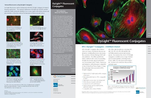
Immunofluorescence using <strong>DyLight</strong> <strong>Conjugates</strong>See bright fluorescence and low background with <strong>KPL</strong> <strong>DyLight</strong> conjugates in immunohistologyapplications. The winning combination of <strong>DyLight</strong> dyes and <strong>KPL</strong> purifiedantibodies enables multicolor labeling of two or more targets with similar intensity andphotostability to Alexa dyes without the limitations of fluorescein and CyDyes.<strong>DyLight</strong> TM <strong>Fluorescent</strong><strong>Conjugates</strong><strong>KPL</strong> Antibodies-What Sets Them ApartMouse primary cortical neuronsMCx WCS stained with <strong>DyLight</strong> 549(red). Synaptophysin stained with<strong>DyLight</strong> 488 (green).Rat hippocampal neuronsWCS stained with; <strong>DyLight</strong> 488 (green).MAP2 stained with <strong>DyLight</strong> 549 (red).In 1979, <strong>KPL</strong> pioneered the productionof large-scale affinity purification and wasthe first company to commercialize affinitypurified secondary antibodies. Rigorousstandards throughout the antibodyproduction process make our antibodiesstandout in the marketplace. Manymanufacturers cut corners by beginningwith inferior serum and extracting theuseful antibody towards the end of theirprocess. <strong>KPL</strong> spends considerable effortdeveloping and purifying its own immunogenformulation to generate the antiserum,because pure immunogen resultsin a more potent and specific antibodyprior to any purification steps.HMVEC-L primary endothelial cellsF-actin detected with DY554-Phalloidin(rendered green). Microtubules stainedwith <strong>DyLight</strong> 649 (red). Nuclei detectedwith DAPI (blue).A549 cells Cytokeratin stainedwith <strong>DyLight</strong> 680 (pseudocoloredwhite). Lamin A stained with<strong>DyLight</strong> 549 (pseudocolored red).Further, ISO 9001:2008-certified qualityprocedures are carried out at morethan six different stages of the antibodyproduction cycle, and material that doesnot meet our high standards for potencyand cross-reactivity is rejected. Ourlate stage purification process has beencontinually refined since 1979 and relieson a one of a kind custom column resinwhich is uniquely suited to our antibodymanufacturing process.<strong>KPL</strong>’s <strong>DyLight</strong> TM <strong>Conjugates</strong> - A Brilliant Choice!<strong>KPL</strong>’s <strong>DyLight</strong> TM conjugates offer a brilliantchoice in a variety of multicolor detection applications,including fluorescence microscopy,flow cytometry, Western blotting, ELISA andarray platforms. Our affinity purified antibodiescombined with a series of outstanding<strong>DyLight</strong> dyes provide superior performanceover conventional CyDye TM fluors, fluoresceinand rhodamine, with performance comparableto that of Alexa Fluor ® dyes (Figure 1). Enjoythese advantages when you switch to <strong>KPL</strong>’s<strong>DyLight</strong> <strong>Conjugates</strong>:<strong>DyLight</strong> TM <strong>Fluorescent</strong> <strong>Conjugates</strong><strong>KPL</strong> offers eight <strong>DyLight</strong> dyes, including 405,488, 549, 594, 633, 649, 680 and 800 withwell-differentiated excitation and emissionspectra. Our extensive line of over 170 <strong>DyLight</strong>conjugates is available across a range of animalspecies immunoglobulin, including human,mouse, rabbit, rat, other species and streptavidin.See back cover to find out what sets <strong>KPL</strong>’santibodies apart.NIH 3T3 cells F-actin detectedwith DY554-Phalloidin (renderedgreen). Microtubules stained with<strong>DyLight</strong> 649 (red). Nuclei detectedwith DAPI (blue).Cells were probed with anti-lamin-Aand rabbit anti-cytokeratin 18. Detectedwith a mixture of <strong>DyLight</strong>405-labeled goat anti-mouse and<strong>DyLight</strong> 633-labeled goat anti-rabbitantibodies.Finally, our process of pooling antiserumfrom multiple animals tempers naturalserum variability, minimizing variancesfrom animal-to-animal. The result ismore standardized large-scale antibodylots with increased consistency. <strong>KPL</strong>’sexperience, innovative processes, andattention to detail result in high- performanceantibodies that are unique.Comparison of <strong>DyLight</strong> 649 and Alexa 647<strong>Conjugates</strong> in a Performance AssayCells were probed with anti-lamin-Aand rabbit anti-cytokeratin18. Detectedwith a mixture of <strong>DyLight</strong> 488-labeledgoat anti-mouse and <strong>DyLight</strong> 405-labeled goat anti-rabbit antibodies.Codetection of mouse anti-tubulinand rabbit anti-lamin B1using <strong>DyLight</strong> 594-labeled goatanti-mouse and <strong>DyLight</strong> 488-labeledgoat anti-rabbit antibodies.To order or for more information on <strong>KPL</strong>’s line of unlabeled and conjugatedaffinity purified antibodies, contact us at 800.638.3167/301.948.7755, fax301.948.0169 or visit www.kpl.com.ML356-05For research use only.<strong>DyLight</strong> is a trademark of Thermo Fisher Scientific Inc. and its subsidiaries. Cy and Cy Dye are trademarks of GE Healthcare.Alexa Fluor is a registered trademark of Invitrogen.©2009 <strong>KPL</strong>, Inc. All rights reserved.www.seracare.com800.676.1881508.244.6400www.kpl.com800.638.3167301.948.7755ISO 9001 Registered• High sensitivity, bright signal enable conjugateconservation• More photostable than Cy, FITC; comparableto Alexa• High signal-to-noise ratios; low background• Wide selection of specific antibodies• Consistent, lot-to-lot performance• Narrow emission spectra enable specific,multicolor analysis• Compatible with a variety of buffers.Antigen Concentration (ng/mL)Figure 1. <strong>KPL</strong> <strong>DyLight</strong> 649 conjugates demonstrate comparablefluorescense intensity and photostability to Alexa 647 conjugatesin a FLISA.Image above: HMVEC-L primary endothelial cells. F-actin detected with DY554-Phalloidin (rendered green). Microtubules detected with anti-tubulinantibody and <strong>DyLight</strong> 649 conjugated goat anti-mouse IgG (red). Nuclei detected with DAPI (blue)
<strong>DyLight</strong> TM <strong>Fluorescent</strong><strong>Conjugates</strong>Near Infrared Fluorophores <strong>DyLight</strong> 680 and 800<strong>Conjugates</strong> Offer Sensitive Multicolor Imaging inWestern Blotting<strong>DyLight</strong> Conjugated Affinity PurifiedAntibodies and Streptavidin<strong>KPL</strong> <strong>DyLight</strong> conjugates provide a choice of outstanding fluorescentconjugates with absorption spectra ranging from 405nm to 800 nm. Their emission profiles correspond to those ofother commonly used fluorophores such as Alexa, FITC andthe Cy dyes. Narrow, defined emission spectra enable the useof multiple fluorescence labeling and simultaneous identificationof several target molecules in the same sample. Light outputis comparable to IRDye and Alexa and more intense thanCy Dyes, FITC or TRITC. <strong>DyLight</strong> dyes demonstrate resistanceto photobleaching, resulting in excellent photostability as wellas high solubility in aqueous solutions across a range of pHvalues.Ordering InformationEmission <strong>DyLight</strong> Dye Ex/Em(nm) ReplacesBlueGreenYellowOrangeRedRedNear IRInfrarednm = nanometersIR = infrared<strong>DyLight</strong> 405 400/420 Alexa 405 andCascade Blue<strong>DyLight</strong> 488 493/518 Alexa 488 and FITC<strong>DyLight</strong> 549 550/568 Alexa 546, Alexa 555,Cy3, TRITC<strong>DyLight</strong> 594 593/618 Alexa 594, Texas Red<strong>DyLight</strong> 633 638/658 Alexa 633<strong>DyLight</strong> 649 646/674 Alexa 647, Cy5<strong>DyLight</strong> 680 682/715 Alexa 680, Cy5.5<strong>DyLight</strong> 800 770/794 IRDye 800Description <strong>DyLight</strong> 405 <strong>DyLight</strong> 488 <strong>DyLight</strong> 549 <strong>DyLight</strong> 594 <strong>DyLight</strong> 633 <strong>DyLight</strong> 649 <strong>DyLight</strong> 680 <strong>DyLight</strong> 800Anti-Mouse IgG (), HSA 072-03-18-02 072-04-18-02 072-05-18-02Anti-Mouse IgG (HL), HSA 072-08-18-06 072-03-18-06 072-04-18-06 072-09-18-06 072-10-18-06 072-05-18-06 072-06-18-06 072-07-18-06Anti-Mouse IgG (HL), HSA, 0.1 mg 042-08-18-06 042-03-18-06 042-04-18-06 042-09-18-06 042-10-18-06 042-05-18-06 042-06-18-06 042-07-18-06Anti-Mouse IgG (HL), RbSA, HSA 072-03-18-18 072-04-18-18 072-05-18-18 072-06-18-18 072-07-18-18Anti-Mouse IgM (), HSA 072-03-18-03 072-04-18-03 072-05-18-03Anti-Mouse IgG+IgM (HL), HSA 072-03-18-09 072-04-18-09 072-05-18-09F(ab’) 2 Anti-Mouse IgG (), HSA 202-03-18-02 202-04-18-02 202-05-18-02F(ab’) 2 Anti-Mouse IgG (H+L), HSA 202-08-18-06 202-03-18-06 202-04-18-06 202-09-18-06 202-10-18-06 202-05-18-06 202-06-18-06 202-07-18-06Anti-Human IgG (H+L) 072-08-10-06 072-03-10-06 072-04-10-06 072-09-10-06 072-10-10-06 072-05-10-06 072-06-10-06 072-07-10-06Anti-Human IgG (H+L), 0.1 mg 042-08-10-06 042-03-10-06 042-04-10-06 042-09-10-06 042-10-10-06 042-05-10-06 042-06-10-06 042-07-10-06Anti-Human IgG () 072-03-10-02 072-04-10-02 072-05-10-02Anti-Human IgM () 072-03-10-03 072-04-10-03 072-05-10-03F(ab’) 2 Anti-Human IgG () 202-03-10-02 202-04-10-02 202-05-10-02F(ab’) 2 Anti-Human IgM () 202-03-10-03 202-04-10-03 202-05-10-03F(ab’) 2 Anti-Human IgG (H+L) 202-08-10-06 202-03-10-06 202-04-10-06 202-09-10-06 202-10-10-06 202-05-10-06 202-06-10-06 202-07-10-06Anti-Rabbit IgG (HL) 072-08-15-06 072-03-15-06 072-04-15-06 072-09-15-06 072-10-15-06 072-05-15-06 072-06-15-06 072-07-15-06Anti-Rabbit IgG (HL), 0.1 mg 042-08-15-06 042-03-15-06 042-04-15-06 042-09-15-06 042-10-15-06 042-05-15-06 042-06-15-06 042-07-15-06Anti-Rabbit IgG (HL), HSA 072-03-15-16 072-04-15-16 072-05-15-16 072-06-15-16 072-07-15-16F(ab’) 2 Anti-Rabbit IgG (H+L), HSA 202-08-15-16 202-03-15-16 202-04-15-16 202-09-15-16 202-10-15-16 202-05-15-16 202-06-15-16 202-07-15-16Anti-Rat IgG (HL) 072-08-16-06 072-03-16-06 072-04-16-06 072-09-16-06 072-10-16-06 072-05-16-06 072-06-16-06 072-07-16-06Anti-Rat IgG (HL), 0.1 mg 042-03-16-06Anti-Guinea Pig IgG (H+L) 072-03-17-06 072-04-17-06 072-05-17-06 072-06-17-06 072-07-17-06Anti-Chicken IgG (H+L) 072-08-24-06 072-03-24-06 072-04-24-06 072-09-24-06 072-10-24-06 072-05-24-06 072-06-24-06 072-07-24-06Anti-Dog IgG (H+L) 072-03-19-06 072-04-19-06 072-05-19-06 072-06-19-06 072-07-19-06Anti-Goat IgG (H+L) 072-08-13-06 072-03-13-06 072-04-13-06 072-09-13-06 072-10-13-06 072-05-13-06 072-06-13-06 072-07-13-06Anti-Horse IgG (H+L) 072-03-21-06 072-04-21-06 072-05-21-06 072-06-21-06 072-07-21-06Rabbit Anti-Sheep IgG (H+L) 072-03-23-06 072-04-23-06 072-05-23-06 072-06-23-06 072-07-23-06Anti-Swine IgG (H+L) 072-03-14-06 072-04-14-06 072-05-14-06 072-06-14-06 072-07-14-06Streptavidin 072-08-30-00 072-03-30-00 072-04-30-00 072-09-30-00 072-10-30-00 072-05-30-00 072-06-30-00 072-07-30-00Streptavidin, 0.1 mg 042-08-30-00 042-03-30-00 042-04-30-00 042-09-30-00 042-10-30-00 042-05-30-00 042-06-30-00 042-07-30-00<strong>DyLight</strong> Dyes Emission SpectraHSA = human serum adsorbedRbSA = rabbit serum adsorbed<strong>DyLight</strong> antibody conjugates are made in goat exceptanti-goat and anti-sheep antibodies are made in rabbit.Supplied in 1.0 mg lyophilized form except select 0.1mg sizes.<strong>DyLight</strong> 680 and 800 dyes emit in the near infrared andinfrared ranges of the light spectrum respectively andare ideal for multicolor protein detection in Westernblotting. Unlike fluorescent conjugates that emit in thevisible range, <strong>DyLight</strong> 680 and 800 conjugates provide aunique set of advantages:• Secondary antibodies with defined specificity andsensitivity• Brighter signal than visible fluorescence• Virtually no background autofluorescence frommembranes or most biological specimensEffective Alternative to Chemiluminescence• Broader dynamic range than chemiluminescence• Quantitation accuracy superior to traditionalmethods• Easy to assay multiple proteins simultaneously onone Western blot• Cost-effective - eliminates need for chemiluminescencesubstrates, film and darkroom• Clean results - no bleeding from consecutive lanesNear Infrared <strong>Fluorescent</strong> Imaging with SecondaryAntibodies labeled with <strong>DyLight</strong> 680 and 800Figure 2. <strong>DyLight</strong> 680 anti-mouse IgG and <strong>DyLight</strong>800 anti-rabbit IgG secondary antibody conjugatesprovide low background and high signal in two-colorWestern blot detection of tubulin and TNF. Membranewas imaged with the LI-COR Odyssey InfraredImaging System.Power Your Immunoassay 1.800.638.3167 www.kpl.com